Observational Study
. 2021 Nov 1;6(11):1257-1266.
doi: 10.1001/jamacardio.2021.3055.

Association of Statin Treatment With Progression of Coronary Atherosclerotic Plaque Composition

Affiliations
Observational Study

Association of Statin Treatment With Progression of Coronary Atherosclerotic Plaque Composition

Alexander R van Rosendael et al. JAMA Cardiol. .

Abstract

Importance: The density of atherosclerotic plaque forms the basis for categorizing calcified and noncalcified morphology of plaques.

Objective: To assess whether alterations in plaque across a range of density measurements provide a more detailed understanding of atherosclerotic disease progression.

Design, setting, and participants: This cohort study enrolled 857 patients who underwent serial coronary computed tomography angiography 2 or more years apart and had quantitative measurements of coronary plaques throughout the entire coronary artery tree. The study was conducted from 2013 to 2016 at 13 sites in 7 countries.

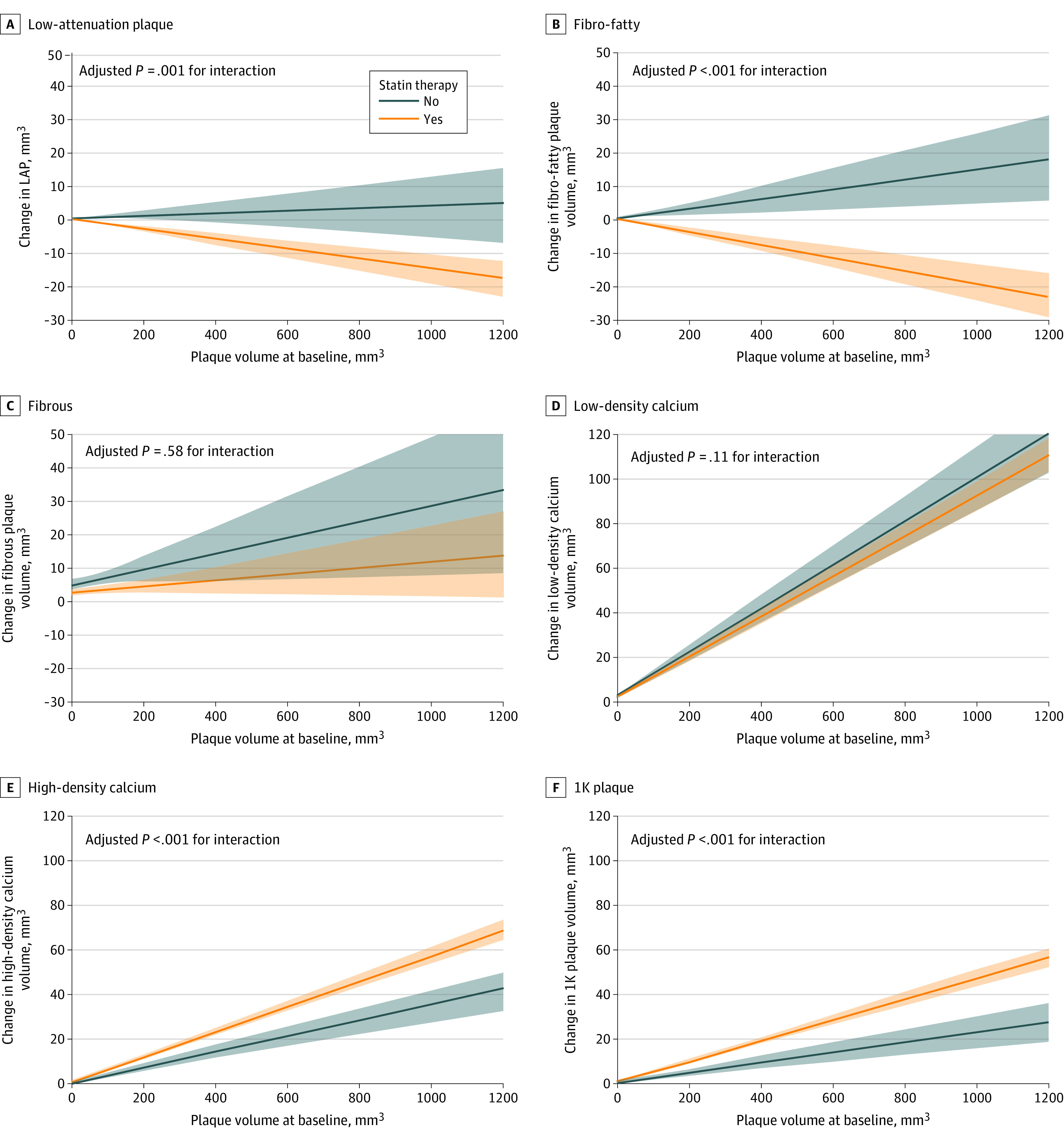
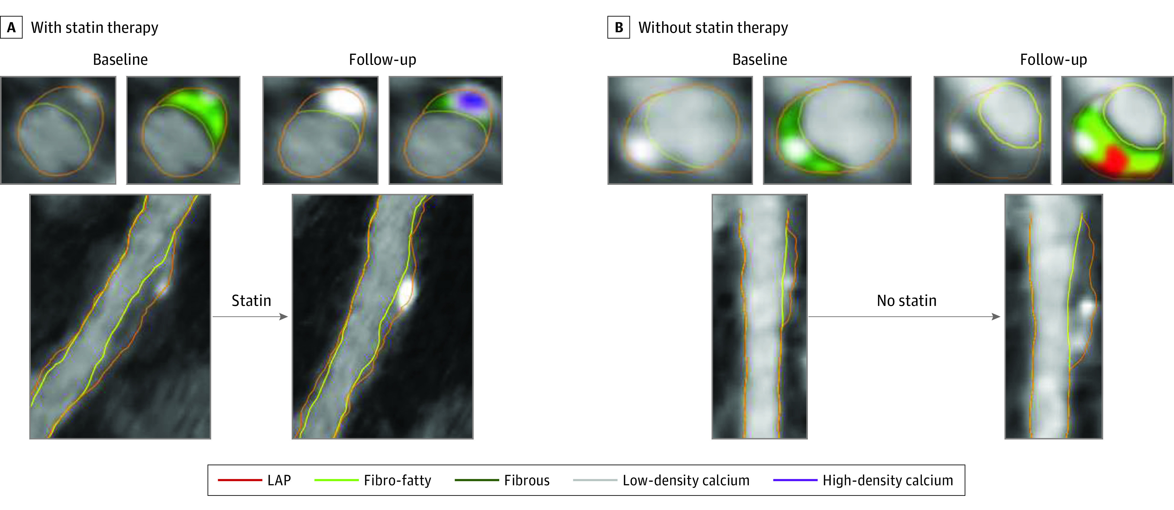
Main outcomes and measures: The main outcome was progression of plaque composition of individual coronary plaques. Six plaque composition types were defined on a voxel-level basis according to the plaque attenuation (expressed in Hounsfield units [HU]): low attenuation (-30 to 75 HU), fibro-fatty (76-130 HU), fibrous (131-350 HU), low-density calcium (351-700 HU), high-density calcium (701-1000 HU), and 1K (>1000 HU). The progression rates of these 6 compositional plaque types were evaluated according to the interaction between statin use and baseline plaque volume, adjusted for risk factors and time interval between scans. Plaque progression was also examined based on baseline calcium density. Analysis was performed among lesions matched at baseline and follow-up. Data analyses were conducted from August 2019 through March 2020.

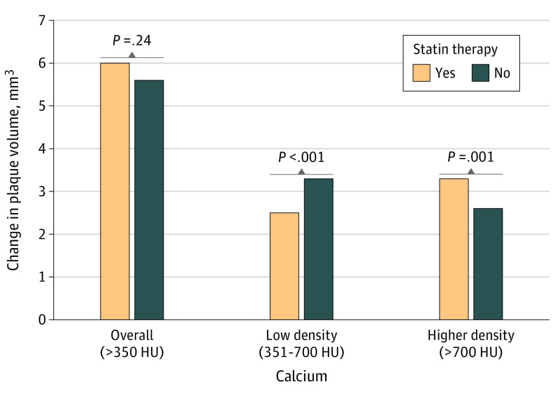
Results: In total, 2458 coronary lesions in 857 patients (mean [SD] age, 62.1 [8.7] years; 540 [63.0%] men; 548 [63.9%] received statin therapy) were included. Untreated coronary lesions increased in volume over time for all 6 compositional types. Statin therapy was associated with volume decreases in low-attenuation plaque (β, -0.02; 95% CI, -0.03 to -0.01; P = .001) and fibro-fatty plaque (β, -0.03; 95% CI, -0.04 to -0.02; P < .001) and greater progression of high-density calcium plaque (β, 0.02; 95% CI, 0.01-0.03; P < .001) and 1K plaque (β, 0.02; 95% CI, 0.01-0.03; P < .001). When analyses were restricted to lesions without low-attenuation plaque or fibro-fatty plaque at baseline, statin therapy was not associated with a change in overall calcified plaque volume (β, -0.03; 95% CI, -0.08 to 0.02; P = .24) but was associated with a transformation toward more dense calcium. Interaction analysis between baseline plaque volume and calcium density showed that more dense coronary calcium was associated with less plaque progression.

Conclusions and relevance: The results suggest an association of statin use with greater rates of transformation of coronary atherosclerosis toward high-density calcium. A pattern of slower overall plaque progression was observed with increasing density. All findings support the concept of reduced atherosclerotic risk with increased densification of calcium.


Conflict of interest statement

Conflict of Interest Disclosures: Dr van Rosendael reported receiving grants from the Leading Foreign Research Institute Recruitment Program through the National Research Foundation of Korea funded by the Ministry of Science and ICT during the conduct of the study. Dr Budoff reported receiving grants from General Electric outside the submitted work. Dr Chinnaiyan reported receiving grants from HeartFlow outside the submitted work and being on the medical advisory board of HeartFlow. Dr Leipsic reported receiving grants from Abbott, Boston Scientific, Core Lab, Edwards LifeSciences, and Medtronic; receiving personal fees from HeartFlow and from Circle CVI during the conduct of the study; and receiving personal fees from Philips and from GE Healthcare outside the submitted work. Dr Virmani reported receiving grants from 480 Biomedical, 4C Medical, 4Tech Inc, Accumedical, Alivas, Amgen, Biosensors, Cardiac Implants, Canon U.S.A., Claret Medical, Concept Medical, Cordis, DuNing Inc, Endotronix, Emboline, Endotronix, Envision Scientific, Gateway, Leducq Foundation, Lifetech, Limflo, MedAlliance, Mercator, Mitra assist, Microport Medical, Microvention, Mitraalign, the National Institutes of Health, NIPRO, NAMSA, Nanova, Neovasc, Phenox, Novogate, Occulotech, Protembis, Profusa, Shockwave, Qool, Senseonics, Symic, Spectranetics, and Vesper; and receiving personal fees from Abbott Vascular, Boston Scientific, Celonova, Cook Medical, CSI, Edwards LifeSciences, Lutonix/Bard, Medtronic, OrbusNeich Medical, ReCor Medical, Sinomed Medical, Surmodics, Terumo, W.L. Gore, and Xeltis outside the submitted work. Dr Samady reported receiving grants from Medtronic; receiving personal fees from Abbott and from Philips during the conduct of the study; and being a co-founder and equity holder in Covanos outside the submitted work. Dr Blankstein reported receiving grants from Amgen and from Astellas Inc; and receiving personal fees from Amgen outside the submitted work. Dr Min reported having an equity interest and being an employee of Cleerly Inc; and being a medical advisory board member of Arineta during the conduct of the study. Dr Lin reported receiving grants from GE Healthcare outside the submitted work. Dr Shaw reported having an equity interest in Cleerly Inc and being a scientific advisory board member for Covanos Inc. No other disclosures were reported.

Figures

Figure 1.
Figure 1.. Compositional Plaque Changes According to Baseline Plaque Volume and Statin Use
Estimated changes in low-attenuation plaque (LAP), fibro-fatty plaque, fibrous plaque, low-density calcium, high-density calcium, and 1K plaque are presented according to baseline plaque volume and statin use. The estimated changes (bold lines) and 95% CIs (shaded areas) are derived from a generalized linear model, including baseline plaque volume, statin use, and the interaction term. The P values for interaction are derived from Table 2. Without statin therapy, increasing trends for all noncalcified and calcified density subgroups are observed, whereas with statin therapy, significant decreases in LAP and fibro-fatty plaque and larger increases in high-density calcium and 1K plaque are observed.
Figure 2.
Figure 2.. Calcium Densification With Statin Therapy
Data are restricted to lesions without low-attenuation and fibro-fatty plaque. Estimated changes in overall calcium, low-density calcium, and higher-density calcium according to statin therapy. Estimated changes in plaque volume are shown for the mean baseline plaque volume, the mean of other continuous variables, and the geometric mean of categorical variables. P values represent the interaction between plaque volume and statin use, adjusted for age, sex, diabetes, hypertension, smoking status, body mass index, and computed tomography interval, derived from linear mixed models. HU indicates Hounsfield units.
Figure 3.
Figure 3.. Coronary Atherosclerosis Progression With or Without Statin Therapy
Progression of plaque over time in coronary lesions of patients treated with or without statins. For each of the 4 time points, 1 multiplanar reconstruction is provided with 2 cross-sectional views obtained at the same site at baseline and follow-up. A, Statin therapy is associated with the reduction of fibro-fatty (light green) plaque together with densification of the calcification (from low-density calcium [gray] to high-density calcium [purple]). B, Plaque expansion of both noncalcified and calcified plaque is observed. LAP indicates low-attenuation plaque.

References

    1. Min JK, Shaw LJ, Devereux RB, et al. . Prognostic value of multidetector coronary computed tomographic angiography for prediction of all-cause mortality. J Am Coll Cardiol. 2007;50(12):1161-1170. doi:10.1016/j.jacc.2007.03.067 - DOI - PubMed
    1. Arbab-Zadeh A, Fuster V. The risk continuum of atherosclerosis and its implications for defining CHD by coronary angiography. J Am Coll Cardiol. 2016;68(22):2467-2478. doi:10.1016/j.jacc.2016.08.069 - DOI - PubMed
    1. Burke AP, Farb A, Malcom GT, Liang YH, Smialek J, Virmani R. Coronary risk factors and plaque morphology in men with coronary disease who died suddenly. N Engl J Med. 1997;336(18):1276-1282. doi:10.1056/NEJM199705013361802 - DOI - PubMed
    1. Puri R, Nicholls SJ, Shao M, et al. . Impact of statins on serial coronary calcification during atheroma progression and regression. J Am Coll Cardiol. 2015;65(13):1273-1282. doi:10.1016/j.jacc.2015.01.036 - DOI - PubMed
    1. Lee SE, Chang HJ, Sung JM, et al. . Effects of statins on coronary atherosclerotic plaques: the PARADIGM study. JACC Cardiovasc Imaging. 2018;11(10):1475-1484. doi:10.1016/j.jcmg.2018.04.015 - DOI - PubMed

Publication types

Substances