. 2022 Jul;84(1):182-197.
doi: 10.1007/s00248-021-01809-5. Epub 2021 Aug 18.

FISH-TAMB, a Fixation-Free mRNA Fluorescent Labeling Technique to Target Transcriptionally Active Members in Microbial Communities

Affiliations

FISH-TAMB, a Fixation-Free mRNA Fluorescent Labeling Technique to Target Transcriptionally Active Members in Microbial Communities

Rachel L Harris et al. Microb Ecol. 2022 Jul.

Abstract

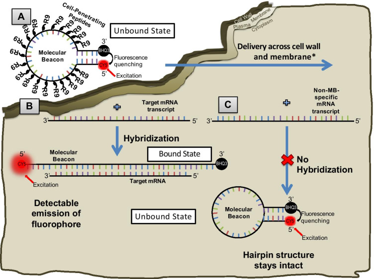
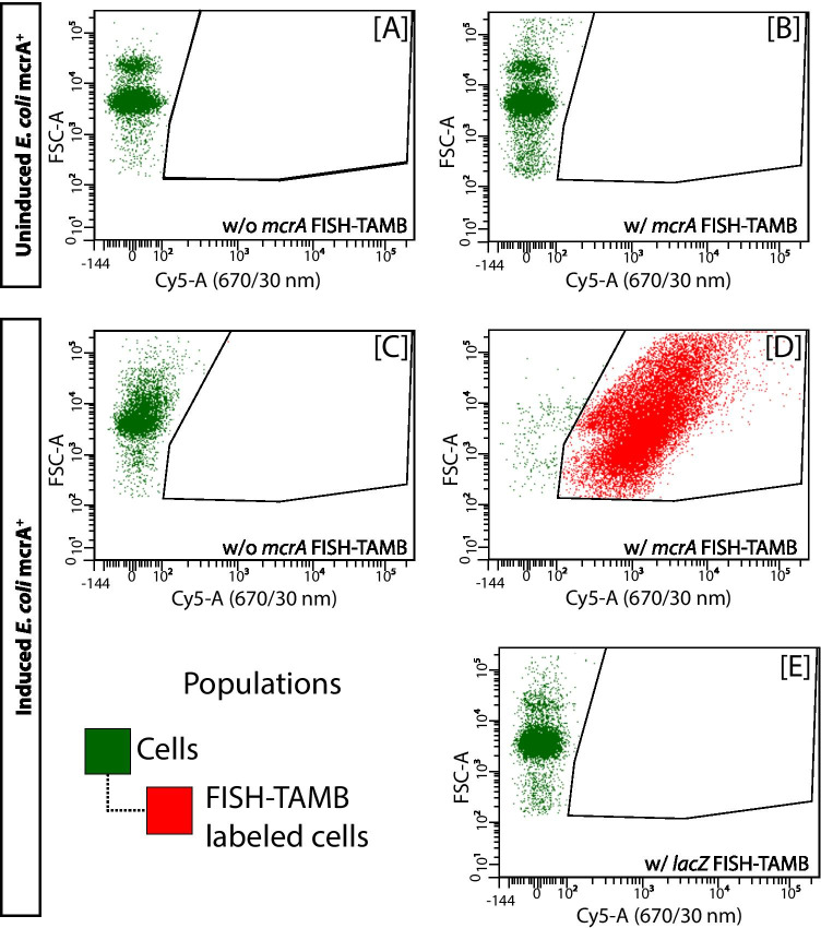
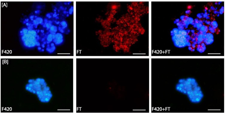
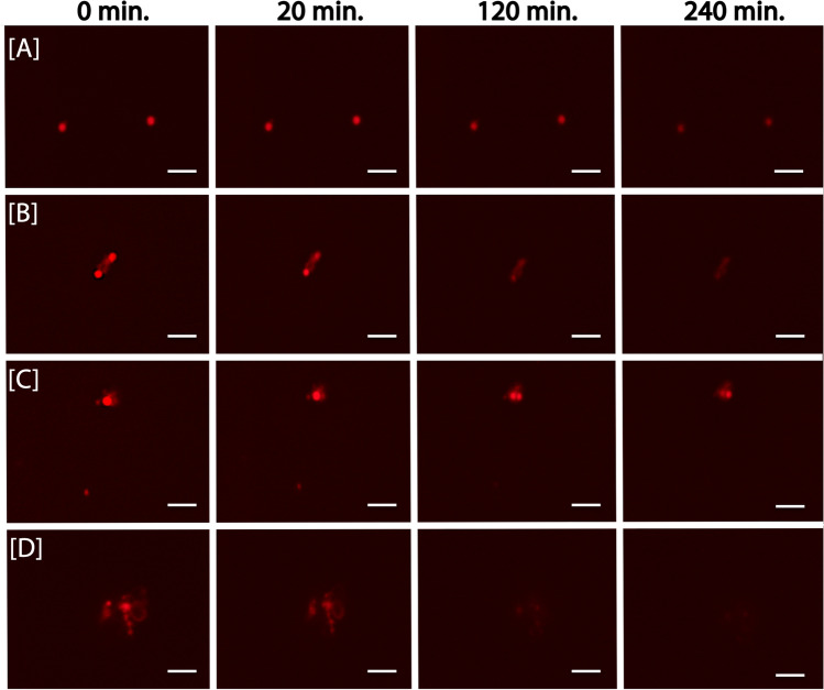
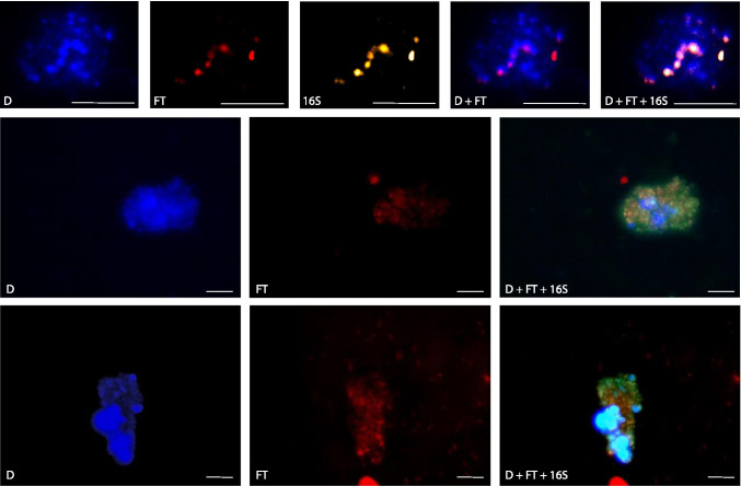
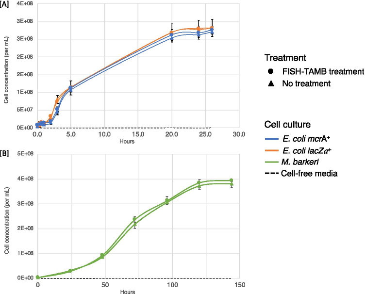
Keystone species or ecological engineers are vital to the health of an ecosystem; however, often, their low abundance or biomass present challenges for their discovery, identification, visualization and selection. We report the development of fluorescent in situ hybridization of transcript-annealing molecular beacons (FISH-TAMB), a fixation-free protocol that is applicable to archaea and bacteria. The FISH-TAMB method differs from existing FISH methods by the absence of fixatives or surfactants in buffers, the fast hybridization time of as short as 15 min at target cells' growth temperature, and the omission of washing steps. Polyarginine cell-penetrating peptides are employed to deliver molecular beacons (MBs) across prokaryotic cell walls and membranes, fluorescently labeling cells when MBs hybridize to target mRNA sequences. Here, the detailed protocol of the preparation and application of FISH-TAMB is presented. To demonstrate FISH-TAMB's ability to label intracellular mRNA targets, differentiate transcriptional states, detect active and rare taxa, and keep cell viability, labeling experiments were performed that targeted the messenger RNA (mRNA) of methyl-coenzyme M reductase A (mcrA) expressed in (1) Escherichia coli containing a plasmid with a partial mcrA gene of the methanogen Methanosarcina barkeri (E. coli mcrA+); (2) M. barkeri; and (3) an anaerobic methanotrophic (ANME) enrichment from a deep continental borehole. Although FISH-TAMB was initially envisioned for mRNA of any functional gene of interest without a requirement of prior knowledge of 16S ribosomal RNA (rRNA)-based taxonomy, FISH-TAMB has the potential for multiplexing and going beyond mRNA and thus is a versatile addition to the molecular ecologist's toolkit, with potentially widespread application in the field of environmental microbiology.

Keywords: ANMEs; Cell-penetrating peptides; FISH; Methanogens; Molecular beacons; mRNA.


Conflict of interest statement

The authors declare no competing interests.

Figures

Fig. 1
Fig. 1
FISH-TAMB probe conformation and hybridization to encountered messenger RNAs. A An oligomer comprised of a 24 base-long complementary mcrA mRNA sequence is flanked by 5 reverse complement nucleotides to form a molecular beacon (MB) loop and stem structure. Cell-penetrating peptides (CPPs) comprising 9 arginine sequences (R9) are non-covalently bound to the MB sequence and are responsible for its delivery across the cell wall and plasma membrane. B Fluorescence of Cy5 fluorophore covalently bound to the 5′ end of the MB sequence remains quenched by BHQ3 bound to the 3′ terminus until the MB hybridizes to a target transcript sequence. Hybridization results in the linearization of the MB, subsequently unquenching Cy5 from BHQ3, allowing the fluorophore’s emission upon excitation by a source in the red bandwidth of the visible light spectrum. C If the MB encounters an mRNA transcript that is not its intended target, it will retain its hairpin conformation, and fluorescence of Cy5 will remain quenched by BHQ3. Images not to scale
Fig. 2
Fig. 2
Flow cytometry data of FISH-TAMB targeting mRNA in E. coli grown in the absence (uninduced) or presence (induced) of IPTG, which triggers the transcription of the lac operon containing this gene. Events with optical properties similar to as E. coli cells are gated in green as cells. FISH-TAMB targeting mcrA mRNA in induced E. coli mcrA+ is indicated by the population gated in red. Cy5 was excited at 640 nm and emitted fluorescence collected via 670/30 nm bandpass filter. FSC-A stands for the area of forward-scattered density. A Uninduced E. coli mcrA+ without FISH-TAMB treatment. B Uninduced E. coli mcrA+ treated with FISH-TAMB probes targeting mcrA mRNA. C IPTG-induced E. coli mcrA+ without FISH-TAMB treatment. D IPTG-induced E. coli mcrA+ treated with FISH-TAMB probes targeting mcrA mRNA. E IPTG-induced E. coli mcrA+ treated with FISH-TAMB probes targeting lacZ α mRNA
Fig. 3
Fig. 3
FISH-TAMB sensitivity to mcrA transcription in Methanosarcina barkeri during A exponential phase, and B following overnight exposure to air. F420, F420 autofluorescence; FT, Cy5 fluorescence from FISH-TAMB labeling; F420 + FT, composite image of F420 and FT micrographs. Scale bar 10 µm
Fig. 4
Fig. 4
Snapshots of time-lapse microscopy of ANME enrichment culture labeled by mcrA FISH-TAMB probes. BE326 BH2 ANME enrichments were incubated anaerobically with 1 µM FISH-TAMB probes targeting mcrA mRNA and subsequently imaged via spinning-disk fluorescence confocal microscopy. Micrographs were snapped every minute for 14 h with an exposure of 100 ms. Composite micrographs of brightfield and Cy5 channel represent the first 4-h observation of A single cells; B and C physically associated cells; and D cell aggregate. Scale bar 5 µm
Fig. 5
Fig. 5
Co-labeling of active ANME-2 cells in ANME enrichment cultures by FISH-TAMB and 16S rRNA FISH. D, DAPI; FT, Cy5 fluorescence from FISH-TAMB labeling, 16S, Atto 565 fluorescence from 16S rRNA FISH labeling; D + FT + 16S, composite of D, FT, and 16S micrographs. Scale bar 10 µm
Fig. 6
Fig. 6
FISH-TAMB viability assessment by growth curve analysis. Pure cultures of A E. coli mcrA+ and E. coli lac+ and B M. barkeri (~ 106 cells mL−1) were incubated with 1 µM FISH-TAMB probes and inoculated into their respective growth media

References

    1. Banerjee S, Schlaeppi K, van der Heijden MGA. Keystone taxa as drivers of microbiome structure and functioning. Nat Rev Microbiol. 2018;16:567–576. doi: 10.1038/s41579-018-0024-1. - DOI - PubMed
    1. Cottee-Jones HEW, Whittaker RJ (2012) perspective: The keystone species concept: a critical appraisal. Front Biogeogr 4:117–127. 10.21425/f5fbg12533
    1. Kietäväinen R, Purkamo L. The origin, source, and cycling of methane in deep crystalline rock biosphere. Front Microbiol. 2015;6:725. doi: 10.3389/fmicb.2015.00725. - DOI - PMC - PubMed
    1. Chivian D, Brodie EL, Alm EJ, et al. Environmental genomics reveals a single-species ecosystem deep within Earth. Science. 2008;322:275–278. doi: 10.1126/science.1155495. - DOI - PubMed
    1. Onstott TC. Deep life: The hunt for the hidden biology of Earth, Mars, and beyond. Princeton: Princeton University Press; 2017.

LinkOut - more resources