. 2021 Oct 13;95(21):e0081721.
doi: 10.1128/JVI.00817-21. Epub 2021 Aug 18.

Redondovirus Diversity and Evolution on Global, Individual, and Molecular Scales

Affiliations

Redondovirus Diversity and Evolution on Global, Individual, and Molecular Scales

Louis J Taylor et al. J Virol. .

Abstract

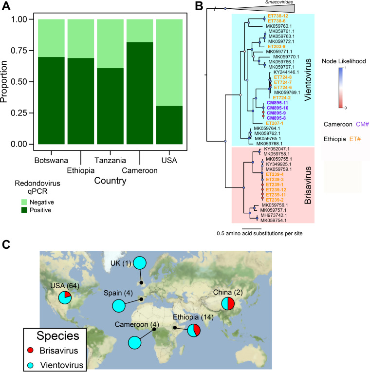
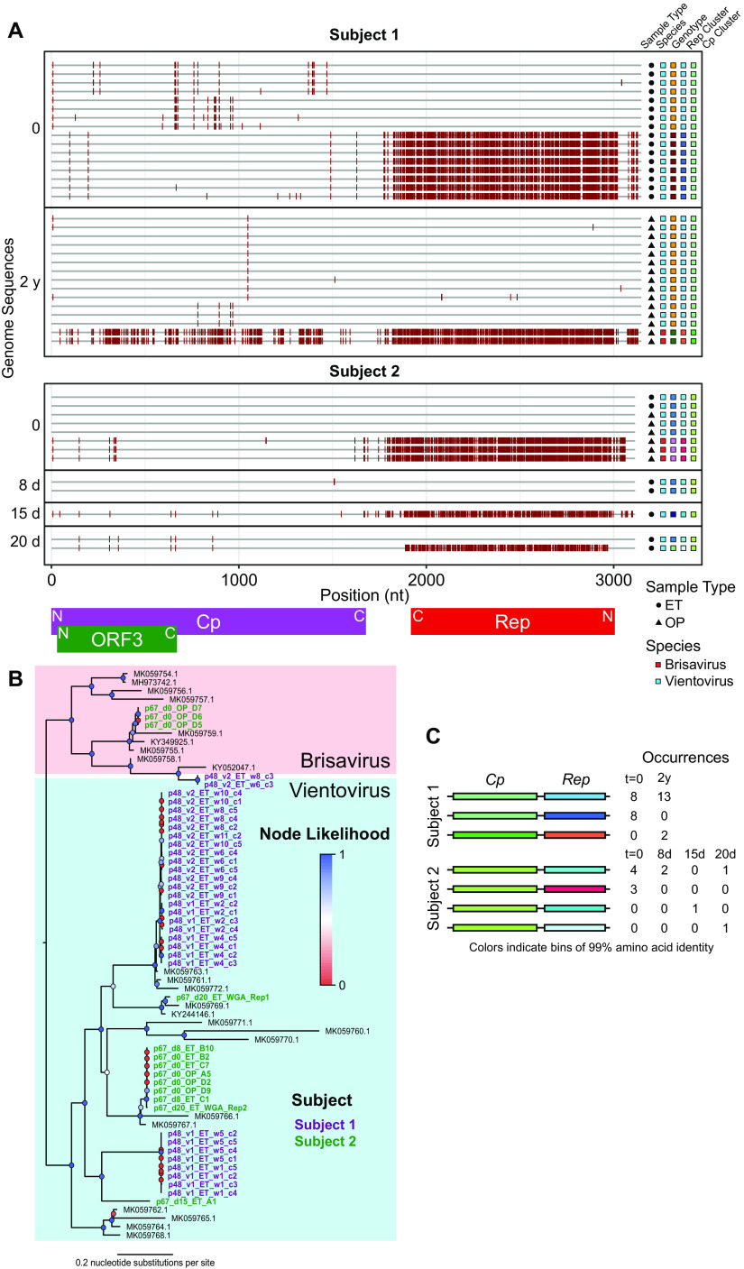
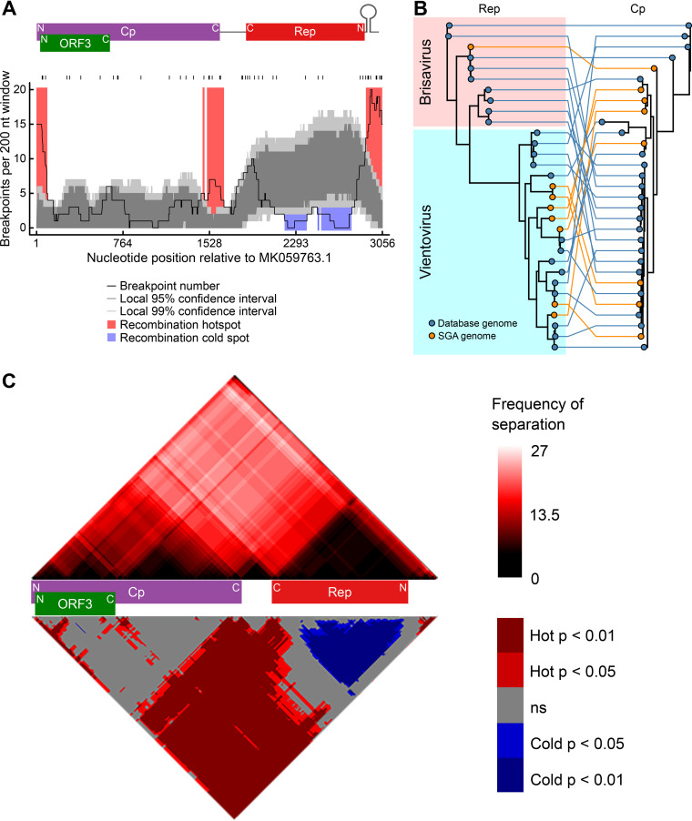
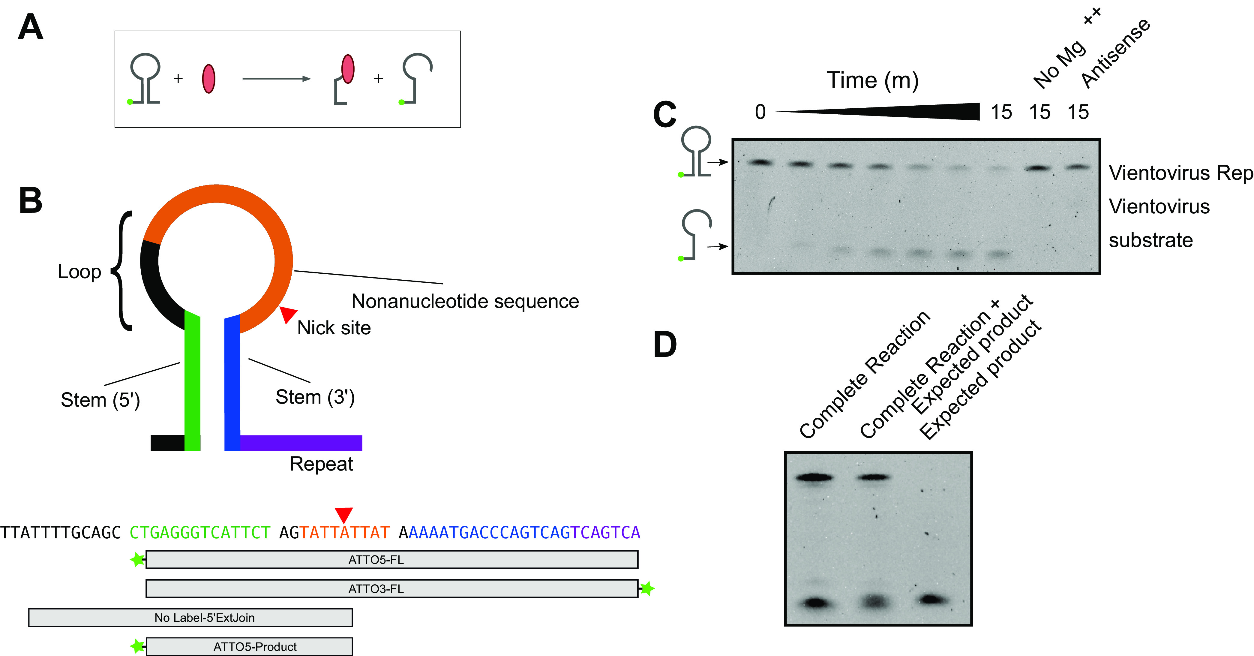
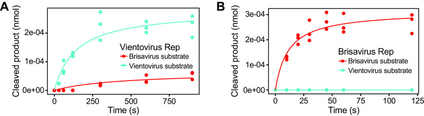
Redondoviridae is a newly established family of circular Rep-encoding single-stranded (CRESS) DNA viruses found in the human ororespiratory tract. Redondoviruses were previously found in ∼15% of respiratory specimens from U.S. urban subjects; levels were elevated in individuals with periodontitis or critical illness. Here, we report higher redondovirus prevalence in saliva samples: four rural African populations showed 61 to 82% prevalence, and an urban U.S. population showed 32% prevalence. Longitudinal, limiting-dilution single-genome sequencing revealed diverse strains of both redondovirus species (Brisavirus and Vientovirus) in single individuals, persistence over time, and evidence of intergenomic recombination. Computational analysis of viral genomes identified a recombination hot spot associated with a conserved potential DNA stem-loop structure. To assess the possible role of this site in recombination, we carried out in vitro studies which showed that this potential stem-loop was cleaved by the virus-encoded Rep protein. In addition, in reconstructed reactions, a Rep-DNA covalent intermediate was shown to mediate DNA strand transfer at this site. Thus, redondoviruses are highly prevalent in humans, found in individuals on multiple continents, heterogeneous even within individuals and encode a Rep protein implicated in facilitating recombination. IMPORTANCERedondoviridae is a recently established family of DNA viruses predominantly found in the human respiratory tract and associated with multiple clinical conditions. In this study, we found high redondovirus prevalence in saliva from urban North American individuals and nonindustrialized African populations in Botswana, Cameroon, Ethiopia, and Tanzania. Individuals on both continents harbored both known redondovirus species. Global prevalence of both species suggests that redondoviruses have long been associated with humans but have remained undetected until recently due to their divergent genomes. By sequencing single redondovirus genomes in longitudinally sampled humans, we found that redondoviruses persisted over time within subjects and likely evolve by recombination. The Rep protein encoded by redondoviruses catalyzes multiple reactions in vitro, consistent with a role in mediating DNA replication and recombination. In summary, we identify high redondovirus prevalence in humans across multiple continents, longitudinal heterogeneity and persistence, and potential mechanisms of redondovirus evolution by recombination.

Keywords: CRESS viruses; Redondoviridae; brisavirus; evolution; genetic recombination; redondovirus; rep protein; vientovirus.


Figures

FIG 1
FIG 1
Redondoviruses are globally distributed and highly prevalent in multiple African countries. (A) DNA extracted from saliva samples collected from subjects in different countries was subjected to redondovirus-selective whole-genome amplification, followed by qPCR. The proportion of samples positive by qPCR for each country is shown on the y axis. (B) Phylogenetic tree of single-genome-derived redondovirus Rep protein amino acid sequences along with sequences from databases. Rep proteins from SGS genomes are marked as circles, with colors representing the country of origin (purple for Cameroon and yellow for Ethiopia), while database redondovirus Reps are shown as black squares. Colored circles at each node show branch likelihood as determined by the approximate-likelihood ratio test. Clades representing the two redondovirus species are marked by colors: Vientovirus in cyan and Brisavirus in pink. (C) Global distribution of redondovirus species. The fraction of sequenced genomes in each of the redondovirus species in a particular country is shown in the pie charts. The number in parentheses after the country name represents the total number of redondovirus genomes sequenced from subjects in that country.
FIG 2
FIG 2
Longitudinal analysis of within-subject redondovirus diversity. (A) Highlighter plot of redondovirus genome nucleotide sequences from each subject analyzed after limiting-dilution SGS. The x axis represents the genomic position in a multiple-sequence alignment. Each gray horizontal line represents a sequenced redondoviral genome. Sequences are grouped by subject and then by time point, shown to the left of the plot. Nucleotide positions in each isolate that do not match the global consensus sequence are indicated by a red tick. Approximate positions of redondovirus ORFs are displayed below the plot. N, amino-terminal coding region; C, carboxyl-terminal coding region. The shapes to the right of the plot indicate the sample type: endotracheal aspirate (ET) or oropharyngeal (OP). The colored boxes indicate, from left to right, the redondovirus species to which each genome belongs, the genotype (cluster at 99% genomic nucleotide sequence identity) to which each sequence belongs, and the cluster (at 99% amino acid sequence identity) to which the Rep and Cp proteins belong. (B) Phylogenetic tree of Rep amino acid sequences from SGS redondovirus genomes sequenced from longitudinally sampled subjects, along with database sequences. Sequences from subject 1 are shown in purple, sequences from subject 3 are in green, and database sequences are in black. Colored circles at each node indicate branch likelihood as indicated by the approximate likelihood ratio test. Colored boxes delineate the two redondovirus species, Vientovirus (cyan) and Brisavirus (pink). (C) Schematic showing pairings between unique redondovirus Rep and Cp clusters. Each cluster is indicated by a different color. The table to the right of the schematic indicates the number of genomes sequenced at each time point belonging to each Cp-Rep pair.
FIG 3
FIG 3
Recombination favors redondovirus intergenic regions and occurs most frequently near the predicted origin of replication. (A) Plot showing the number of inferred recombination breakpoints in a 200-nucleotide (nt) sliding window across the redondovirus genome. The black ticks above the plot indicate observed recombination breakpoints. The positions of the redondovirus open reading frames and the stem-loop sequence are shown above the plot. N, amino-terminal coding region; C, carboxyl-terminal coding region. The black line indicates the count of breakpoints per 200-nt sliding window. The dark and light gray shaded areas represent local 95% and 99% confidence thresholds, respectively, for recombination hot and cold spots. Recombination hot spots (areas with more recombinants than expected by chance) are shaded in red; recombination cold spots (areas with fewer recombination breakpoints than expected by chance) are shown in blue. (B) Tanglegram showing relationships between Rep and Cp phylogenies. A Rep amino acid phylogeny is shown on the left; Cp amino acid phylogeny is shown to the right. Rep and Cp proteins originating from the same virus are connected by colored lines. The color code indicates whether each viral sequence was from a database (blue) or our SGS (orange). (C) Cp and Rep coding sequences are frequently separated by recombination events. Recombination region count matrix showing the frequency (top) and P value (bottom) of a recombination event separating any two nucleotide positions. In the top matrix, the color intensity represents the number of times each pair of nucleotides was separated. The bottom matrix shows nucleotide pairs that are most (red) and least (blue) frequently separated. The positions of redondovirus ORFs are displayed between the matrices.
FIG 4
FIG 4
Redondovirus Rep cleaves at the DNA stem-loop recombination hot spot. (A) Reaction schematic. Rep is shown in red. Rep nicks a DNA stem-loop, which is shown fluorescently labeled on the DNA 5′ end (green ball). The cleavage reaction produces a shorter labeled oligonucleotide and a covalent Rep-DNA intermediate. (B) (Top) Diagram of the DNA stem-loop sequences and reaction substrates. The Rep nick site is indicated by a red triangle. (Bottom) Sequence of the Vientovirus FB stem-loop with boxes showing the regions included in each oligonucleotide. Fluorescent ATTO488 labels (Integrated DNA Technologies) are indicated by green stars. All sequences are shown from 5′ to 3′ (left to right). (C) Results of electrophoresis on a TBE-urea acrylamide gel showing fluorescent products after reacting Vientovirus FB Rep with the labeled stem-loop from Vientovirus FB. The expected products are diagrammed to the left of the gel. “No Mg++” indicates a complete reaction with magnesium omitted; “antisense” indicates a substrate that is antisense in sequence compared to the expected correct orientation of the stem-loop. (D) Specificity of cleavage by the Vientovirus Rep protein. The product of the complete reaction (left) was mixed with the hypothetical product (middle) to demonstrate coelectrophoresis. The hypothetical product alone is shown in the right lane.
FIG 5
FIG 5
Specificity of cleavage by Vientovirus and Brisavirus Rep proteins. Quantification of cleavage products produced in the presence of Vientovirus Rep (A) and Brisavirus Rep (B). Results with the two substrates are color coded (Vientovirus substrate in blue, Brisavirus substrate in red). Time is on the x axis, and the amount of cleavage product is on the y axis.
FIG 6
FIG 6
Redondovirus Rep forms a covalent DNA-protein intermediate and catalyzes re-joining to de novo-introduced DNA strands. (A) (Top) Schematic of Rep-DNA covalent intermediate formation. Rep nicks a 3′-end-labeled oligonucleotide, forming a fluorescently labeled protein-DNA covalent complex. (Bottom) Reaction products electrophoresed on an SDS-PAGE gel and visualized by fluorescence detection (left); purified Rep visualized by Coomassie staining (right). Locations of Rep and Rep-DNA are indicated on the far left. (B) (Top) Reaction schematic. Rep is mixed with 3′-end-labeled oligonucleotide and, after 2 min, chased with a 100× molar excess of longer, unlabeled substrate. Rep joining activity in trans results in the production of a longer, fluorescently labeled oligonucleotide species. (Bottom) The products of this reaction were electrophoresed on a TBE-urea acrylamide gel and then visualized by fluorescence detection.

References

    1. Cui L, Wu B, Zhu X, Guo X, Ge Y, Zhao K, Qi X, Shi Z, Zhu F, Sun L, Zhou M. 2017. Identification and genetic characterization of a novel circular single-stranded DNA virus in a human upper respiratory tract sample. Arch Virol 162:3305–3312. 10.1007/s00705-017-3481-3. - DOI - PubMed
    1. Abbas A, Taylor LJ, Dothard MI, Leiby JS, Fitzgerald AS, Khatib LA, Collman RG, Bushman FD. 2019. Redondoviridae, a family of small, circular DNA viruses of the human oro-respiratory tract associated with periodontitis and critical illness. Cell Host Microbe 25:719–729. 10.1016/j.chom.2019.04.001. - DOI - PMC - PubMed
    1. Abbas A, Taylor LJ, Collman RG, Bushman FD. 2020. ICTV virus taxonomy profile: Redondoviridae. J Gen Virol. 102:jgv001526. 10.1099/jgv.0.001526. - DOI - PMC - PubMed
    1. Spezia PG, Macera L, Mazzetti P, Curcio M, Biagini C, Sciandra I, Turriziani O, Lai M, Antonelli G, Pistello M, Maggi F. 2020. Redondovirus DNA in human respiratory samples. J Clin Virol 131:104586. 10.1016/j.jcv.2020.104586. - DOI - PMC - PubMed
    1. Rosario K, Duffy S, Breitbart M. 2012. A field guide to eukaryotic circular single-stranded DNA viruses: insights gained from metagenomics. Arch Virol 157:1851–1871. 10.1007/s00705-012-1391-y. - DOI - PubMed

Publication types

MeSH terms

LinkOut - more resources