. 2022 Mar;16(6):1290-1308.
doi: 10.1002/1878-0261.13084. Epub 2021 Aug 29.

A novel anti-c-Kit antibody-drug conjugate to treat wild-type and activating-mutant c-Kit-positive tumors

Affiliations

A novel anti-c-Kit antibody-drug conjugate to treat wild-type and activating-mutant c-Kit-positive tumors

Jin-Ock Kim et al. Mol Oncol. 2022 Mar.

Abstract

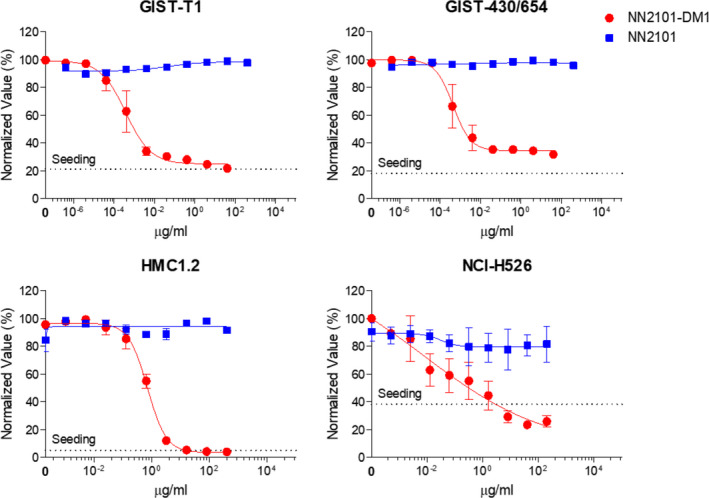
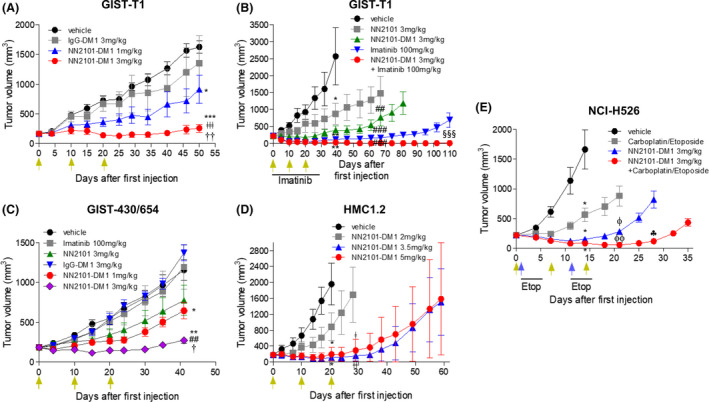
c-Kit overexpression and activating mutations, which are reported in various cancers, including gastrointestinal stromal tumor (GIST), small-cell lung cancer (SCLC), acute myeloid leukemia, acral melanoma, and systemic mastocytosis (SM), confer resistance to tyrosine kinase inhibitors (TKIs). To overcome TKI resistance, an anti-c-Kit antibody-drug conjugate was developed in this study to treat wild-type and mutant c-Kit-positive cancers. NN2101, a fully human IgG1, was conjugated to DM1, a microtubule inhibitor, through N-succinimidyl-4-(N-maleimidomethyl) cyclohexane-1-carboxylate (SMCC) (to give NN2101-DM1). The antitumor activity of NN2101-DM1 was evaluated in vitro and in vivo using various cancer cell lines. NN2101-DM1 exhibited potent growth-inhibitory activities against c-Kit-positive cancer cell lines. In a mouse xenograft model, NN2101-DM1 exhibited potent growth-inhibitory activities against imatinib-resistant GIST and SM cells. In addition, NN2101-DM1 exhibited a significantly higher anti-cancer effect than carboplatin/etoposide against SCLC cells where c-Kit does not mediate cancer pathogenesis. Furthermore, the combination of NN2101-DM1 with imatinib in imatinib-sensitive GIST cells induced complete remission compared with treatment with NN2101-DM1 or imatinib alone in mouse xenograft models. These results suggest that NN2101-DM1 is a potential therapeutic agent for wild-type and mutant c-Kit-positive cancers.

Keywords: antibody-drug conjugate; c-Kit; imatinib-resistant cancer; stem cell factor; tyrosine kinase inhibitor.


Conflict of interest statement

The authors declare no conflict of interest.

Figures

Fig. 1
Fig. 1
NN2101 inhibits SCF‐mediated signal transduction in vitro. Cancer cell lines were serum‐starved for 6 h, followed by pretreatment with NN2101 at the indicated concentration for 1 h and stimulation with human SCF for 5–10 min. Cell lysates were prepared and subjected to western blotting to examine the phosphorylation status of c‐Kit, ERK, and Akt. NN2101 dose‐dependently inhibited the phosphorylation of c‐Kit at various tyrosine residues. However, NN2101 partially inhibited the phosphorylation of c‐Kit in the GIST‐430/654 cells, which can be attributed to the presence of heterozygous c‐Kit genotypes (wild‐type and V560‐L576; del/V654A). Tubulin was used as a loading control. All experiments were independently repeated at least three times.
Fig. 2
Fig. 2
NN2101 is internalized into cancer cells. (A, B) GIST cells were incubated with cycloheximide (75 µg·mL−1) and blocked with Fc blocker for 10 min to inhibit the Fc receptor‐mediated internalization. The cells were incubated in the presence or absence of NN2101 (1 µg·mL−1) at 4 or 37 °C for 1–4 h and subjected to flow cytometry. The fluorescent signal of the NN2101/c‐Kit complex on the cell surface decreased after incubation at 37 °C. (C, D) GIST cancer cells were blocked with Fc blocker for 10 min and treated with phycoerythrin‐conjugated NN2101 (10 µg·mL−1) at 4 °C for 1 h. The cells were washed and incubated at 37 °C for the indicated time. Next, the cells were fixed with 4% paraformaldehyde at RT for 10 min. After washing, the cells were permeabilized with 0.1% Triton X‐100 at RT for 15 min and stained with allophycocyanin‐conjugated anti‐human 107a (LAMP‐1) antibody and 4′,6‐diamidino‐2‐phenylindole (100 nm) at RT for 1 h. The fluorescence images were captured under a confocal microscope. NN2101 co‐localized with LAMP‐1 (a lysosomal marker) after 15 min of incubation (yellow signal). All experiments were independently repeated at least three times.
Fig. 3
Fig. 3
Characterization of NN2101‐DM1. (A) Optical absorbance at 252 nm of NN2101‐DM1 was compared with that of NN2101. (B) Drug‐antibody ratio was visualized as DAR and determined as described in the Materials and Methods section. The binding affinity of NN2101 and NN2101‐DM1 to c‐Kit was compared using ELISA (C) and flow cytometry (D). The c‐Kit binding affinities of NN2101 and NN2101‐DM1 were similar. COS7 cells were used to examine the non‐specific binding of c‐Kit ADC to c‐Kit negative cells. The cells were treated with 10 µg·mL−1 of NN2101 or NN2101‐DM1 and subjected to flow cytometry analysis. All experiments were independently repeated at least three times.
Fig. 4
Fig. 4
NN2101‐DM1 induces cell cycle arrest and apoptosis. (A–E) Cells were seeded into 96‐well plates and incubated with NN2101 (1 µg·mL−1), IgG‐DM1 (1 µg·mL−1), or NN2101‐DM1 (1 µg·mL−1) for 24 and 48 h. Next, the cells were fixed with 80% ice‐cold ethanol at 4 °C for 2 h and washed with cold PBS. The cells were then stained with propidium iodide and analyzed using a Celigo Imaging Cytometer (#, ##, and ### vs. their respective corresponding control, NN2101, and IgG‐DM1). NN2101‐DM1 increased the proportion of cells at the G2/M and S phases at 24 h. Additionally, NN2101‐DM1 increased the proportion of cells at the subG1 phase at 48 h. The results represent mean ± standard error of mean (SEM) from at least three independent experiments. (F) For apoptosis assays, cells were seeded into 96‐well plates and incubated with vehicle, NN2101 (1 µg·mL−1), IgG‐DM1 (1 µg·mL−1), or NN2101‐DM1 (1 µg·mL−1) for 72 h. The cells were stained with caspase 3/7 reagent (2 µm) and Hoechst 33342 (10 µm) and analyzed using a Celigo Imaging Cytometer (## and ### vs. their respective corresponding control, NN2101, and IgG‐DM1). NN2101‐DM1 increased the proportion of caspase 3/7‐positive cells, which indicated that it increased apoptosis. The results represent mean ± SEM from at least three independent experiments. The means were compared using an one‐way analysis of variance, followed by Tukey's post hoc test. # P < 0.05, ## P < 0.01, and ### P < 0.001.
Fig. 5
Fig. 5
NN2101 exerts cytotoxic effects against cancer cells in vitro. Cells were seeded into 96‐well plates and incubated with diluted doses of NN2101 or NN2101‐DM1 for 3–5 days. Live cells were stained with Hoechst 33342 (10 µm) or Calcein AM (1.2 µg·mL−1) at 37 °C for 30 min and quantitated using a Celigo Imaging Cytometer. NN2101‐DM1 dose‐dependently decreased cell viability. The results represent mean ± standard error of mean from at least three independent experiments.
Fig. 6
Fig. 6
Antitumor activity of NN2101‐DM1 in in vivo xenograft mouse models. Cancer cells were implanted into immunodeficient mice. Mice with established tumors were randomized into different treatment groups when the volume of the tumors reached ~ 200 mm3 (n = 6 or 7). Animals were intravenously administered vehicle, NN2101, IgG‐DM1, or NN2101‐DM1. Additionally, the animals were orally administered imatinib (100 mg·kg−1) or a combination of NN2101‐DM1 and imatinib once daily or intraperitoneally administered carboplatin (60 mg·kg−1·day−1) on days 1 and 11 and etoposide (3 mg·kg−1·day−1 on days 1–5 and days 11–15 or a combination of NN2101‐DM1 and carboplatin/etoposide. Tumor volumes were plotted against days after drug administration. Green arrows indicate the administration of vehicle, NN2101, IgG‐DM1, or NN2101‐DM1, while the blue arrows indicate the administration of carboplatin. (*, **, and *** vs. their respective corresponding vehicle; ## and ### vs. their respective corresponding NN2101 (3 mg·kg−1); ǂ and ǂǂ vs. NN2101‐DM1 (2 mg·kg−1); ǂǂǂ vs. IgG‐DM1; † and †† vs. NN2101‐DM1 (1 mg·kg−1); §§§ vs. imatinib; ϕ and ϕϕ vs. carboplatin/etoposide; ♣ vs. NN2101‐DM1 (3 mg·kg−1). The results represent the mean ± SEM of at least three independent experiments. The means were compared using an unpaired Student's two‐sided t‐test. *P < 0.05, **P < 0.01, ***P < 0.001, ## P < 0.01, ### P < 0.001, ǂ P < 0.05, ǂǂ P < 0.01, ǂǂǂ P < 0.001, P < 0.05, †† P < 0.01, §§§ P < 0.001, ϕ P < 0.05, ϕϕ P < 0.01, and P < 0.05.

Similar articles

Cited by

References

    1. Yasuda A, Sawai H, Takahashi H, Ochi N, Matsuo Y, Funahashi H, Sato M, Okada Y, Takeyama H & Manabe T (2007) Stem cell factor/c‐kit receptor signaling enhances the proliferation and invasion of colorectal cancer cells through the PI3K/Akt pathway. Dig Dis Sci 52, 2292–2300. - PubMed
    1. Sun J, Pedersen M & Ronnstrand L (2009) The D816V mutation of c‐Kit circumvents a requirement for Src family kinases in c‐Kit signal transduction. J Biol Chem 284, 11039–11047. - PMC - PubMed
    1. Broudy VC (1997) Stem cell factor and hematopoiesis. Blood 90, 1345–1364. - PubMed
    1. Lennartsson J & Ronnstrand L (2012) Stem cell factor receptor/c‐Kit: from basic science to clinical implications. Physiol Rev 92, 1619–1649. - PubMed
    1. Bougherara H, Subra F, Crepin R, Tauc P, Auclair C & Poul MA (2009) The aberrant localization of oncogenic kit tyrosine kinase receptor mutants is reversed on specific inhibitory treatment. Mol Cancer Res 7, 1525–1533. - PubMed

Publication types

MeSH terms