. 2021 Nov 15:242:118479.
doi: 10.1016/j.neuroimage.2021.118479. Epub 2021 Aug 15.

Laminar dynamics of high amplitude beta bursts in human motor cortex

Affiliations

Laminar dynamics of high amplitude beta bursts in human motor cortex

James J Bonaiuto et al. Neuroimage. .

Abstract

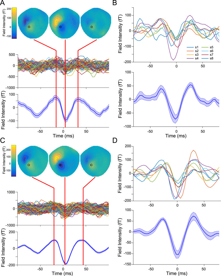
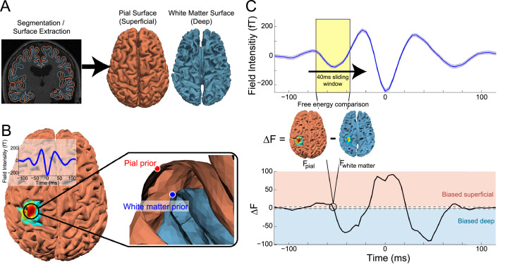
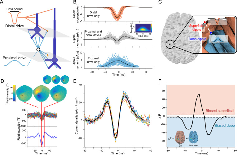
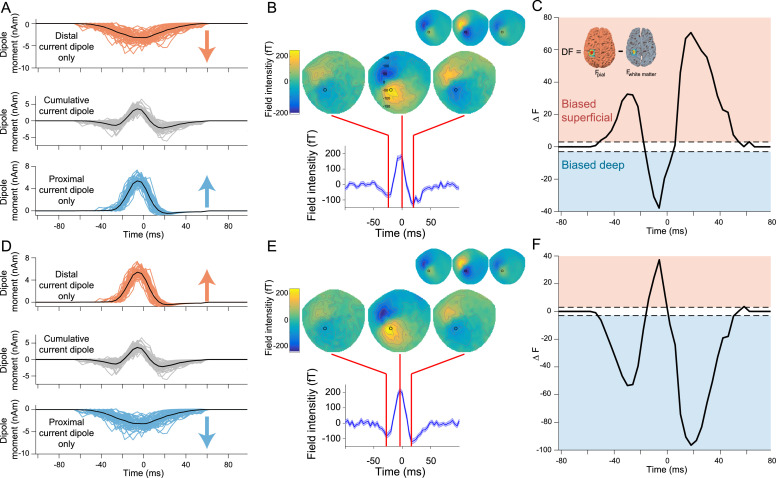
Motor cortical activity in the beta frequency range is one of the strongest and most studied movement-related neural signals. At the single trial level, beta band activity is often characterized by transient, high amplitude, bursting events rather than slowly modulating oscillations. The timing of these bursting events is tightly linked to behavior, suggesting a more dynamic functional role for beta activity than previously believed. However, the neural mechanisms underlying beta bursts in sensorimotor circuits are poorly understood. To address this, we here leverage and extend recent developments in high precision MEG for temporally resolved laminar analysis of burst activity, combined with a neocortical circuit model that simulates the biophysical generators of the electrical currents which drive beta bursts. This approach pinpoints the generation of beta bursts in human motor cortex to distinct excitatory synaptic inputs to deep and superficial cortical layers, which drive current flow in opposite directions. These laminar dynamics of beta bursts in motor cortex align with prior invasive animal recordings within the somatosensory cortex, and suggest a conserved mechanism for somatosensory and motor cortical beta bursts. More generally, we demonstrate the ability for uncovering the laminar dynamics of event-related neural signals in human non-invasive recordings. This provides important constraints to theories about the functional role of burst activity for movement control in health and disease, and crucial links between macro-scale phenomena measured in humans and micro-circuit activity recorded from animal models.

Keywords: Beta bursts; High precision MEG; Laminar comparison.


Figures

Fig. 1
Fig. 1
Sensor-level pre- and post-movement beta bursts. A) Single subject sensor-level broadband data aligned to the peak of pre-movement motor beta bursts. The mean spatial topography (top) indicates a field reversal over left sensorimotor cortex centered on the burst peak. Individual pre-movement burst waveforms (middle), taken from the preprocessed (not beta bandpass filtered) sensor data indicated by a black circle in the spatial topography (MLP34), exhibit a stereotyped, wavelet-like shape that becomes more apparent when averaged over all pre-movement bursts (shaded area indicates the standard error of the burst waveform across all bursts). B) Mean pre-movement burst waveforms from all subjects (n = 8; 2614 total bursts) after alignment with a Woody filter (top) and averaged over subjects (bottom). The shaded area indicates the standard error of the mean burst waveform across subjects. Post-movement motor beta bursts have the same spatial and temporal pattern as pre-movement bursts at the single subject (C) and group level (D; 5780 total bursts).
Fig. 2
Fig. 2
Sliding time window laminar source inversion. A) Pial and white matter surfaces are extracted from quantitative maps of proton density and T1 times from a multi-parameter mapping MRI protocol. B) Source inversion over the entire burst time course was used to localize the average beta burst sensor signal (inlay). The peak pial surface vertex and the corresponding vertex on the white matter surface were used as priors in the following sliding window inversion. C) Sliding time window source inversion was performed using a 40 ms wide window. For each iteration, source inversion was run using a pial generative model with the pial vertex from the localization inversion as a prior, and using a white matter generative model with the corresponding white matter vertex as a prior. The difference in free energy between the two models (Fpial – Fwhite matter) was used to determine the laminar locus of dominant activity as the window advanced along the average time series of the beta burst (example data shown from a single human subject).
Fig. 3
Fig. 3
A biophysical model of beta burst generation predicts a bilaminar time course of beta burst dynamics. A) Beta bursts were generated by a model with a broad proximal excitatory synaptic drive temporally aligned with a strong distal synaptic drive. B) The model was run with just the distal drive (top) and just the proximal drive (bottom), and the resulting cumulative dipole moments were used as source signals to generate the simulated MEG sensor data. Each line shows the time series from a single burst simulation (n = 50) and the average is shown as a black line. The middle panel shows the cumulative dipole moment generated from the model with both drives, exhibiting the same waveform and time-frequency (inlay) features observed in the human MEG data. C) The generative model included two oppositely oriented currents positioned at corresponding locations on the pial and white matter surfaces in motor cortex, which represent the superficial and deep cortical layers, with source activity given by the model run with the distal and proximal synaptic drives, respectively. D) Simulated sensor data generated by the model has the same spatial and temporal features as beta bursts observed in the human subject MEG data used to determine the simulated current locations and orientations (inlay). E) Time course of source current density resulting from the localizer source inversion on the simulated sensor datasets. Each simulation (n = 10) is shown as a colored line and the black line corresponds to the average over all simulations. F) The sliding window source inversion correctly identifies that the simulated bursts were generated by activity predominately in deep layers at the beginning and end of the burst, with stronger superficial layer activity at the peak of the burst. The dashed lines (at ΔF ​= ±3) show the point at which one laminar model is 20 times more likely than the other model.
Fig. 4
Fig. 4
Alternate synthetic models with reversed proximal and distal dipole moment waveforms or polarities generate different predictions. A) Synthetic model with reversed distal (top) and proximal (bottom) dipole moment waveforms from which simulated MEG data were generated. B) The synthetic model with reversed dipole moment waveforms generated simulated sensor data with oppositely oriented spatial and temporal features, compared to beta bursts observed in the human MEG data used to determine the simulated current locations and orientations (inlay). C) The sliding window source inversion correctly identifies that the bursts generated by the reversed dipole moment waveforms model were generated by activity predominately in superficial layers at the beginning and end of the burst, with stronger deep layer activity at the peak of the burst. D) Alternative synthetic model with reversed dipole polarities. E) Similar to the reversed dipole moment waveforms model, the reversed dipole polarity model generated simulated sensor data with oppositely oriented spatial and temporal features, compared to bursts in human MEG data (inlay). F) The sliding time window source inversion can determine the laminar source of dominant activity in the reversed dipole polarity model.
Fig. 5
Fig. 5
High precision MEG data enables localization of laminar dynamics. A) At SNR levels below −30 dB (dashed vertical line), it is impossible to detect laminar biases during the tail ends and peak of simulated bursts (shaded area shows standard error). Above −30 dB the full time course of laminar activity can be resolved. B) When co-registration error is less than 2 mm and 2° (dashed vertical line), it is possible to resolve the deep and superficial biases during the tails and peak of simulated beta bursts (shaded area shows standard error). Above 2 mm and 2°, laminar bias detection is unreliable.
Fig. 6
Fig. 6
Pre- and post-movement motor beta bursts exhibit the predicted bilaminar dynamics. A) The aligned pre-movement beta burst source level current density time courses averaged across subjects (shaded area shows standard error). B) Activity at the beginning and end of pre-movement bursts localized to deep cortical layers, whereas activity at the peak of the bursts localized superficially, in agreement with the results from the biophysical model (inset, reproduced from Fig. 3F). This was not true for spatially randomized (blue) surrogate data which yielded a flat Bayes factor time course that was not biased to either surface (shared area shows standard error). Post-movement beta bursts had similar source level temporal (C) and laminar (D) characteristics as pre-movement bursts (inset demonstrates how ΔF is computed).

References

    1. Adams R.A., Shipp S., Friston K.J. Predictions not commands: active inference in the motor system. Brain Struct. Funct. 2013;218:611–643. doi: 10.1007/s00429-012-0475-5. - DOI - PMC - PubMed
    1. Adjamian P., Barnes G.R., Hillebrand A., Holliday I.E., Singh K.D., Furlong P.L., Harrington E., Barclay C.W., Route P.J.G. Co-registration of magnetoencephalography with magnetic resonance imaging using bite-bar-based fiducials and surface-matching. Clin. Neurophysiol. 2004;115:691–698. doi: 10.1016/j.clinph.2003.10.023. - DOI - PubMed
    1. Alegre M., Alvarez-Gerriko I., Valencia M., Iriarte J., Artieda J. Oscillatory changes related to the forced termination of a movement. Clin. Neurophysiol. 2008;119:290–300. doi: 10.1016/j.clinph.2007.10.017. - DOI - PubMed
    1. Alegre M., Labarga A., Gurtubay I.G., Iriarte J., Malanda A., Artieda J. Beta electroencephalograph changes during passive movements: sensory afferences contribute to beta event-related desynchronization in humans. Neurosci. Lett. 2002;331:29–32. doi: 10.1016/S0304-3940(02)00825-X. - DOI - PubMed
    1. Arikuni T., Watanabe K., Kubota K. Connections of area 8 with area 6 in the brain of the macaque monkey. J. Comp. Neurol. 1988;277:21–40. - PubMed

Publication types