. 2021 Oct;41(10):e468-e479.
doi: 10.1161/ATVBAHA.121.316725. Epub 2021 Aug 19.

Apolipoprotein M and Sphingosine-1-Phosphate Receptor 1 Promote the Transendothelial Transport of High-Density Lipoprotein

Affiliations

Apolipoprotein M and Sphingosine-1-Phosphate Receptor 1 Promote the Transendothelial Transport of High-Density Lipoprotein

Srividya Velagapudi et al. Arterioscler Thromb Vasc Biol. 2021 Oct.

Abstract

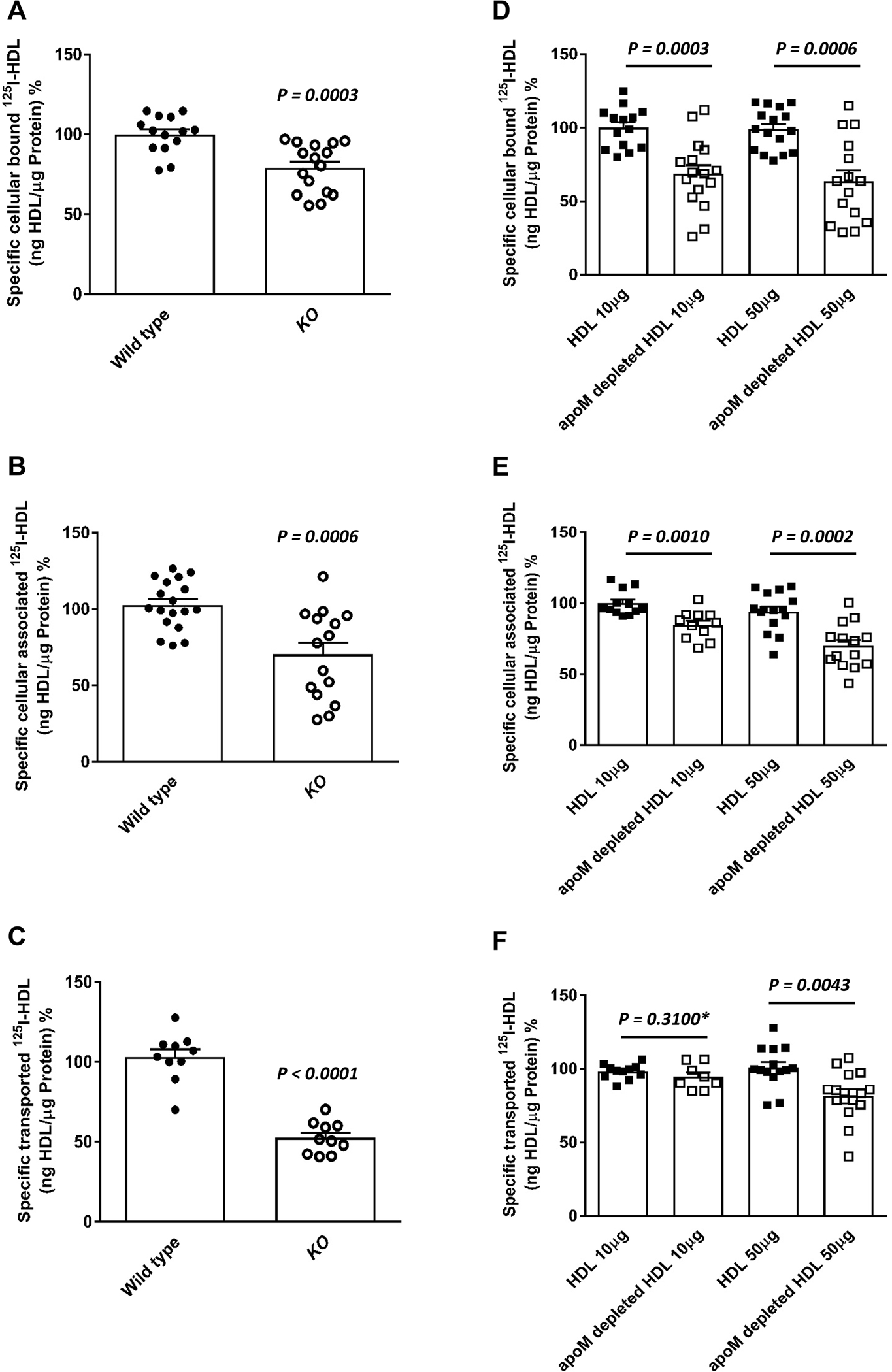
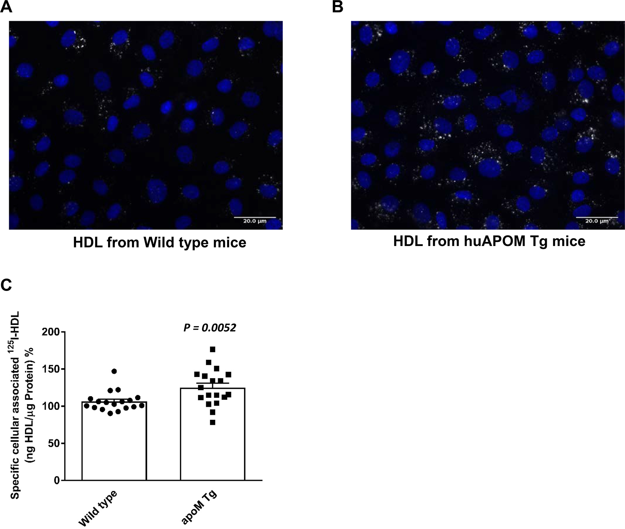
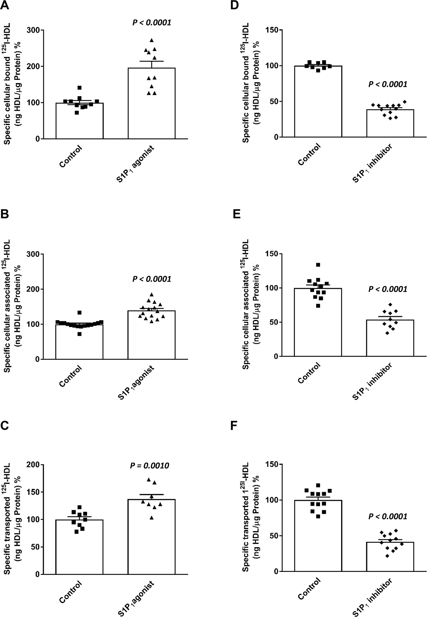
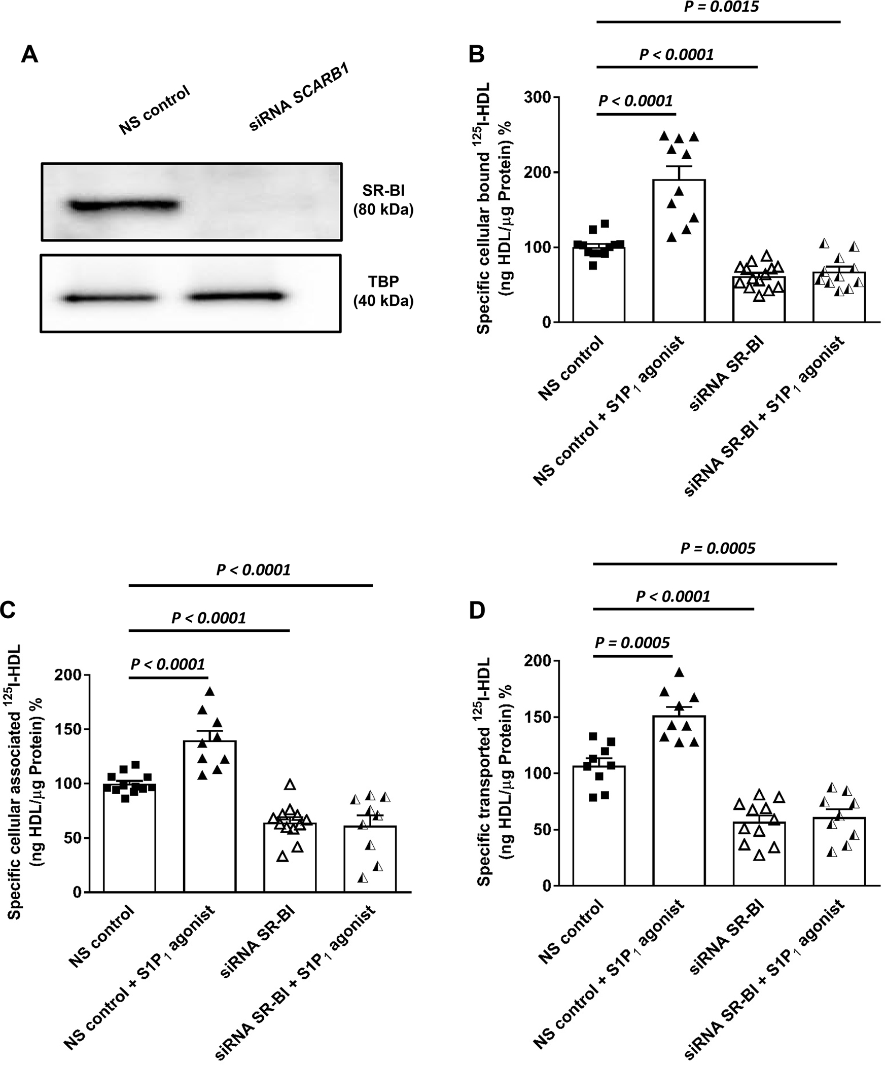
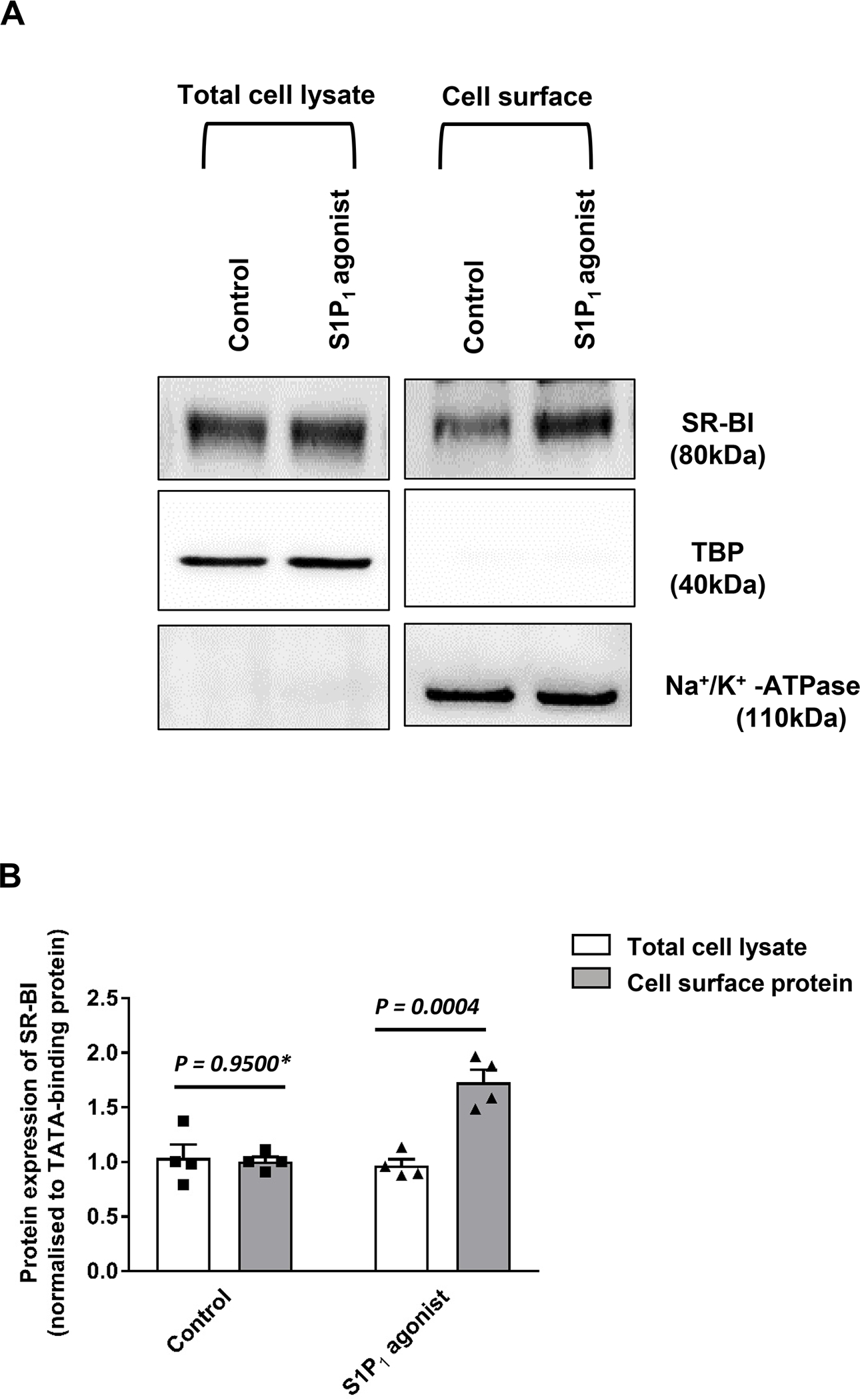
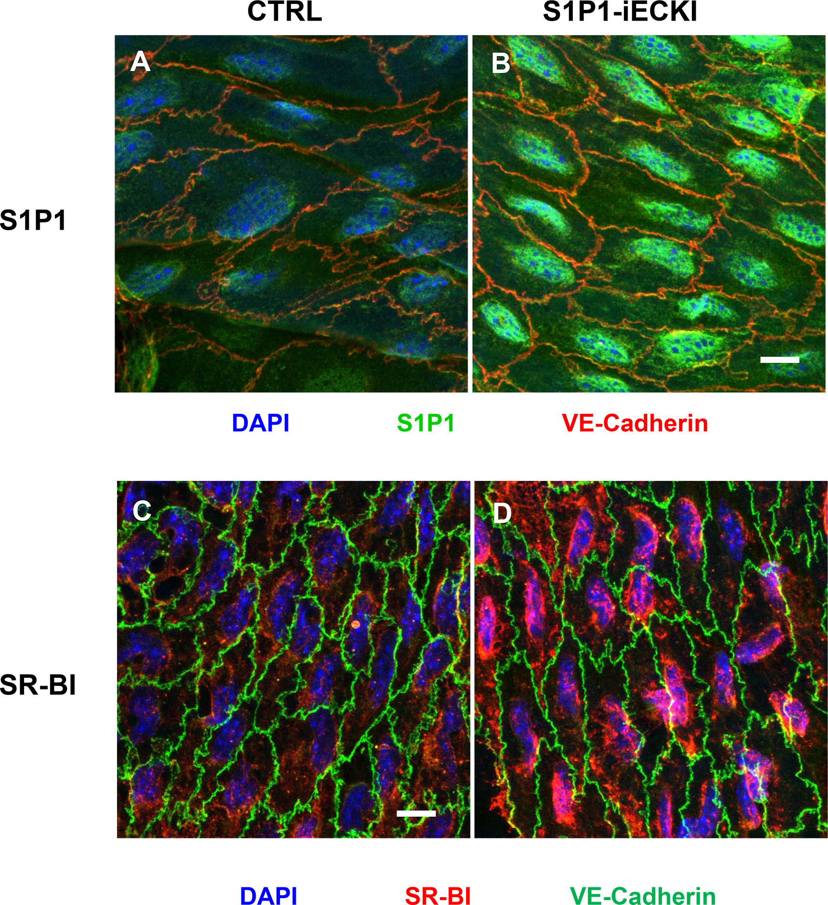
Objective: ApoM enriches S1P (sphingosine-1-phosphate) within HDL (high-density lipoproteins) and facilitates the activation of the S1P1 (S1P receptor type 1) by S1P, thereby preserving endothelial barrier function. Many protective functions exerted by HDL in extravascular tissues raise the question of how S1P regulates transendothelial HDL transport. Approach and Results: HDL were isolated from plasma of wild-type mice, Apom knockout mice, human apoM transgenic mice or humans and radioiodinated to trace its binding, association, and transport by bovine or human aortic endothelial cells. We also compared the transport of fluorescently-labeled HDL or Evans Blue, which labels albumin, from the tail vein into the peritoneal cavity of apoE-haploinsufficient mice with (apoE-haploinsufficient mice with endothelium-specific knockin of S1P1) or without (control mice, ie, apoE-haploinsufficient mice without endothelium-specific knockin of S1P1) endothelium-specific knockin of S1P1. The binding, association, and transport of HDL from Apom knockout mice and human apoM-depleted HDL by bovine aortic endothelial cells was significantly lower than that of HDL from wild-type mice and human apoM-containing HDL, respectively. The binding, uptake, and transport of 125I-HDL by human aortic endothelial cells was increased by an S1P1 agonist but decreased by an S1P1 inhibitor. Silencing of SR-BI (scavenger receptor BI) abrogated the stimulation of 125I-HDL transport by the S1P1 agonist. Compared with control mice, that is, apoE-haploinsufficient mice without endothelium-specific knockin of S1P1, apoE-haploinsufficient mice with endothelium-specific knockin of S1P1 showed decreased transport of Evans Blue but increased transport of HDL from blood into the peritoneal cavity and SR-BI expression in the aortal endothelium. Conclusions: ApoM and S1P1 promote transendothelial HDL transport. Their opposite effect on transendothelial transport of albumin and HDL indicates that HDL passes endothelial barriers by specific mechanisms rather than passive filtration.

Keywords: apolipoprotein; endothelium; lipoprotein; mice; sphingosine-1-phosphate.


Conflict of interest statement

Disclosure

MS is a scientific advisory board member of Alnylam Pharmaceuticals. The other authors have no conflict of interest to disclose with respect to the work described except the funding mentioned above

Figures

Figure 1:
Figure 1:. Presence of ApoM enhances binding binding, association and transendothelial transport of HDL in bovine aortic endothelial cells (BAECs).
BAECs were cultured for 72 hours before they were incubated for 1 hour with 10μg/mL of 125I-HDL from wild type mice or apom knock-out mice (A- C) or radioioidinated total HDL or radioioidinated apoM-depleted HDL from humans at indicated amounts (D-F) in the absence (total) or in the presence of 40-fold excess of unlabeled HDL, to record nonspecific interactions. Specific binding, association and transport were calculated by subtracting nonspecific values from total values. Specific binding was measured by incubating cells with 125I-HDL (A, D) at 4 °C. To measure specific cell association, cells were incubated with 125I-HDL (B, E) at 37 °C. For the measurement of transport, BAECs were cultured on inserts. The transport of 125I-HDL (C, F) from the apical to basolateral compartment was measured at 37 °C. The results are presented as means ± SEM of three to six independent triplicate experiments (n=3). P was calculated by unpaired Mann Whitney U-test
Figure 2:
Figure 2:. Enhanced uptake of HDL from human APOM transgenic mice by bovine aortic endothelial cells (BAECs).
For fluorescence microscopy (A), BAECs were cultured until confluence on cover slips and incubated for 60min at 37° with 50 μg/ml 594Atto-HDL, fixed with 3.75% formaldehyde and counter-stained with DAPI for nuclear staining. (B) BAECs were incubated with 125I-HDL of APOM transgenic mice at 37 °C for 1 hour in the absence (total) or in the presence of 40-fold excess of unlabeled HDL. Specific association was calculated by subtracting unspecific values from total values. The results are presented as means ± SEM of six independent triplicate experiments (n=3). P was calculated by unpaired Mann Whitney U-test.
Figure 3:
Figure 3:. Agonists (a-c) and inhibitors (d-f) of S1P1 regulate binding, association and transendothelial transport of HDL in human aortic endothelial cells (HAECs).
HAECs were cultured for 72h. Cells were then treated with S1P1 agonist (SEW2871, 20nM; A-C) or S1P1 inhibitor (W146, 20nM, D-F) for 30 minutes, at 37 °C as indicated. To study cellular binding, association and transport, HAECs were incubated with 10μg/mL of 125I-HDL for 1 hour in the absence (total) or in the presence of 40-fold excess of unlabeled HDL, to record nonspecific interactions. Specific binding, association and transport were calculated by subtracting nonspecific values from total values. Specific binding was measured by incubating cells with 125I-HDL (A, D) at 4°C. To measure specific cell association, cells were incubated with 125I-HDL (B, E) at 37°C. For the measurement of transport, HAECs were cultured on inserts. The transport of 125I-HDL (C, F) from the apical to basolateral compartment was measured at 37°C. The results are presented as means ± SEM of three independent triplicate experiments (n=3). P was calculated by unpaired Mann Whitney U-test.
Figure 4:
Figure 4:. SR-BI is involved in the S1P1 regulated binding, association and transport of HDL by human aortic endothelial cells (HAECs).
HAECs were transfected with a specific siRNA against SCARB1 or with non-silencing control siRNA (NS control). Assays were performed 72 hours post-transfection. (A) representative Western blot showing the efficacy of the silencing relative to the non-silencing siRNA (NS control) and TATA-binding protein (TBP) used as the loading control. Cells were then treated with S1P1 agonist (SEW2871, 20nM) for 30 minutes at 37 °C. (B) cellular binding of 125I-HDL was measured at 4 °C after pre-treating cells with the S1P1 agonist. (C) cellular association of 125I-HDL was measured at 37 °C. (D) for the measurement of transport of 125I-HDL, HAECs were cultured on inserts. The transport of 125I-HDL was measured after pre-treatment with the S1P1 agonist from the apical to basolateral compartment was measured at 37 °C. The results are presented as means ± SEM of three independent triplicate experiments (n=3). P was calculated by unpaired Mann Whitney U-test).
Figure 5:
Figure 5:. Effects of S1P1 activation on the cell surface abundance of SR-BI.
HAECs were cultured for 72 hours. Cells were then treated with 20nM SEW2871 for 30 minutes at 37 °C. Cell surface expression of SR-BI in HAECs was measured using Western blot analysis in total cell lysates (left) and on the cell surface (right). The western blots were probed with anti-SR-BI (82kDa), anti-TBP (40kDa, used as a control for intracellular protein expression) and Na+/K+-ATPase (110 kDa, used as a loading control for cell surface protein expression). (A) shows a representative western blot. (B) shows the summary of four independent experiments quantified by densitometry. P was calculated by unpaired Mann Whitney U-test.
Figure 6:
Figure 6:. Demonstration of S1P1 (A, B) or SR-BI (C, D) in the endothelium of aortas from Apoe haploinsufficient mice without (CTRL: A, C) or with overexpression of S1P1 (S1P1-iECKI; B, D).
Figure shows en-face prepared aortic immunostainings. Aortas were quickly cleaned of adventitial tissue, opened longitudinally and incubated with primary and secondary antibodies conjugated with green or red fluorescent dyes, as indicated. Nuclei were counterstained with DAPI. Images were captured by confocal microscope and z-axis projections of 14 scanned planes are shown. Scale bar = 10μm. Original micrographs are shown as supplemental figures IV and V.

References

    1. Marz W, Kleber ME, Scharnagl H, Speer T, Zewinger S, Ritsch A, Parhofer KG, von Eckardstein A, Landmesser U, Laufs U. Hdl cholesterol: Reappraisal of its clinical relevance. Clin Res Cardiol. 2017;106:663–675 - PMC - PubMed
    1. Annema W, von Eckardstein A. High-density lipoproteins. Multifunctional but vulnerable protections from atherosclerosis. Circ J. 2013;77:2432–2448 - PubMed
    1. Cardner M, Yalcinkaya M, Goetze S, Luca E, Balaz M, Hunjadi M, Hartung J, Shemet A, Krankel N, Radosavljevic S, Keel M, Othman A, Karsai G, Hornemann T, Claassen M, Liebisch G, Carreira E, Ritsch A, Landmesser U, Krutzfeldt J, Wolfrum C, Wollscheid B, Beerenwinkel N, Rohrer L, von Eckardstein A. Structure-function relationships of hdl in diabetes and coronary heart disease. JCI Insight. 2020;5 - PMC - PubMed
    1. Cartier A, Hla T. Sphingosine 1-phosphate: Lipid signaling in pathology and therapy. Science. 2019;366 - PMC - PubMed
    1. Christoffersen C, Obinata H, Kumaraswamy SB, Galvani S, Ahnstrom J, Sevvana M, Egerer-Sieber C, Muller YA, Hla T, Nielsen LB, Dahlback B. Endothelium-protective sphingosine-1-phosphate provided by hdl-associated apolipoprotein m. Proc Natl Acad Sci U S A. 2011;108:9613–9618 - PMC - PubMed

Publication types

MeSH terms