. 2021 Aug 18;11(1):16767.
doi: 10.1038/s41598-021-96184-z.

Secretome screening reveals immunomodulating functions of IFNα-7, PAP and GDF-7 on regulatory T-cells

Affiliations

Secretome screening reveals immunomodulating functions of IFNα-7, PAP and GDF-7 on regulatory T-cells

Mei Ding et al. Sci Rep. .

Abstract

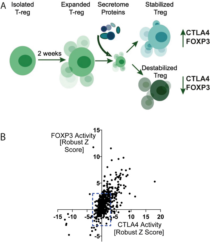
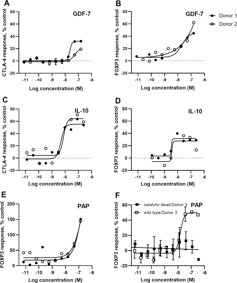
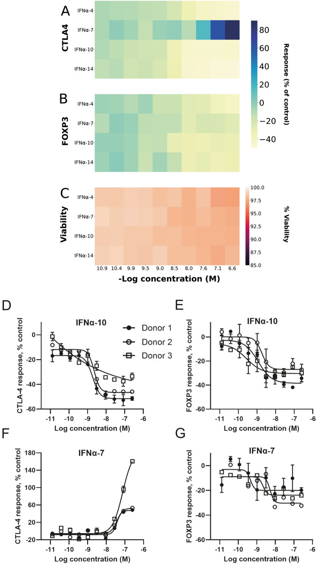
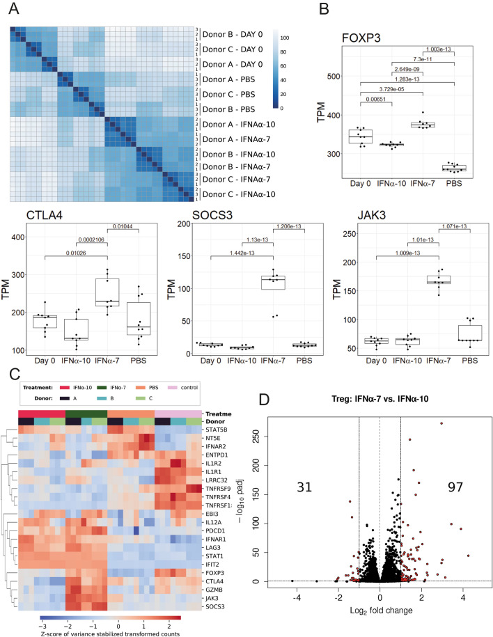
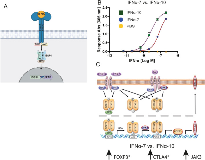
Regulatory T cells (Tregs) are the key cells regulating peripheral autoreactive T lymphocytes. Tregs exert their function by suppressing effector T cells. Tregs have been shown to play essential roles in the control of a variety of physiological and pathological immune responses. However, Tregs are unstable and can lose the expression of FOXP3 and suppressive functions as a consequence of outer stimuli. Available literature suggests that secreted proteins regulate Treg functional states, such as differentiation, proliferation and suppressive function. Identification of secreted proteins that affect Treg cell function are highly interesting for both therapeutic and diagnostic purposes in either hyperactive or immunosuppressed populations. Here, we report a phenotypic screening of a human secretome library in human Treg cells utilising a high throughput flow cytometry technology. Screening a library of 575 secreted proteins allowed us to identify proteins stabilising or destabilising the Treg phenotype as suggested by changes in expression of Treg marker proteins FOXP3 and/or CTLA4. Four proteins including GDF-7, IL-10, PAP and IFNα-7 were identified as positive regulators that increased FOXP3 and/or CTLA4 expression. PAP is a phosphatase. A catalytic-dead version of the protein did not induce an increase in FOXP3 expression. Ten interferon proteins were identified as negative regulators that reduced the expression of both CTLA4 and FOXP3, without affecting cell viability. A transcriptomics analysis supported the differential effect on Tregs of IFNα-7 versus other IFNα proteins, indicating differences in JAK/STAT signaling. A conformational model experiment confirmed a tenfold reduction in IFNAR-mediated ISG transcription for IFNα-7 compared to IFNα-10. This further strengthened the theory of a shift in downstream messaging upon external stimulation. As a summary, we have identified four positive regulators of FOXP3 and/or CTLA4 expression. Further exploration of these Treg modulators and their method of action has the potential to aid the discovery of novel therapies for both autoimmune and infectious diseases as well as for cancer.


Conflict of interest statement

MD, RM, TO, JB, UG, EB, BM, DRT, II, KFS, LMM, RD and LHS are employees of AstraZeneca. The authors have no additional financial interests. ML, AM, HT, SH, MU and JR declare no competing financial interests.

Figures

Figure 1
Figure 1
Identification of secreted proteins that stabilise/destabilise Treg markers. (A) Schematic describing the concept of the Treg assay where FOXP3 and CTLA4 protein expression was used as an indicator of Treg stability. (B) Effect of proteins screened on FOXP3 and CTLA4 expression in Tregs at top concentration tested. The effect was measured as FOXP3 and CTLA4 Median Fluorescence Intensity (MFI) of live cells and normalized to Robust Z Score based on the on-plate PBS neutral controls. The blue dashed lines indicate the threshold for identifying hits based on ≥ 3 × Robust Z-score, or ≤ -3 × Robust Z-score of FOXP3 or CTLA4 expression.
Figure 2
Figure 2
GDF-7, IL-10 and PAP stabilise the Treg markers. The concentration dependent effects on FOXP3 and CTLA4 protein expression induced by GDF-7, IL-10 and PAP are shown in (A,B) for GDF-7; (C,D) for IL-10; and (E,F) for PAP. The effect on FOXP3 and CTLA4 expression was normalized to % activity as compared to the MFI of live cells treated with the on-plate PBS neutral controls. These proteins were tested in human Treg cells from multiple donors (Donor 1 filled circle ●, Donor 2 empty circle ○, Donor 3 empty square □ for wild type PAP, Donor 3 filled square ■ for catalytic dead mutant PAP [H12A]).
Figure 3
Figure 3
IFNα-4, IFNα-10 and IFNα-14 destabilise Treg markers whereas IFNα-7 stabilises CTLA4 expression. (A) Heatmap over mean effect on CTLA4 protein expression, (B) heatmap over mean effect on FOXP3 protein expression, (C) Heatmap over mean effect on cell viability, (DE) Effect of IFNα-10 treatment on CTLA4 and FOXP3 expression, (FG) Effect of IFNα-7 treatment on CTLA4 and FOXP3 expression. Tregs were from multiple donors (Donor 1 filled circle ●, Donor 2 empty circle ○, Donor 3 empty square □). The effect on FOXP3 and CTLA4 expression was normalized to % activity as compared to the MFI of live cells treated with the on-plate PBS neutral controls.
Figure 4
Figure 4
Transcriptomics reveal distinct activation profile for IFNα-7 compared to IFNα-10. (A) Heatmap of the sample-to-sample euclidean distances for the RNA samples, based on variance stabilised counts data. (B) Expression analysis (TPM) shows that FOXP3, CTLA4, SOCS3 and JAK3 genes were upregulated when comparing IFNα-7 treated to IFNα-10 treated cells. (C) Heatmap showing differences in expression between samples for genes mentioned in the results section. Values were based on transformed count data (variance stabilising transformed) extracted from DESeq2 with its vst function and with blind set to FALSE. Z-scores were subsequently applied to accentuate differences between samples. (D) Differentially expressed genes (twofold change and FDR < 0.05) when comparing IFNα-7 and IFNα-10 treatments of Treg cells; 97 genes were significantly upregulated and 31 genes were downregulated with this threshold. Heatmap of the sample-to-sample euclidean distances for the RNA samples, based on variance stabilized counts data. Treg cells from 3 individual donors were analysed in the transcriptomics experiments in 3 replicates from each individual donor.
Figure 5
Figure 5
Suggested model of IFNα-7 vs IFNα-10 strengthens difference in ISG transcription signaling. (A) Schematic representation of HEK293 reporter cell line for IFNAR1/2 mediated JAK STAT1/2 signaling leading to ISG transcription of secreted embryonic alkaline phosphatase. (B) Dose–response curves of HEK293 reporter cell line upon treatment with either IFNα-7 (blue circles) or IFNα-10 (green squares), SEM error bars based on n = 2, duplicate measurements and subtracted baseline signal (PBS). (C) A pathway map showing signaling pathways for the IFNAR1/IFNAR2 receptor and the IL-2 receptor. Genes in red are upregulated when comparing IFNα7-treated versus IFNα10-treated cells. JAK3 is significantly upregulated (FDR < 0.05 and > twofold upregulation). * indicates additional phenotypic readouts at a protein level in the primary assay. The observed differences between IFNα-7 and IFNα-10 are further discussed in the main text.

References

    1. Adeegbe DO, Nishikawa H. Natural and induced T regulatory cells in cancer. Front. Immunol. 2013;4:190. doi: 10.3389/fimmu.2013.00190. - DOI - PMC - PubMed
    1. Rudensky AY. Regulatory T cells and Foxp3. Immunol. Rev. 2011;241:260–268. doi: 10.1111/j.1600-065X.2011.01018.x. - DOI - PMC - PubMed
    1. Sakaguchi S, Yamaguchi T, Nomura T, Ono M. Regulatory T cells and immune tolerance. Cell. 2008;133:775–787. doi: 10.1016/j.cell.2008.05.009. - DOI - PubMed
    1. Grant CR, Liberal R, Mieli-Vergani G, Vergani D, Longhi MS. Regulatory T-cells in autoimmune diseases: Challenges, controversies and—yet—unanswered questions. Autoimmun. Rev. 2015;14:105–116. doi: 10.1016/j.autrev.2014.10.012. - DOI - PubMed
    1. Kanamori M, Nakatsukasa H, Okada M, Lu Q, Yoshimura A. Induced regulatory T cells: Their development, stability, and applications. Trends Immunol. 2016;37:803–811. doi: 10.1016/j.it.2016.08.012. - DOI - PubMed

Publication types

MeSH terms