. 2021 Aug 18;4(1):982.
doi: 10.1038/s42003-021-02496-1.

The development and structure of the mesentery

Affiliations

The development and structure of the mesentery

Kevin G Byrnes et al. Commun Biol. .

Abstract

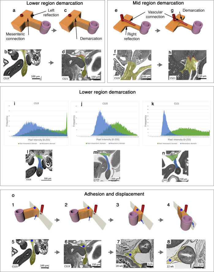
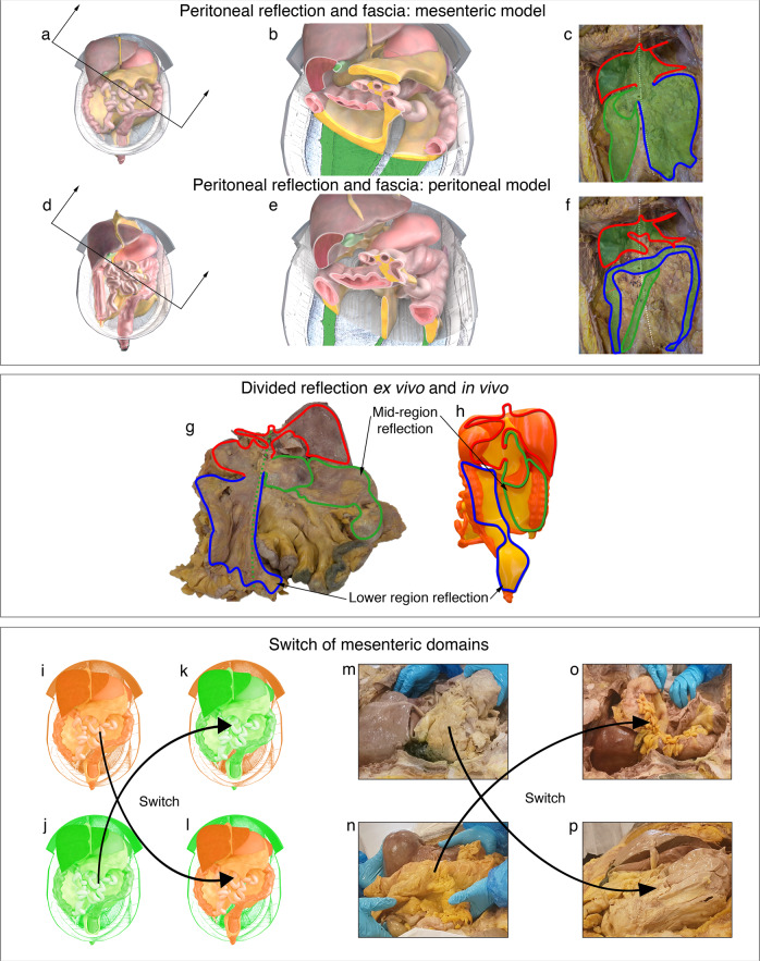
The position of abdominal organs, and mechanisms by which these are centrally connected, are currently described in peritoneal terms. As part of the peritoneal model of abdominal anatomy, there are multiple mesenteries. Recent findings point to an alternative model in which digestive organs are connected to a single mesentery. Given that direct evidence of this is currently lacking, we investigated the development and shape of the entire mesentery. Here we confirm that, within the abdomen, there is one mesentery in which all abdominal digestive organs develop and remain connected to. We show that all abdominopelvic organs are organised into two, discrete anatomical domains, the mesenteric and non-mesenteric domain. A similar organisation occurs across a range of animal species. The findings clarify the anatomical foundation of the abdomen; at the foundation level, the abdomen comprises a visceral (i.e. mesenteric) and somatic (i.e. musculoskeletal) frame. The organisation at that level is a fundamental order that explains the positional anatomy of all abdominopelvic organs, vasculature and peritoneum. Collectively, the findings provide a novel start point from which to systemically characterise the abdomen and its contents.


Conflict of interest statement

The authors declare no competing interests.

Figures

Fig. 1
Fig. 1. Developing mesentery.
a Photograph of a digitally reconstructed mesentery (yellow) at Carnegie stage (CS) 13. a14 Photomicrographs at levels indicated. b13 Photographs of the reconstructed mesentery at CS 16. b2, 4 Photomicrographs of sections at levels indicated. c14 Photographs and photomicrographs of mesenteric regions at CS16. d12 Photographs and photomicrographs of the anterior aspect of the mid-region fold. Photographs demonstrating anterior (e) and posterior (f) aspects of the mid-region at CS 16. gl Photographs showing anterior and posterior aspects of the developing mid-region. mo Schematic depiction of the mid-region switch.
Fig. 2
Fig. 2. The mesentery and posterior abdominal wall.
ad Photographs and corresponding photomicrographs demonstrating continuity between lower region mesentery and posterior abdominal wall. eh Photographs and corresponding photomicrographs demonstrating continuity between mid-region mesentery and posterior abdominal wall. ik Histograms demonstrating brightness values of developing mesentery and posterior abdominal wall. ln Photomicrographs from which histograms were generated. o14 Panel demonstrating the relationships between the mesentery (*) and posterior abdominal wall during development. The mesothelial junction between both is indicated (arrow). o58 Photomicrographs demonstrating histological correlates for o1-4.
Fig. 3
Fig. 3. Developing and adult mesentery.
a Panel demonstrating the developing mesentery, stomach and pancreas. Each row corresponds to a developmental stage and reads from left to right. b Photographs demonstrating the anterior aspect of (b) the ex vivo mesentery and of a 3D printed ex vivo mesentery (c). Photographs demonstrating the posterior aspect of (d) the ex vivo mesentery and of the 3D print used in (c, e). Illustrations demonstrating the right anterolateral (f) and left posterolateral (g) aspects of a digital version of the ex vivo mesentery. hk Illustrations of the same digital reconstruction with individual regions highlighted.
Fig. 4
Fig. 4. Mid region of the mesentery.
Photographs demonstrating the anterior aspect of the central zone (encircled) of the mid-region in (a) a digital and (b) ex vivo mesentery. In (c) a schematic has been superimposed on (b) explaining the underlying anatomy. colour code: pancreas (yellow), right mesocolon (orange), hepatic flexure (green), splenic flexure (red). Photographs demonstrating the anterior aspect of the peripheral zone (blue boundary) in (d) a digital model and (e) the ex vivo mesentery. In (f) a schematic has been superimposed on (e) explaining the underlying anatomy. colour code: original left side of the peripheral zone (red), original right side of the peripheral zone (orange).
Fig. 5
Fig. 5. Mesenteric based anatomy.
Illustrations of the mesenteric model of the abdomen before (a), after (b) sectioning. c Photograph of the cadaveric non-mesenteric domain (schematic indicates the distribution of the reflection). Illustrations of the peritoneal model before (d), and after (e) sectioning. f Schematic indicating peritoneal based description of the mesentery/peritoneum. Photographs showing the posterior aspect of the ex vivo mesentery (g) and 3D printed mesentery (h) and reflection schematically superimposed. colour code: upper region (red), mid-region (green), lower region (blue). ip Panel demonstrating switching of the mesenteric domain between cadavers in schematic (il) and cadaveric (mp) formats.

References

    1. Coffey, J. C. & Dockery, P. Ch 62. Peritoneum, mesentery and peritoneal cavity. in Gray’s Anatomy: The Anatomical Basis of Clinical Practice(42nd edition) 1150–1160 (2020).
    1. Coffey JC, O’Leary DP. The mesentery: structure, function, and role in disease. Lancet Gastroenterol. Hepatol. 2016;1:238–247. doi: 10.1016/S2468-1253(16)30026-7. - DOI - PubMed
    1. Spencer SP, Sonnenburg JL. When gut microbiota creep into fat, the fat creeps back. Cell. 2020;183:589–591. doi: 10.1016/j.cell.2020.10.008. - DOI - PMC - PubMed
    1. Kurokawa Y, et al. Bursectomy versus omentectomy alone for resectable gastric cancer (JCOG1001): a phase 3, open-label, randomised controlled trial. Lancet Gastroenterol. Hepatol. 2018;3:460–468. doi: 10.1016/S2468-1253(18)30090-6. - DOI - PubMed
    1. Toldt C. Bau und Wachsthumsveränderungen der Gekröse des menschlichen Darmkanales. Denkschrdmathnaturwissensch. 1879;41:56.

MeSH terms