

Disentangling the association between retinal non-perfusion and anti-VEGF agents in diabetic retinopathy
- PMID: 34408316
- PMCID: PMC8956693
- DOI: 10.1038/s41433-021-01750-4
Disentangling the association between retinal non-perfusion and anti-VEGF agents in diabetic retinopathy
Abstract
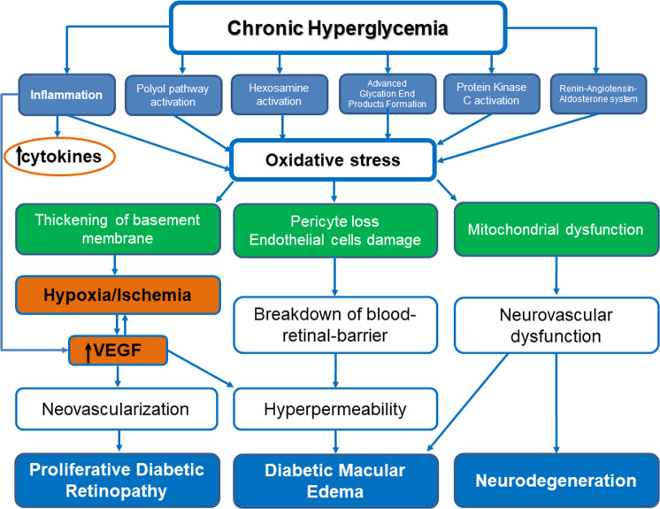
Diabetic retinopathy (DR) is the most common microvascular complication of diabetes mellitus (DM) and the leading cause of blindness in patients with DM. In the pathogenesis of DR, chronic hyperglycemia leads to biochemical and structural alterations in retinal blood vessels' wall, resulting in hyperpermeability and non-perfusion. Since vascular endothelial growth factor (VEGF) has been found to play a significant role in the pathogenesis of DR, this review sheds light on the effect of intravitreal anti-VEGF agents on retinal non-perfusion in patients with DR. Based on the existing literature, anti-VEGF agents have been shown to improve DR severity, although they cannot reverse retinal ischemia. The results of the published studies are controversial and differ based on the location of retinal non-perfusion, as well as the imaging modality used to assess retinal non-perfusion. In cases of macular non-perfusion, most of studies showed no change in both fundus fluorescein angiography (FFA) and optical coherence tomography (OCTA) in patients with DR treated with intravitreal anti-VEGF agents, while few studies reported worsening of non-perfusion with enlargement of foveal avascular zone (FAZ). Regarding peripheral ischemia, studies using wide-field-FFA demonstrated an improvement or stability in non-perfusion areas after anti-VEGF treatment. However, the use of wide-field-OCTA revealed no signs of re-perfusion of retinal vessels post anti-VEGF treatment. Further prospective studies with long follow-up and large sample size are still needed to draw solid conclusions.
摘要: 糖尿病视网膜病变 (DR) 是糖尿病 (DM) 最常见的微血管并发症, 也是DM患者致盲的主要原因。在DR的发病机制中, 慢性高血糖会引起视网膜血管壁的生化和结构发生改变, 并导致高渗和无灌注。由于血管内皮生长因子 (VEGF) 在DR的发病机制中起着重要作用, 本文综述了玻璃体内抗VEGF药物对DR患者视网膜非灌注区的影响。根据现有文献, 抗VEGF药物已被证明可以改善DR的严重程度, 但仍不能逆转视网膜缺血。目前已发表的研究结果存在争议, 并根据视网膜无灌注的位置以及用于评估视网膜无灌注的成像方式而有所不同。在黄斑区无灌注的病例中, 大多数研究报道了玻璃体内抗VEGF药物治疗后的DR患者, 其眼底荧光素血管造影(FFA)和光学相干断层成像(OCTA)均无变化, 而少数研究则报道了无灌注区域的加重伴随中央凹无血管区(FAZ)的增大。关于外周缺血, 使用广角FFA的研究表明, 抗VEGF治疗后的无灌注区有一定的改善或稳定。然而, 在抗VEGF治疗后, 使用广角OCTA并未发现视网膜血管有再灌注的迹象。未来还需要进一步的长期随访和大样本的前瞻性研究才能得出可靠的结论。.
© 2021. The Author(s), under exclusive licence to The Royal College of Ophthalmologists.
Conflict of interest statement
The authors declare no competing interests.
Figures
Comment in
-
Comment on: Disentangling the association between retinal non-perfusion and anti-VEGF agents in diabetic retinopathy.Eye (Lond). 2022 Aug;36(8):1705. doi: 10.1038/s41433-021-01800-x. Epub 2021 Oct 6. Eye (Lond). 2022. PMID: 34616003 Free PMC article. No abstract available.
References
-
- International Diabetes Federation. IDF Diabetes Atlas. 7th ed. Brussels, Belgium: International Diabetes Federation; 2015.
-
- Forbes JM, Cooper ME. Mechanisms of diabetic complications. Physiol Rev. 2013;93:137–88. - PubMed
-
- Antonetti DA, Klein R, Gardner TW. Diabetic retinopathy. New Engl J Med. 2012;366:1227–39. - PubMed
Publication types
MeSH terms
Substances
LinkOut - more resources
Full Text Sources
Medical
