. 2021 Jul 22;3(3):fcab168.
doi: 10.1093/braincomms/fcab168. eCollection 2021.

Spectrum, risk factors and outcomes of neurological and psychiatric complications of COVID-19: a UK-wide cross-sectional surveillance study

Affiliations

Spectrum, risk factors and outcomes of neurological and psychiatric complications of COVID-19: a UK-wide cross-sectional surveillance study

Amy L Ross Russell et al. Brain Commun. .

Abstract

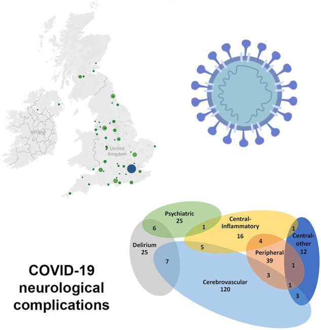
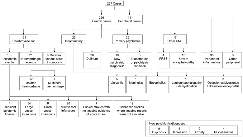
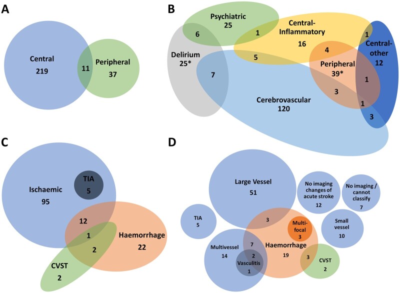
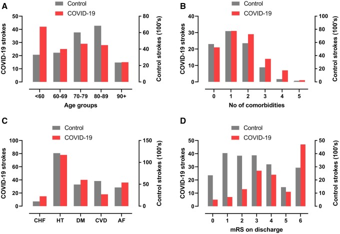
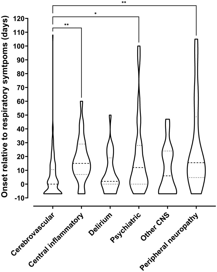
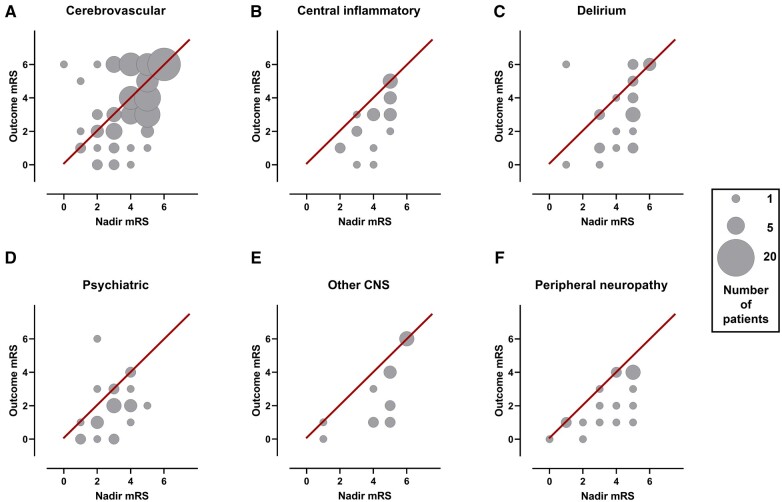
SARS-CoV-2 is associated with new-onset neurological and psychiatric conditions. Detailed clinical data, including factors associated with recovery, are lacking, hampering prediction modelling and targeted therapeutic interventions. In a UK-wide cross-sectional surveillance study of adult hospitalized patients during the first COVID-19 wave, with multi-professional input from general and sub-specialty neurologists, psychiatrists, stroke physicians, and intensivists, we captured detailed data on demographics, risk factors, pre-COVID-19 Rockwood frailty score, comorbidities, neurological presentation and outcome. A priori clinical case definitions were used, with cross-specialty independent adjudication for discrepant cases. Multivariable logistic regression was performed using demographic and clinical variables, to determine the factors associated with outcome. A total of 267 cases were included. Cerebrovascular events were most frequently reported (131, 49%), followed by other central disorders (95, 36%) including delirium (28, 11%), central inflammatory (25, 9%), psychiatric (25, 9%), and other encephalopathies (17, 7%), including a severe encephalopathy (n = 13) not meeting delirium criteria; and peripheral nerve disorders (41, 15%). Those with the severe encephalopathy, in comparison to delirium, were younger, had higher rates of admission to intensive care and a longer duration of ventilation. Compared to normative data during the equivalent time period prior to the pandemic, cases of stroke in association with COVID-19 were younger and had a greater number of conventional, modifiable cerebrovascular risk factors. Twenty-seven per cent of strokes occurred in patients <60 years. Relative to those >60 years old, the younger stroke patients presented with delayed onset from respiratory symptoms, higher rates of multi-vessel occlusion (31%) and systemic thrombotic events. Clinical outcomes varied between disease groups, with cerebrovascular disease conferring the worst prognosis, but this effect was less marked than the pre-morbid factors of older age and a higher pre-COVID-19 frailty score, and a high admission white cell count, which were independently associated with a poor outcome. In summary, this study describes the spectrum of neurological and psychiatric conditions associated with COVID-19. In addition, we identify a severe COVID-19 encephalopathy atypical for delirium, and a phenotype of COVID-19 associated stroke in younger adults with a tendency for multiple infarcts and systemic thromboses. These clinical data will be useful to inform mechanistic studies and stratification of patients in clinical trials.

Keywords: COVID-19; SARS-CoV-2; encephalopathy; neurology; stroke.


Figures

Graphical Abstract
Graphical Abstract
Figure 1
Figure 1
Classification of main neurological diagnoses.
Figure 2
Figure 2
Magnetic resonance imaging demonstrating the range of neurological complications seen in this study. (A) Territorial infarct, secondary to internal carotid artery (ICA) dissection in a middle-aged previously fit male: Axial fluid-attenuated inversion recovery image (i) showing a right middle cerebral artery (MCA) territory infarct following decompressive craniectomy for malignant MCA syndrome despite treatment with thrombolysis. Reformatted images from a CT angiogram (ii) showing irregularity of the extracranial segment of both internal carotid arteries, consistent with dissection (arrows), with tight stenosis of the true lumen on the right (arrowhead). (B) Multiple territorial infarcts in a female >60 years old with hypertension and dyslipidaemia: Diffusion-weighted images (DWI) demonstrate recent infarcts in the right medial occipital lobe and lentiform nucleus, involving the territories of the right posterior cerebral artery and lenticulo-striate perforators of the right MCA respectively. (C) Acute lacunar infarcts due to small vessel vasculopathy in a male > 60 years old, with a background of hypertension and type 2 diabetes: B1000 images (i, ii) and corresponding apparent diffusion coefficient (ADC) maps (iii, iv) from DWI showing multiple tiny foci of restricted diffusion. (D) Vasculitis in a male >60 years old, with a background of type 2 diabetes, hypertension and hypercholesterolaemia: T1-weighted SPACE vessel wall imaging of both distal ICAs and proximal MCAs, with curved multiplanar coronal reconstructions along the course of both proximal MCAs (first column) and perpendicular to the right MCA (second column, at the position of the dotted line). Pre-treatment pre-contrast (i, ii) and post-contrast images (iii, iv) demonstrate abnormal concentric, long segment vessel wall enhancement (arrows) of both proximal MCAs. Post-contrast images after treatment with prednisolone and tocilizumab (v, vi) demonstrate treatment response with resolution of the previous abnormal mural MCA enhancement (arrows). (E) Acute encephalomyelitis with haemorrhage in a middle-aged male, with a history of chronic obstructive pulmonary disease, who required intensive care and haemofiltration: Coronal FLAIR (i) and axial gradient echo (ii) images showing focal heterogeneous signal abnormality and swelling of the splenium of the corpus callosum, with peripheral low signal indicative of haemosiderin staining (arrows). Confluent high signal is present in periventricular and deep white matter of the parieto-occipital region. (F) Typical imaging appearances of posterior reversible encephalopathy syndrome in a normotensive middle-aged female: Axial T2 image (i) demonstrating hyperintense signal in subcortical white matter of both occipital lobes, with B1000 image (ii) and ADC map (iii) from DWI showing no corresponding restricted diffusion.
Figure 3
Figure 3
Venn diagrams showing overlap of diagnostic groups. The numbers shown here are when all diagnoses were considered, in addition to the primary neurological diagnosis. The total numbers for several groups are larger in this Figure than the primary diagnosis flowchart (Fig. 1) due to coexisting diagnoses. (A) Central and peripheral nervous system disease. (B) Primary diagnostic categories (*two cases of Guillain–Barré syndrome with delirium were not possible to accommodate on this diagram). (C) Stroke group subtypes. (D) Specific stroke group subtypes. CVST, cerebral venous sinus thrombosis; TIA, transient ischaemic attack.
Figure 4
Figure 4
COVID-19 strokes versus historical controls. Comparison between strokes associated with COVID-19 in this study and strokes from a national UK audit in 2019. (A, B) total number of co-morbidities which are risk factors for stroke (atrial fibrillation, hypertension, diabetes mellitus, congestive heart failure and previous TIA or stroke). (C) Age distributions. (D) mRS (modified Rankin scale) scores on discharge from hospital (or death).
Figure 5
Figure 5
Timing of onset of neurology. Violin plot demonstrating distributions of time intervals in days between onset of respiratory symptoms and onset of neurological symptoms for each primary diagnostic category. Patients whose neurological symptoms preceded COVID-19 symptoms were arbitrarily assigned a value of minus seven days. The Kruskal–Wallis test was used to determine any significant difference in time intervals between groups (P < 0.0001). Dunn’s multiple group comparison test showed a significant difference between stroke and central inflammatory primary diagnostic groups (P = 0.001), stroke and psychiatric groups (P = 0.037), and stroke and peripheral groups (P = 0.003).
Figure 6
Figure 6
Recovery from neurological condition. Bubble plots displaying the relationship between mRS (modified Rankin scale) at nadir of illness whilst in hospital and mRS at outcome assessment, within individual diagnostic categories. Bubble area corresponds to patient number. Line of equivalence is shown in red: cases below the line improved, cases above the line got worse, while cases on the line stayed the same.

References

    1. Mao L, Jin H, Wang M, et al.Neurologic manifestations of hospitalized patients with coronavirus disease 2019 in Wuhan, China. JAMA Neurol. 2020;77(6):683–690. - PMC - PubMed
    1. Ellul MA, Benjamin L, Singh B, et al.Neurological associations of COVID-19. Lancet Neurol. 2020;19(9):767–783. - PMC - PubMed
    1. Lechien JR, Chiesa-Estomba CM, Place S, et al.COVID-19 Task Force of YO-IFOS. Clinical and epidemiological characteristics of 1420 European patients with mild-to-moderate coronavirus disease 2019. J Intern Med. 2020;288(3):335–344. - PMC - PubMed
    1. Cagnazzo F, Arquizan C, Derraz I, et al.Neurological manifestations of patients infected with the SARS-CoV-2: A systematic review of the literature. J Neurol. 2021;268(8):2656–2665. - PMC - PubMed
    1. Romero-Sánchez CM, Díaz-Maroto I, Fernández-Díaz E, et al.Neurologic manifestations in hospitalized patients with COVID-19: The ALBACOVID registry. Neurology. 2020;95(8):e1060–e1070. - PMC - PubMed

LinkOut - more resources