. 2022 Apr;28(7-8):312-329.
doi: 10.1089/ten.TEA.2021.0137. Epub 2022 Jan 4.

Photoreactive Hydrogel Stiffness Influences Volumetric Muscle Loss Repair

Affiliations

Photoreactive Hydrogel Stiffness Influences Volumetric Muscle Loss Repair

Ivan M Basurto et al. Tissue Eng Part A. 2022 Apr.

Abstract

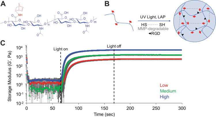
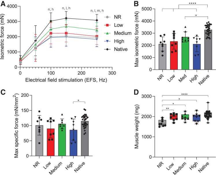
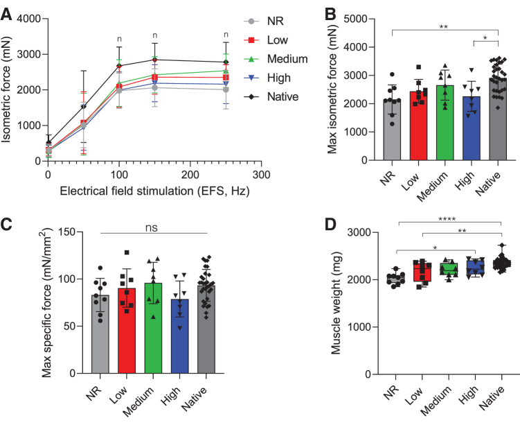
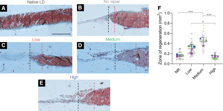
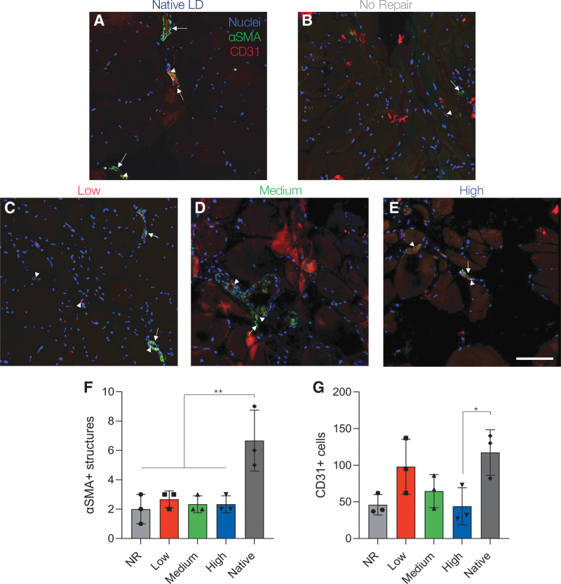
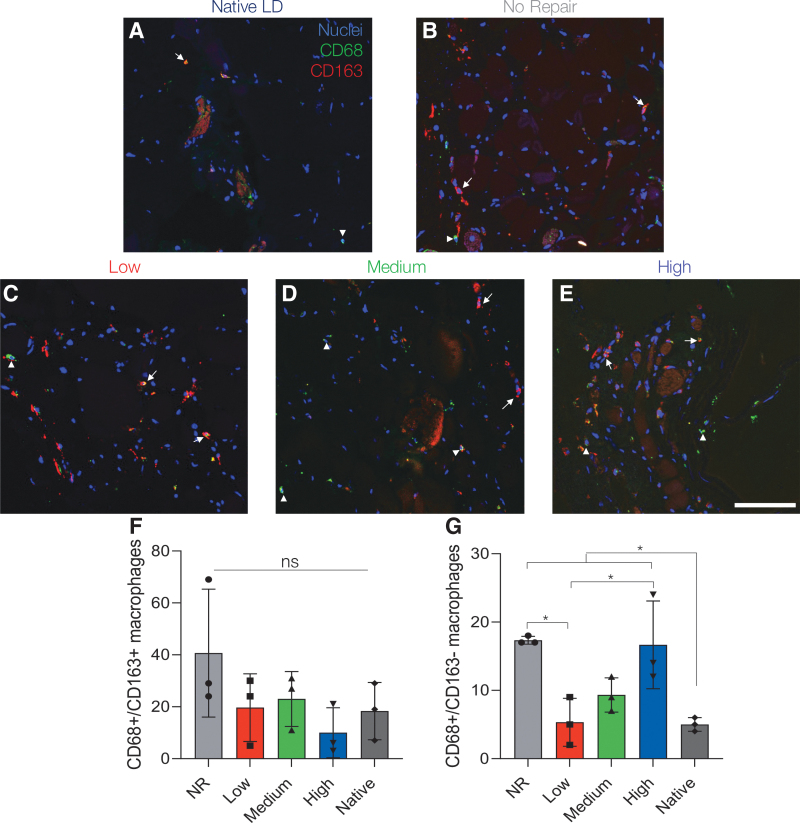
Volumetric muscle loss (VML) injuries are characterized by permanent loss of muscle mass, structure, and function. Hydrogel biomaterials provide an attractive platform for skeletal muscle tissue engineering due to the ability to easily modulate their biophysical and biochemical properties to match a range of tissue characteristics. In this work, we successfully developed a mechanically tunable hyaluronic acid (HA) hydrogel system to investigate the influence of hydrogel stiffness on VML repair. HA was functionalized with photoreactive norbornene groups to create hydrogel networks that rapidly crosslink through thiol-ene click chemistry with tailored mechanics. Mechanical properties were controlled by modulating the amount of matrix metalloproteinase-degradable peptide crosslinker to produce hydrogels with increasing elastic moduli of 1.1 ± 0.002, 3.0 ± 0.002, and 10.6 ± 0.006 kPa, mimicking a relevant range of developing and mature muscle stiffnesses. Functional muscle recovery was assessed following implantation of the HA hydrogels by in situ photopolymerization into rat latissimus dorsi (LD) VML defects at 12 and 24 weeks postinjury. After 12 weeks, muscles treated with medium stiffness (3.0 kPa) hydrogels produced maximum isometric forces most similar to contralateral healthy LD muscles. This trend persisted at 24 weeks postinjury, suggestive of sustained functional recovery. Histological analysis revealed a significantly larger zone of regeneration with more de novo muscle fibers following implantation of medium stiffness hydrogels in VML-injured muscles compared to other experimental groups. Lower (low and medium) stiffness hydrogels also appeared to attenuate the chronic inflammatory response characteristic of VML injuries, displaying similar levels of macrophage infiltration and polarization to healthy muscle. Together these findings illustrate the importance of hydrogel mechanical properties in supporting functional repair of VML injuries. Impact statement This report defines the role hydrogel mechanical properties play in the repair of volumetric muscle loss (VML) injuries. We show that an intermediate hydrogel stiffness (3 kPa) more compliant than adult muscle tissue facilitated improved and sustained regenerative outcomes up to 24 weeks postinjury in a rat latissimus dorsi model of VML. Muscles treated with 3 kPa hydrogels showed enhanced myogenesis and attenuation of the chronic inflammatory response characteristic of VML injuries. These results should help guide the future design of hydrogels for skeletal muscle tissue engineering and regeneration.

Keywords: hydrogel; regenerative medicine; volumetric muscle loss.


Conflict of interest statement

No competing financial interests exist.

Figures

FIG. 1.
FIG. 1.
Hydrogel mechanical characterization. (A) Chemical structure of NorHA. (B) Schematic of reaction between NorHA (HA: blue, norbornene: red) and MMP-degradable dithiol peptide by UV light-mediated thiol-ene click chemistry. (C) Oscillatory shear rheology showed that increasing the concentration of MMP-degradable peptide crosslinker resulted in hydrogels of increasing stiffness. Low (1 kPa), medium (3 kPa), and high (10 kPa) elastic modulus hydrogels were developed to approximate a range of physiological muscle tissue stiffnesses. Data presented as mean ± SD, n = 3 hydrogels per experimental group. HA, hyaluronic acid; LAP, lithium acylphosphinate; MMP, matrix metalloproteinase; NorHA, norbornene-modified hyaluronic acid; SD, standard deviation; UV, ultraviolet. Color images are available online.
FIG. 2.
FIG. 2.
In situ photopolymerization enables facile and reproducible hydrogel delivery to rat LD VML defects. (A) NorHA hydrogels were photopolymerized in situ and (B) conformed to the injury dimensions, completely filling the defect area. (C) Surgical dissections resulted in reproducible injuries of 148.4 ± 10.4 mg and (D) 2.44 ± 0.22 cm2, which were not statistically different between experimental groups. Data presented as box plots of interquartile range (line: median) with whiskers showing minimum and maximum values, n = 16 animals per experimental group. LD, latissimus dorsi; ns, not significant; VML, volumetric muscle loss. Color images are available online.
FIG. 3.
FIG. 3.
Medium stiffness hydrogels support muscle force generation most similar to native tissue. (A) Dose/response curve and (B) maximum isometric contraction force in response to electrical field stimulation at 12 weeks postsurgery. Statistically significant differences from native (p < 0.05) are denoted by n (NR), l (low), m (medium), and h (high). (C) Specific force (specific P0), calculated by normalizing maximal isometric force (P0) to PCSA, was similar to contralateral control values across all treatment groups, except muscles treated with the high stiffness (10 kPa) hydrogels. (D) Muscle weight at the time of explant shows NR muscles were significantly lighter than other experimental groups. Data presented as mean ± SD (A–C), while panel D data are presented as box plots of interquartile range (line: median) with whiskers showing minimum and maximum values. *p < 0.05, **p < 0.01, ****p < 0.0001. n = 8 muscles per experimental group and n = 32 for the native contralateral control. NR, no repair; PCSA, physiological cross-sectional area. Color images are available online.
FIG. 4.
FIG. 4.
Lower stiffness hydrogels enable similar force generation to native muscle at 24 weeks. (A) Dose/response curve and (B) maximum isometric contraction force in response to electrical field stimulation at 24 weeks postsurgery. Statistically significant differences from native (p < 0.05) are denoted by n (NR). (C) Specific P0 was similar to contralateral control values across all treatment groups at the time of explant. (D) Explanted muscle weight shows NR and low stiffness hydrogel-treated muscles were significantly lighter than native muscle. Data presented as mean ± SD (A–C), while panel D data are presented as box plots of interquartile range (line: median) with whiskers showing minimum and maximum values. *p < 0.05, **p < 0.01, ***p < 0.001, ****p < 0.0001. n = 8 muscles per experimental group and n = 33 for the native contralateral control. Color images are available online.
FIG. 5.
FIG. 5.
Histological analysis of LD muscles shows improved myogenesis in medium stiffness hydrogel-treated muscles at 24 weeks. (A–E) Tissue and cell morphology at the interface between native muscle and the VML injury were visualized by Masson's trichrome staining (muscle tissue in red, collagen deposition in blue, and nuclei in black). The area between the dashed red and black lines represents the zone of regeneration characterized by small disorganized myofibers. The red line denotes the interface between native muscle and VML injury site and the black line represents the last point at which detectable myofibers were present. (F) Quantification of the zone of regeneration at 24 weeks postinjury indicated improved myogenesis in medium stiffness hydrogel-treated muscles. Colors denote different experimental muscles (biological replicates, n = 3 per group) with solid shapes representing the mean and translucent shapes representing individual sections per muscle (technical replicates, n = 10). Data presented as mean ± SD. *p < 0.05, ***p < 0.001. Scale bar: 1 mm. Color images are available online.
FIG. 6.
FIG. 6.
Medium stiffness hydrogels support increased number of muscle fibers. (A) Quantification of the number of muscle fibers in the zone of regeneration per muscle section revealed that medium stiffness hydrogels supported increased myogenesis. (B) Median minimum Feret diameter (dashed lines) was significantly reduced in NR treated muscle compared to native muscle (dotted lines, first and third quartiles). (C) Minimum Feret diameter frequency distribution curve shows a leftward shift toward smaller muscle fibers compared to uninjured muscle regardless of treatment type. Statistically significant differences from native (p < 0.05) are denoted by n (NR), l (low), and h (high). Data presented as mean ± SD (A, C). *p < 0.05. n = 3 muscles per experimental group. Color images are available online.
FIG. 7.
FIG. 7.
Lower stiffness hydrogels support modest vascularization 24 weeks post-VML. (A–E) Representative images of vascular staining at 24 weeks post-VML. Images were taken in the zone of regeneration showing αSMA structures (green, arrow) and CD31 cells (red, arrowhead) at the defect-tissue interface. (F) Quantification of αSMA structures indicative of arteries and veins was significantly reduced across all experimental groups compared to native muscle. (G) Lower stiffness hydrogels (1 and 3 kPa) supported statistically similar levels of CD31+ cells to native muscle. Data presented as mean ± SD. *p < 0.05, **p < 0.01. n = 3 muscles per experimental group. Scale bar: 100 μm. αSMA, α-smooth muscle actin. Color images are available online.
FIG. 8.
FIG. 8.
Lower stiffness hydrogels support similar macrophage polarization profiles to native muscle. (A–E) Representative images of macrophage infiltration at 24 weeks post-VML. Images were taken in the zone of regeneration showing CD68 (green, arrowhead) and CD163 (red, arrow) at the defect-tissue interface. (F) Quantification of CD68+/CD163+ cells indicative of M2 macrophages showed no statistically significant difference across experimental groups. (G) CD68+/CD163- M1 macrophages remained significantly elevated in NR and high stiffness hydrogel-treated muscle indicating a prolonged inflammatory response. Data presented as mean ± SD. *p < 0.05. n = 3 muscles per experimental group. Scale bar: 100 μm. Color images are available online.

References

    1. Carlson, B., and Faulkner, J.. The regeneration of skeletal muscle fibers following injury: a review. Med Sci Sports Exerc 15, 187, 1983. - PubMed
    1. Gorgan, B., and Hsu, J.R.. Volumetric muscle loss. J Am Acad Orthop Surg 19, 35, 2011. - PubMed
    1. Passipieri, J.A., Hu, X., Mintz, E., et al. . In silico and in vivo studies detect functional repair mechanisms in a volumetric muscle loss injury. Tissue Eng Part A 25, 1272, 2019. - PMC - PubMed
    1. Goldberg, M.S. Death and injury rates of U.S. military personnel in Iraq. Mil Med 175, 220, 2010. - PubMed
    1. Corona, B.T., Rivera, J.C., Owens, J.G., Wenke, J.C., and Rathbone, C.R.. Volumetric muscle loss leads to permanent disability following extremity trauma. J Rehabil Res Dev 52, 785, 2015. - PubMed

Publication types