

Evaluation of Iodine-123 and Iodine-131 SPECT activity quantification: a Monte Carlo study
- PMID: 34410539
- PMCID: PMC8377107
- DOI: 10.1186/s40658-021-00407-1
Evaluation of Iodine-123 and Iodine-131 SPECT activity quantification: a Monte Carlo study
Abstract
Purpose: The quantitative accuracy of Nuclear Medicine images, acquired for both planar and SPECT studies, is influenced by the isotope-collimator combination as well as image corrections incorporated in the iterative reconstruction process. These factors can be investigated and optimised using Monte Carlo simulations. This study aimed to evaluate SPECT quantification accuracy for 123I with both the low-energy high resolution (LEHR) and medium-energy (ME) collimators and 131I with the high-energy (HE) collimator.
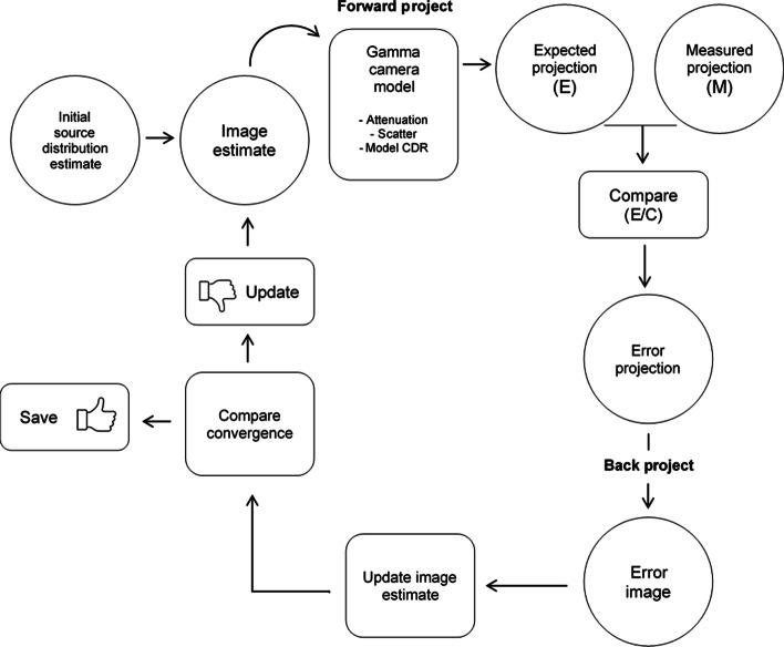
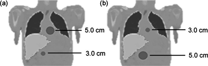
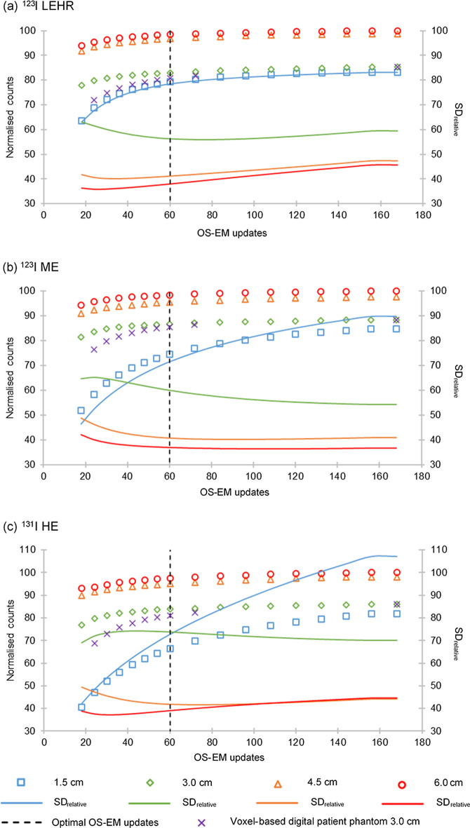
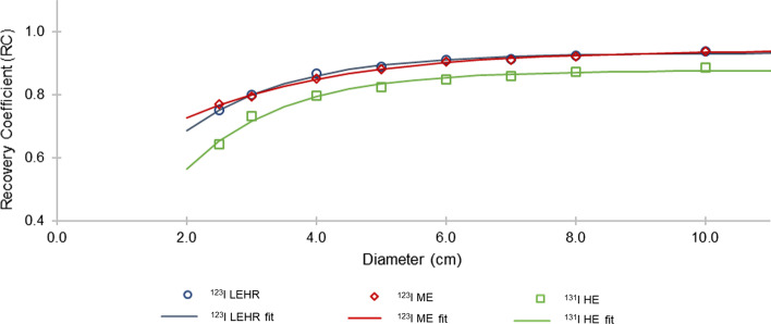
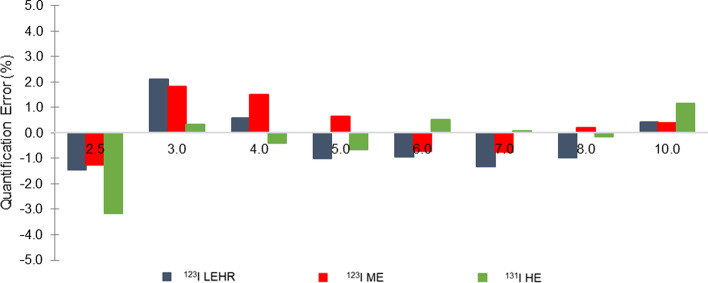
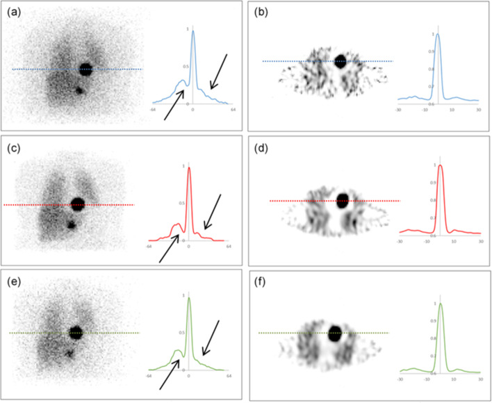
Methods: Simulated SPECT projection images were reconstructed using the OS-EM iterative algorithm, which was optimised for the number of updates, with appropriate corrections for scatter, attenuation and collimator detector response (CDR), including septal scatter and penetration compensation. An appropriate calibration factor (CF) was determined from four different source geometries (activity-filled: water-filled cylindrical phantom, sphere in water-filled (cold) cylindrical phantom, sphere in air and point-like source), investigated with different volume of interest (VOI) diameters. Recovery curves were constructed from recovery coefficients to correct for partial volume effects (PVEs). The quantitative method was evaluated for spheres in voxel-based digital cylindrical and patient phantoms.
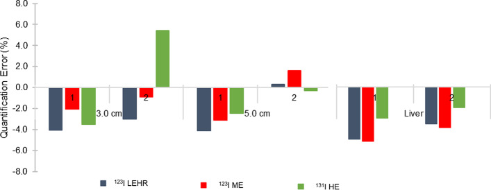
Results: The optimal number of OS-EM updates was 60 for all isotope-collimator combinations. The CFpoint with a VOI diameter equal to the physical size plus a 3.0-cm margin was selected, for all isotope-collimator geometries. The spheres' quantification errors in the voxel-based digital cylindrical and patient phantoms were less than 3.2% and 5.4%, respectively, for all isotope-collimator combinations.
Conclusion: The study showed that quantification errors of less than 6.0% could be attained, for all isotope-collimator combinations, if corrections for; scatter, attenuation, CDR (including septal scatter and penetration) and PVEs are performed. 123I LEHR and 123I ME quantification accuracies compared well when appropriate corrections for septal scatter and penetration were applied. This can be useful in departments that perform 123I studies and may not have access to ME collimators.
Keywords: 123I; 131I; Monte Carlo simulations; Quantification accuracy; SIMIND; SPECT.
© 2021. The Author(s).
Conflict of interest statement
The authors declare that they have no competing interests.
Figures
References
-
- Wieland DM, Wu J, Brown LE, Mangner TJ, Swanson DP. Radiolabeled adrenergic neuron-blocking agents : adrenomedullary imaging with [131I ] iodobenzylguanidine. J Nucl Med. 1980;21(4):349–353. - PubMed
-
- Sjögreen K, Ljungberg M, Strand S, Library PM. An activity quantification method based on registration of CT and whole-body scintillation camera images, with application to I131. J Nucl Med. 2002;43(7):972–982. - PubMed
Grants and funding
LinkOut - more resources
Full Text Sources
