

A deep learning algorithm for automatic detection and classification of acute intracranial hemorrhages in head CT scans
- PMID: 34411910
- PMCID: PMC8377493
- DOI: 10.1016/j.nicl.2021.102785
A deep learning algorithm for automatic detection and classification of acute intracranial hemorrhages in head CT scans
Abstract
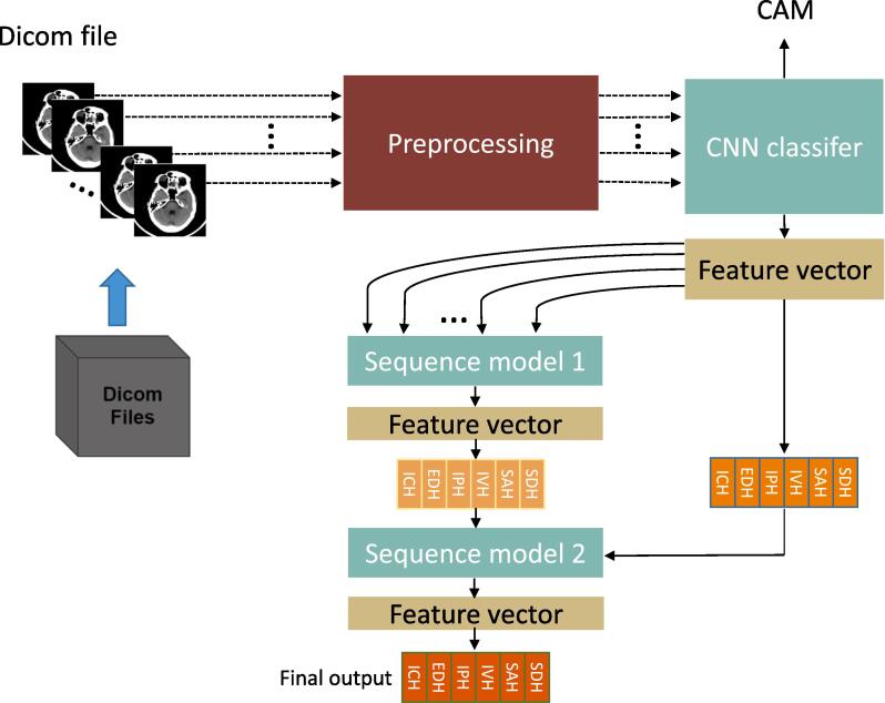
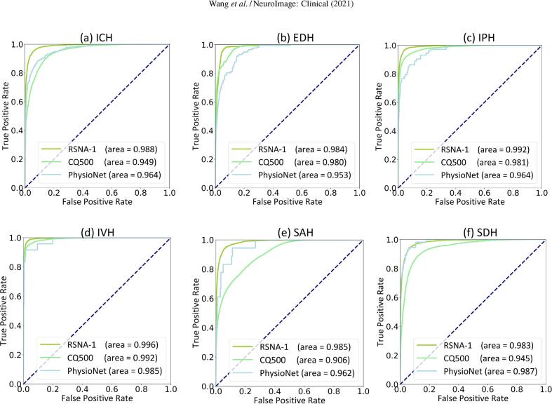
Acute Intracranial hemorrhage (ICH) is a life-threatening disease that requires emergency medical attention, which is routinely diagnosed using non-contrast head CT imaging. The diagnostic accuracy of acute ICH on CT varies greatly among radiologists due to the difficulty of interpreting subtle findings and the time pressure associated with the ever-increasing workload. The use of artificial intelligence technology may help automate the process and assist radiologists for more prompt and better decision-making. In this work, we design a deep learning approach that mimics the interpretation process of radiologists, and combines a 2D CNN model and two sequence models to achieve accurate acute ICH detection and subtype classification. Being developed using the extensive 2019-RSNA Brain CT Hemorrhage Challenge dataset with over 25000 CT scans, our deep learning algorithm can accurately classify the acute ICH and its five subtypes with AUCs of 0.988 (ICH), 0.984 (EDH), 0.992 (IPH), 0.996 (IVH), 0.985 (SAH), and 0.983 (SDH), respectively, reaching the accuracy level of expert radiologists. Our method won 1st place among 1345 teams from 75 countries in the RSNA challenge. We have further evaluated our algorithm on two independent external validation datasets with 75 and 491 CT scans, respectively, and our method maintained high AUCs of 0.964 and 0.949 for acute ICH detection. These results have demonstrated the high performance and robust generalization ability of our proposed method, which makes it a useful second-read or triage tool that can facilitate routine clinical applications.
Keywords: Deep learning; Head CT; Image classification; Intracranial hemorrhage (ICH); Sequence model.
Copyright © 2021 The Author(s). Published by Elsevier Inc. All rights reserved.
Figures
References
-
- Aslanian H.R. Nurse observation during colonoscopy increases polyp detection: a randomized prospective study. Gastrointestinal Endoscopy. 2013;108:166–172. - PubMed
-
- Bello H.R. Skull base–related lesions at routine head CT from the emergency department: pearls, pitfalls, and lessons learned. RadioGraphics. 2019;39:1161–1182. - PubMed
-
- Bishop, C.M., 2006. Pattern recognition and machine learning, Springer.
-
- Charlotte Intracerebral haemorrhage: current approaches to acute management. The Lancet. 2018;392:1257–1268. - PubMed
-
- Chilamkurthy S. Deep learning algorithms for detection of critical findings in head CT scans: a retrospective study. The Lancet. 2018;392:2388–2396. - PubMed
Publication types
MeSH terms
LinkOut - more resources
Full Text Sources
