. 2021 Aug 19;18(1):25.
doi: 10.1186/s12950-021-00291-7.

USP10 alleviates sepsis-induced acute kidney injury by regulating Sirt6-mediated Nrf2/ARE signaling pathway

Affiliations

USP10 alleviates sepsis-induced acute kidney injury by regulating Sirt6-mediated Nrf2/ARE signaling pathway

Fei Gao et al. J Inflamm (Lond). .

Abstract

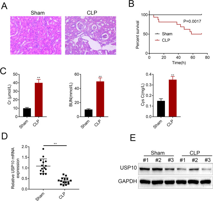
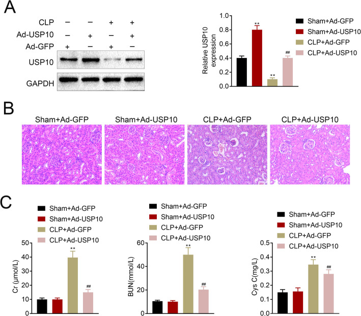
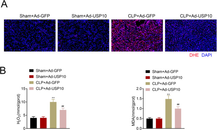
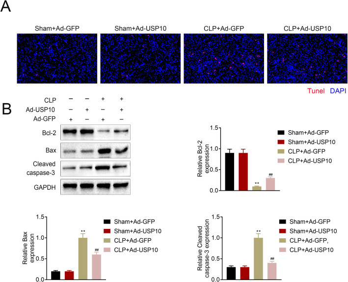
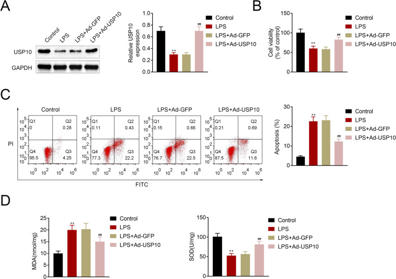
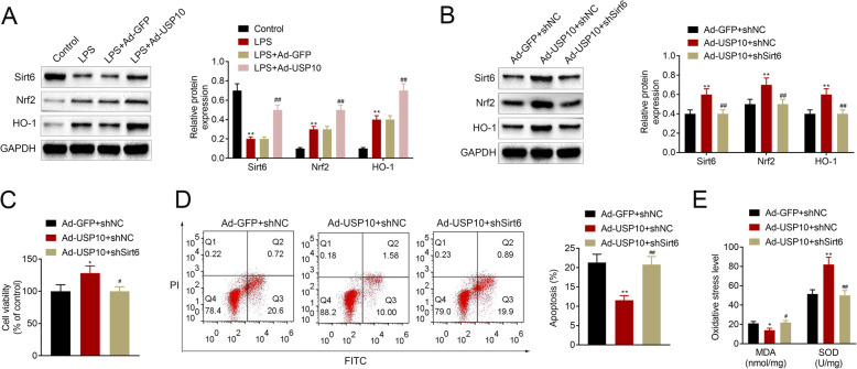
Background: Severe sepsis, a major health problem worldwide, has become one of the leading causes of death in ICU patients. Further study on the pathogenesis and treatment of acute kidney injury (AKI) is of great significance to reduce high mortality rate of sepsis. In this study, the mechanism by which ubiquitin specific peptidase 10 (USP10) reduces sepsis-induced AKI was investigated. Ligation and perforation of cecum (CLP) was employed to establish C57BL/6 mouse models of sepsis. Hematoxylin-eosin (H&E) staining was performed to detect renal injury. The concentrations of serum creatinine (Cr), urea nitrogen (BUN) and cystatin C (Cys C) were determined using a QuantiChrom™ Urea Assay kit. RT-qPCR and western blot were conducted to assess the USP10 expression level. DHE staining was used to detect reactive oxygen species (ROS) levels. H2O2, MDA and SOD levels were assessed using corresponding colorimetric kits. Western blot was used to examine the expression levels of Bcl-2, Bax, cleaved caspase-3, Sirt6, Nrf2 and HO-1. MTT assay was used to determine cell viability, whereas TUNEL staining and flow cytometry were used to assess cell apoptosis.

Results: In this study, we found that USP10 was decreased in CLP-induced mouse renal tissues. We identified that USP10 alleviated renal dysfunction induced by CLP. Moreover, USP10 was found to reduce oxidative stress, and abated LPS-induced renal tubular epithelial cell injury and apoptosis. Finally, we discovered that USP10 promoted activation of the NRF2/HO-1 pathway through SIRT6 and attenuated LPS-induced renal tubular epithelial cell injury.

Conclusions: This study found that USP10 activates the NRF2/ARE signaling through SIRT6. USP10 alleviates sepsis-induced renal dysfunction and reduces renal tubular epithelial cell apoptosis and oxidative stress.

Keywords: AKI; ARE; CLP; NRF2; SIRT6; USP10.


Conflict of interest statement

The authors state that there are no conflicts of interest to disclose.

Figures

Fig. 1
Fig. 1
USP10 is decreased in CLP-induced mouse renal tissues. (A) H&E images show the cell morphology and inflammatory changes in renal tissues of the CLP and sham groups (n = 15). (B) Graph shows 72-hour survival rates. (C) The concentrations of Cr, BUN and Cys C were examined at 24 h after CLP. The mRNA (D) and protein (E) expression levels of USP10 were analyzed in the CLP and sham groups, **p < 0.01. GAPDH was used as an internal control
Fig. 2
Fig. 2
USP10 alleviates renal dysfunction induced by CLP. (A) Western blot images show the transfection efficiency of USP10, **p < 0.01. GAPDH was used as an internal control. (B) H&E images show the cell morphology and inflammatory changes in renal tissues of the Sham + Ad-GFP, Sham + Ad-USP10, CLP + Ad-GFP and CLP + Ad-USP10 groups (n = 15). (C) The concentrations of Cr, BUN and Cys C were examined at 24 h after CLP in the Sham + Ad-GFP, Sham + Ad-USP10, CLP + Ad-GFP and CLP + Ad-USP10 groups, **p < 0.01
Fig. 3
Fig. 3
USP10 attenuates CLP-induced oxidative stress in renal tissues. (A) Representative DHE staining images of the Sham + Ad-GFP, Sham + Ad-USP10, CLP + Ad-GFP and CLP + Ad-USP10 groups demonstrating ROS levels, bar = 20 μm. (B) Comparison of H2O2 and MDA levels in renal tissues (n = 8) of the Sham + Ad-GFP, Sham + Ad-USP10, CLP + Ad-GFP and CLP + Ad-USP10 groups using the colorimetric kits. **p < 0.01
Fig. 4
Fig. 4
USP10 reduces CLP-induced apoptosis in renal tissues. (A) Representative TUNEL staining images of renal tissues of the Sham + Ad-GFP, Sham + Ad-USP10, CLP + Ad-GFP and CLP + Ad-USP10 groups (n = 8) indicating the apoptotic rate, bar = 20 μm. (B) Western blot images demonstrating Bcl-2, Bax, and cleaved caspase-3 expression levels in renal tissues of the Sham + Ad-GFP, Sham + Ad-USP10, CLP + Ad-GFP and CLP + Ad-USP10 groups, **p < 0.01. GAPDH was used as an internal control
Fig. 5
Fig. 5
USP10 attenuates LPS-induced renal tubular epithelial cell injury. (A) Western blot images showing USP10 protein levels in the control, LPS, LPS + Ad-GFP and LPS + Ad-USP10 treated human renal tubular epithelial cell line, HK-2, **p < 0.01. GAPDH was used as an internal control. (B) MTT assay shows cell viability in control, LPS, LPS + Ad-GFP and LPS + Ad-USP10 HK-2 cells, **p < 0.01. (C) Flow cytometry charts show cell apoptosis in control, LPS, LPS + Ad-GFP and LPS + Ad-USP10 HK-2 cells, **p < 0.01. (D) Comparison of MDA and SOD levels in four HK-2 treatment groups using the colorimetric kits, **p < 0.01
Fig. 6
Fig. 6
USP10 promotes activation of the NRF2/HO-1 pathway through SIRT6 and attenuates LPS-induced renal tubular epithelial cell injury. (A-B) Western blot images show the expression levels of Sirt6, Nrf2 and HO-1 in control, LPS, LPS + Ad-GFP, LPS + Ad-Sirt6, Ad-GFP + shNC, Ad-USP10 + shNC and Ad-USP10 + shSirt6 HK-2 cells, **p < 0.01. GAPDH was used as an internal control. (C) MTT assay demonstrating cell viability in Ad-GFP + shNC, Ad-USP10 + shNC and Ad-USP10 + shSirt6 HK-2 cells, *p < 0.05. (D) Flow cytometry charts showing cell apoptosis in Ad-GFP + shNC, Ad-USP10 + shNC and Ad-USP10 + shSirt6 HK-2 cells, **p < 0.01. (E) Comparison of MDA and SOD levels in Ad-GFP + shNC, Ad-USP10 + shNC and Ad-USP10 + shSirt6 HK-2 cells using the colorimetric kits, *p < 0.05, **p < 0.01

References

    1. Rhodes A, Evans LE, Alhazzani W, Levy MM, Antonelli M, Ferrer R, et al. Surviving Sepsis Campaign: International Guidelines for Management of Sepsis and Septic Shock: 2016. Intensive care medicine. 2017;43(3):304–77. doi: 10.1007/s00134-017-4683-6. - DOI - PubMed
    1. Poukkanen M, Wilkman E, Vaara ST, Pettilä V, Kaukonen KM, Korhonen AM, et al. Hemodynamic variables and progression of acute kidney injury in critically ill patients with severe sepsis: data from the prospective observational FINNAKI study. Crit Care (London England) 2013;17(6):R295. doi: 10.1186/cc13161. - DOI - PMC - PubMed
    1. Wang Z, Jin L, Shen T, Zhan S. The Value of Urine NAG, NGAL Combined with Serum Cys-C in Early Diagnosis of Neonatal Hyperbilirubinemia-related Acute Kidney Injury. Signa Vitae. 2020;16(2):109–13.
    1. Basile DP, Yoder MC. Renal endothelial dysfunction in acute kidney ischemia reperfusion injury. Cardiovasc Hematol Disord Drug Targets. 2014;14(1):3–14. doi: 10.2174/1871529X1401140724093505. - DOI - PMC - PubMed
    1. Lee S, Lee SJ, Coronata AA, Fredenburgh LE, Chung SW, Perrella MA, et al. Carbon monoxide confers protection in sepsis by enhancing beclin 1-dependent autophagy and phagocytosis. Antioxid Redox Signal. 2014;20(3):432–42. doi: 10.1089/ars.2013.5368. - DOI - PMC - PubMed