Observational Study
. 2021 Aug 19;6(62):eabl4348.
doi: 10.1126/sciimmunol.abl4348.

X-linked recessive TLR7 deficiency in ~1% of men under 60 years old with life-threatening COVID-19

Takaki Asano #  1 Bertrand Boisson #  2   3   4 Fanny Onodi #  5 Daniela Matuozzo #  3   4 Marcela Moncada-Velez #  1 Majistor Raj Luxman Maglorius Renkilaraj #  3   4 Peng Zhang #  1 Laurent Meertens #  5 Alexandre Bolze #  6 Marie Materna #  3   4 Sarantis Korniotis  7 Adrian Gervais  3   4 Estelle Talouarn  3   4 Benedetta Bigio  1 Yoann Seeleuthner  3   4 Kaya Bilguvar  8 Yu Zhang  9   10 Anna-Lena Neehus  3   4 Masato Ogishi  1 Simon J Pelham  1 Tom Le Voyer  3   4 Jérémie Rosain  3   4 Quentin Philippot  3   4 Pere Soler-Palacín  11   12   13 Roger Colobran  13   14   15 Andrea Martin-Nalda  11   12   13 Jacques G Rivière  11   12   13 Yacine Tandjaoui-Lambiotte  16   17 Khalil Chaïbi  18   19 Mohammad Shahrooei  20   21 Ilad Alavi Darazam  22   23 Nasrin Alipour Olyaei  20   21 Davood Mansouri  24   25   26 Nevin Hatipoğlu  27 Figen Palabiyik  27 Tayfun Ozcelik  28 Giuseppe Novelli  29 Antonio Novelli  30 Giorgio Casari  31   32 Alessandro Aiuti  31   33 Paola Carrera  32 Simone Bondesan  32 Federica Barzaghi  33 Patrizia Rovere-Querini  31   34 Cristina Tresoldi  35 Jose Luis Franco  36 Julian Rojas  36 Luis Felipe Reyes  37 Ingrid G Bustos  37 Andres Augusto Arias  1   36   38 Guillaume Morelle  39 Kyheng Christèle  39 Jesús Troya  40 Laura Planas-Serra  41   42 Agatha Schlüter  41   42 Marta Gut  43 Aurora Pujol  41   42   44 Luis M Allende  45   46 Carlos Rodriguez-Gallego  47   48 Carlos Flores  49   50   51   52 Oscar Cabrera-Marante  45 Daniel E Pleguezuelo  45 Rebeca Pérez de Diego  53 Sevgi Keles  54 Gokhan Aytekin  55 Ozge Metin Akcan  54 Yenan T Bryceson  56 Peter Bergman  57   58 Petter Brodin  59 Daniel Smole  60 C I Edvard Smith  61   58 Anna-Carin Norlin  58 Tessa M Campbell  56 Laura E Covill  56 Lennart Hammarström  62 Qiang Pan-Hammarström  62 Hassan Abolhassani  62   63 Shrikant Mane  64 Nico Marr  65 Manar Ata  65 Fatima Al Ali  65 Taushif Khan  65 András N Spaan  1   66 Clifton L Dalgard  67   68 Paolo Bonfanti  69 Andrea Biondi  70 Sarah Tubiana  71   72 Charles Burdet  71   73 Robert Nussbaum  74 Amanda Kahn-Kirby  74 Andrew L Snow  75 COVID Human Genetic EffortCOVID-STORM CliniciansCOVID CliniciansImagine COVID GroupFrench COVID Cohort Study GroupCoV-Contact CohortAmsterdam UMC Covid-BiobankNIAID-USUHS COVID Study GroupJacinta Bustamante  1   3   4   76 Anne Puel  1   3   4 Stéphanie Boisson-Dupuis  1   3   4 Shen-Ying Zhang  1   3   4 Vivien Béziat  1   3   4 Richard P Lifton #  8   77 Paul Bastard #  3   4 Luigi D Notarangelo #  9   10 Laurent Abel #  1   3   4 Helen C Su #  9   10   78 Emmanuelle Jouanguy #  1   3   4 Ali Amara #  5 Vassili Soumelis #  7   79 Aurélie Cobat #  1   3   4 Qian Zhang #  1 Jean-Laurent Casanova #  2   3   4   80
Collaborators, Affiliations
Observational Study

X-linked recessive TLR7 deficiency in ~1% of men under 60 years old with life-threatening COVID-19

Takaki Asano et al. Sci Immunol. .

Abstract

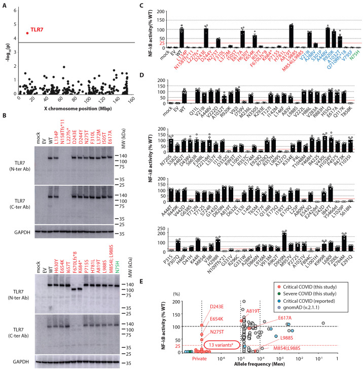
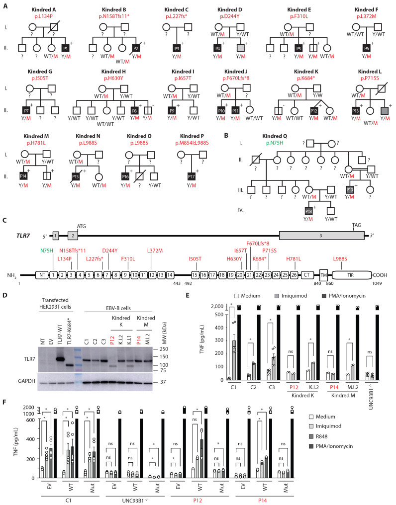
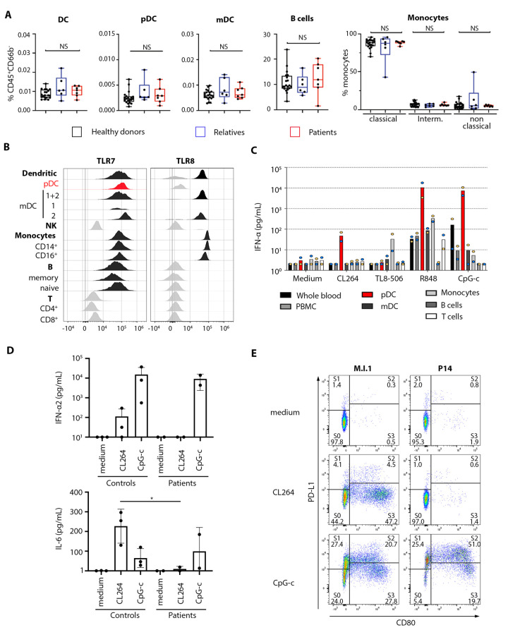
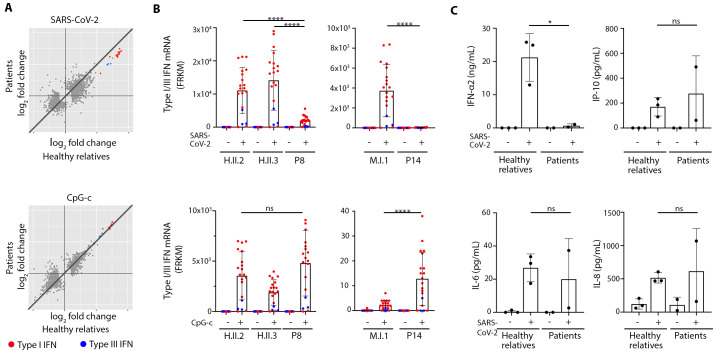
Autosomal inborn errors of type I IFN immunity and autoantibodies against these cytokines underlie at least 10% of critical COVID-19 pneumonia cases. We report very rare, biochemically deleterious X-linked TLR7 variants in 16 unrelated male individuals aged 7 to 71 years (mean: 36.7 years) from a cohort of 1,202 male patients aged 0.5 to 99 years (mean: 52.9 years) with unexplained critical COVID-19 pneumonia. None of the 331 asymptomatically or mildly infected male individuals aged 1.3 to 102 years (mean: 38.7 years) tested carry such TLR7 variants (p = 3.5 × 10⁻⁵). The phenotypes of five hemizygous relatives of index cases infected with SARS-CoV-2 include asymptomatic or mild infection (n=2, 5 and 38 years), or moderate (n=1, 5 years), severe (n=1, 27 years), or critical (n=1, 29 years) pneumonia. Two boys (aged 7 and 12 years) from a cohort of 262 male patients with severe COVID-19 pneumonia (mean: 51.0 years) are hemizygous for a deleterious TLR7 variant. The cumulative allele frequency for deleterious TLR7 variants in the male general population is < 6.5x10⁻⁴ We also show that blood B cell lines and myeloid cell subsets from the patients do not respond to TLR7 stimulation, a phenotype rescued by wild-type TLR7 The patients' blood plasmacytoid dendritic cells (pDCs) produce low levels of type I IFNs in response to SARS-CoV-2. Overall, X-linked recessive TLR7 deficiency is a highly penetrant genetic etiology of critical COVID-19 pneumonia, in about 1.8% of male patients below the age of 60 years. Human TLR7 and pDCs are essential for protective type I IFN immunity against SARS-CoV-2 in the respiratory tract.


Figures

Fig. 1
Fig. 1
Enrichment in rare TLR7 deleterious alleles among men with critical COVID-19 pneumonia. (A) Manhattan plot showing the results of the variant enrichment test for the 190 genes of the X chromosome with at least 5 patients carrying non-synonymous variants. The gray line indicates the corresponding Bonferroni-corrected significance threshold. (B) Western blot of extracts from non-transfected HEK293T cells (mock), HEK293T cells transfected with pCMV6 empty vector (EV), the wild-type (WT) TLR7 allele, or one of the TLR7 variant alleles of interest. All extracts were probed with monoclonal antibodies specific for the leucine-rich repeats to the N terminus (N-ter) or amino-acid 1,000 to the C terminus (C-ter) within the human TLR7 protein. (C) (D) Luciferase assay on HEK293T cells transfected with the pGL4.32 luciferase reporter construct and an expression vector for Renilla luciferase together with no vector (mock), EV, WT, or TLR7 variants: (C) 21 variants found in our cohort and eight previously reported variants, (D) 109 variants found in male individuals from the gnomAD database. After 24 hours, transfected cells were left untreated or were treated by incubation with 1 μg/mL R848 for 24 hours. These data were established from two independent experiments. The y-axis represents NF-κB transcriptional activity as a percentage of the WT. The x-axis indicates the alleles used for transfection. (E) Diagram showing the correlation between allele frequency and NF-κB activity (% of WT). The 20 variants from 21 patients with critical SARS-CoV-2 from our cohort are shown in red, one variant from 2 patients with severe SARS-CoV-2 from our cohort are shown in green, the eight previously reported variants are shown in blue and the 109 variants found in the general population (allele frequency above 10−5 in men) are shown in gray. Activity of all LOF/hypomorphic alleles compared to WT allele were statistically significance (one-way ANOVA with Dunnett’s post hoc test, P < 0.01).
Fig. 2
Fig. 2
X-linked recessive TLR7 deficiency in 16 kindreds. (A) Pedigrees of the 16 kindreds containing 17 patients with life-threatening COVID-19 pneumonia (P1-17) bearing deleterious TLR7 alleles. The mutations are indicated above each pedigree. Solid black symbols indicate patients with critical COVID-19, and solid dark gray symbols indicate severe cases and solid light gray symbols indicate mild/moderate cases. The genotype is indicated under each symbol, with M corresponding to the mutation found in each kindred. ‘+’ and ‘-’ indicate the presence and absence, respectively, of antibodies against SARS-CoV-2 in the serum of the individual. Asymptomatic or paucisymptomatic family members hemizygous for the mutation are indicated by bold vertical lines. (B) Pedigree of one kindred containing two patients with severe COVID-19. (C) Schematic representation of TLR7. The upper part represents the genomic organization of the TLR7 locus, with rectangles for the various exons of the gene, and exon numbers indicated within the rectangle. The bottom part shows the primary structure of TLR7. The N-terminal portion and the leucine-rich repeat containing 26 leucine residues are located in the lumen of the endosome, and TM indicates the transmembrane domain. The Toll/interleukin-1 (IL-1) receptor (TIR) domain is cytoplasmic. The deleterious mutations reported in this study are indicated. (D) TLR7 expression in unstimulated EBV-B cells from two patients with XR TLR7 deficiency (P12 and P14), the fathers of P12 and P14, and the mother of P12, and three healthy donors (Control 1 to 3), determined by Western blotting with detection with a specific TLR7 antibody. (E) TNF production by XR TLR7-deficient EBV-B cells from two independent experiments. Cells were either left untreated or were stimulated with 5 μg/mL imiquimod (gray), or 25 ng/mL PMA and 0.25 μM ionomycin (black) for 24 hours and TNF production were measured by ELISA. (F) TNF production in XR TLR7-deficient EBV-B cells re-expressing WT TLR7 from three independent experiments. EBV-B cells from a control, P12, P14, or an UNC-93B-deficient patient, cultured in the presence of IRAK4 inhibitor (PF06650833- 5 μM) were transduced with lentiviral particles that were empty or contained the WT TLR7 or mutant TLR7 cDNA. The cells were incubated for 24 hours without IRAK4 inhibitor and were then left untreated or were stimulated with 5 μg/mL imiquimod (light gray), 1 μg/mL R848 (dark gray), or 25 ng/mL PMA and 0.25 μM ionomycin (black) for 24 hours, and TNF production were measured by ELISA. Statistical tests were performed using one-way ANOVA with Dunnett’s post hoc test (*: P < 0.05, ns: not significant).
Fig. 3
Fig. 3
Type I IFN responses to TLR7 agonist in TLR7-deficient pDCs and leukocytes. (A) Frequencies of five leukocyte subsets in whole blood, determined by CyTOF. Healthy donors (black rectangles), relatives not carrying deleterious TLR7 alleles (blue rectangles) and hemizygous TLR7 variant carriers (red rectangles) are depicted. (B) TLR7 and TLR8 expression in different leukocyte subsets, determined by flow cytometry for the healthy control (C1). The result for another healthy control (C2) is shown in Figure S5C. Gating strategy for the classification in each cell subset is shown in Data file S6. (C) IFN-α production in purified leukocyte subsets from two healthy donors (blue or yellow dot) with and without stimulation with various TLR7, 8, or 9 agonists (1 μg/mL CL264, 100 ng/mL TL8-506, 1 μg/mL R848, or 2 μM CpG-c) for 24 hours. The y-axis shows IFN-α production on a logarithmic scale. The red bar corresponds to pDCs. (D) pDCs isolated from healthy donors and TLR7-deficient patients (P8, P14) were either left untreated (medium) or were stimulated with CL264 or CpG-c, and the production of IFN-α2 and IL-6 was assessed with CBAs on the supernatant. (E) Dotplot showing pDC diversification into subsets S1, S2, and S3 from magnetically sorted blood. pDCs from a TLR7-deficient patient (P14) and a healthy relative (M.I.1) were cultured for 24 hours with medium alone or with 1 μg/mL CL264 or 2 μM CpG-c. Statistical tests were performed using unpaired two-sample t test (*: P < 0.05).
Fig. 4
Fig. 4. Type I IFN responses to SARS-CoV-2 infection in TLR7-deficient pDCs.
(A) pDCs isolated from healthy relatives and TLR7-deficient patients (P8, P14) were either left untreated or were infected with SARS-CoV-2 for 24 hours. RNA profiles were then determined by RNA-seq. Genes with expression >2.0-fold higher or lower in controls after stimulation or infection are plotted as the fold-change in expression. (B) Induction of the type I and III IFN genes from (A) infected with SARS-CoV-2 for 24 hours (top) or stimulated with CpG-c (bottom). (C) pDCs isolated from healthy relatives and TLR7-deficient patients (P8, P14) were either left untreated or were infected with SARS-CoV-2 for 24 hours and the production of IFN-α2, IP-10, IL-6 and IL-8 was measured with CBAs on the supernatant. Statistical tests were performed using unpaired two-sample t test (*: P < 0.05, ****: P < 0.0001, ns: not significant).

References

    1. Casanova J. L., Su H. C.; COVID Human Genetic Effort , A Global Effort to Define the Human Genetics of Protective Immunity to SARS-CoV-2 Infection. Cell 181, 1194–1199 (2020). 10.1016/j.cell.2020.05.016 - DOI - PMC - PubMed
    1. Zhang Q., Bastard P., Bolze A., Jouanguy E., Zhang S. Y., Cobat A., Notarangelo L. D., Su H. C., Abel L., Casanova J. L.; COVID Human Genetic Effort , Life-Threatening COVID-19: Defective Interferons Unleash Excessive Inflammation. Med (N Y) 1, 14–20 (2020). - PMC - PubMed
    1. O’Driscoll M., Ribeiro Dos Santos G., Wang L., Cummings D. A. T., Azman A. S., Paireau J., Fontanet A., Cauchemez S., Salje H., Age-specific mortality and immunity patterns of SARS-CoV-2. Nature 590, 140–145 (2021). 10.1038/s41586-020-2918-0 - DOI - PubMed
    1. Scully E. P., Haverfield J., Ursin R. L., Tannenbaum C., Klein S. L., Considering how biological sex impacts immune responses and COVID-19 outcomes. Nat. Rev. Immunol. 20, 442–447 (2020). 10.1038/s41577-020-0348-8 - DOI - PMC - PubMed
    1. Ciancanelli M. J., Huang S. X., Luthra P., Garner H., Itan Y., Volpi S., Lafaille F. G., Trouillet C., Schmolke M., Albrecht R. A., Israelsson E., Lim H. K., Casadio M., Hermesh T., Lorenzo L., Leung L. W., Pedergnana V., Boisson B., Okada S., Picard C., Ringuier B., Troussier F., Chaussabel D., Abel L., Pellier I., Notarangelo L. D., García-Sastre A., Basler C. F., Geissmann F., Zhang S. Y., Snoeck H. W., Casanova J. L., Infectious disease. Life-threatening influenza and impaired interferon amplification in human IRF7 deficiency. Science 348, 448–453 (2015). 10.1126/science.aaa1578 - DOI - PMC - PubMed

Publication types