. 2021 Aug 19;4(1):987.
doi: 10.1038/s42003-021-02508-0.

LDL receptor-peptide conjugate as in vivo tool for specific targeting of pancreatic ductal adenocarcinoma

Affiliations

LDL receptor-peptide conjugate as in vivo tool for specific targeting of pancreatic ductal adenocarcinoma

Angélina Acier et al. Commun Biol. .

Abstract

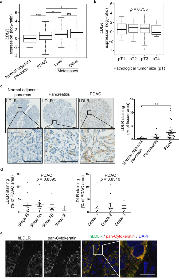
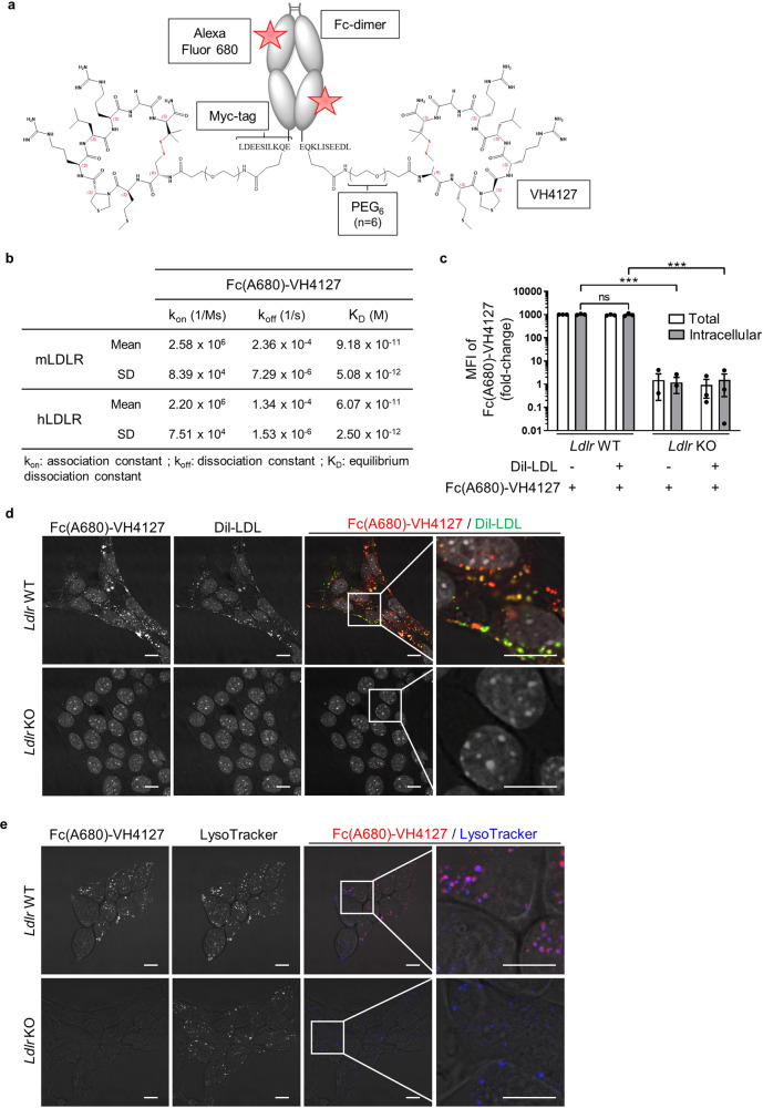
Despite clinical advances in diagnosis and treatment, pancreatic ductal adenocarcinoma (PDAC) remains the third leading cause of cancer death, and is still associated with poor prognosis and dismal survival rates. Identifying novel PDAC-targeted tools to tackle these unmet clinical needs is thus an urgent requirement. Here we use a peptide conjugate that specifically targets PDAC through low-density lipoprotein receptor (LDLR). We demonstrate by using near-infrared fluorescence imaging the potential of this conjugate to specifically detect and discriminate primary PDAC from healthy organs including pancreas and from benign mass-forming chronic pancreatitis, as well as detect metastatic pancreatic cancer cells in healthy liver. This work paves the way towards clinical applications in which safe LDLR-targeting peptide conjugate promotes tumor-specific delivery of imaging and/or therapeutic agents, thereby leading to substantial improvements of the PDAC patient's outcome.


Conflict of interest statement

The authors declare no competing interests.

Figures

Fig. 1
Fig. 1. Human LDLR is a promising candidate for PDAC targeting.
Box-and-whisker plots of human LDLR expression (a) in normal adjacent pancreas (n = 96 biologically independent samples), and in primary (n = 728 biologically independent samples) and metastatic PDAC (n = 76 biologically independent samples, including 35 liver metastases); b in primary PDAC according to pathological tumor size (pT) (pT1: n = 17, pT2: n = 61, pT3: n = 294 and pT4: n = 11 biologically independent samples). a, b Median values of LDLR expression in PDAC and metastasis samples were compared to that of normal adjacent pancreas samples and expressed as log2-ratio. c Representative immunohistochemistry (IHC) and automatic quantitation of LDLR in pancreatitis (acute: n = 1 and chronic: n = 6 biologically independent samples), PDAC (n = 32 biologically independent samples) and normal adjacent pancreas (n = 22 biologically independent samples) from patient pancreatic tissue microarray. Mean value ± s.e.m. of two independent tissue cores from each patient sample was illustrated. LDLR IHC images: ×6.8 and ×20 magnification, scale bar: 100 µm. d LDLR levels in patient PDAC according to tumor stage (left, n = 32 biologically independent samples) and grade (right, n = 27 biologically independent samples). a, d One-way ANOVA with post-hoc Tukey HSD test, ns: no significant difference, *p < 0.05, **p < 0.01, ***p < 0.001. e Representative confocal microscopy images of BxPC-3 orthotopic xenograft sections representing co-staining of human LDLR (in green color) and epithelial tumor cell marker (pan-Cytokeratin in red color). ×60 magnification, scale bar: 10 µm. An enlarged merge image of the indicated part is provided in the inset box. n = 3 mice/group.
Fig. 2
Fig. 2. LDLR-dependent internalization and trafficking of LDL toward lysosomal compartment in pancreatic cancer cells.
a Representative confocal microscopy images of total and/or cell surface LDLR (in green color) in 2D and 3D Ldlr WT cells. ×60 magnification, scale bar: 10 µm. An enlarged merge image of indicated part is provided in inset box. n = 2 independent experiments. b Flow cytometry assessment of total DiI-LDL (i.e., bound and internalized) in Ldlr WT and KO PK4A cells and of intracellular DiI-LDL in cells previously treated with an acid solution. Data are represented as fold-change ± s.e.m. of the mean of fluorescence intensity (MFI) of DiI-LDL and are expressed relative to value obtained in Ldlr WT cells and arbitrarily set to 1. One-way ANOVA with post-hoc Tukey HSD test, ***p < 0.001. n = 3 independent experiments. c Representative confocal microscopy images of DiI-LDL (in red color) and LysoTracker blue (i.e., late acidic endosome and lysosome marker) in live Ldlr WT and KO PK4A cells. x60 magnification, scale bar: 10 µm. An enlarged merge image of indicated part is provided in inset box. n = 3 independent experiments.
Fig. 3
Fig. 3. LDLR-dependent uptake of Fc(A680)-VH4127 conjugate by pancreatic cancer cells.
a Schematic structure of the fluorescent Fc(A680)-VH4127 conjugate. b Kinetic parameters (kon and koff) and equilibrium dissociation constant (KD) of Fc(A680)-VH4127 conjugate. Data are represented as mean ± standard deviation (SD). n = 3 independent experiments. c Flow cytometry analysis of total and internalized Fc(A680)-VH4127 in Ldlr WT and KO PK4A cells with or without DiI-LDL. Data are represented as fold-change ± s.e.m. of the mean of fluorescence intensity (MFI) of Fc(A680)-VH4127 expressed relative to value measured in Ldlr WT cells incubated without DiI-LDL and arbitrarily set to 1000. One-way ANOVA with post-hoc Tukey HSD test, ns: no significant difference, ***p < 0.001. n = 3 independent experiments. d, e Representative confocal microscopy images of Fc(A680)-VH4127 (pseudo-colored red) and DiI-LDL (pseudo-colored green) (d) or LysoTracker blue (e) in live Ldlr WT and KO PK4A cells. ×60 magnification, scale bar: 10 µm. An enlarged merge image of indicated part is provided in inset box. n = 3 independent experiments.
Fig. 4
Fig. 4. The Fc(A680)-VH4127 specifically targets subcutaneous pancreatic tumors.
a Representative merged fluorescence and visible light images of Ldlr WT and Ldlr KO pancreatic tumors grown subcutaneously in immunodeficient mice. Fluorescence acquisitions were performed before intravenous injection (IV) of Fc(A680)-VH4127 conjugate (1 nmole/mouse) and 1, 4, and 24 h after. The kinetic of tumor-specific fluorescence of Fc(A680)-VH4127 (i.e., region of interest (ROI)) is illustrated and expressed as mean (ph/s/cm2/sr) ± s.e.m. b Representative merged fluorescence and visible light images of the tumors (Ldlr WT and Ldlr KO), pancreas (P), liver (L), adrenal glands (Ag) and kidney (K) acquired 24 h after administration of Fc(A680)-VH4127 conjugate. The tissue-specific fluorescence of Fc(A680)-VH4127 was illustrated and expressed as mean (ph/s/cm2/sr) ± s.e.m. a, b One-way ANOVA with post-hoc Tukey HSD test, ns: no significant difference, *p < 0.05, **p < 0.01, ***p < 0.001. n = 8–13 mice at each time-point.
Fig. 5
Fig. 5. The Fc(A680)-VH4127 conjugate discriminates advanced PDAC from inflammatory and healthy pancreas without causing hepatic and renal damages.
a Representative merged fluorescence and visible light images of control (KI), induced-chronic pancreatitis (KI + CP), and spontaneous PDAC-bearing (KIC) mice before, 24 and 48 h after retro-orbital IV injection of the LDLR-targeting conjugate (1 nmole/mouse). Quantification of Fc(A680)-VH4127 fluorescence in abdominal region (bladder excluded) of each experimental group is illustrated (left graph). Receiver operating characteristic (ROC) curve established from Fc(A680)-VH4127 fluorescence values obtained at 48 h post-injection in each experimental group is shown in right graph. b, c Representative merged fluorescence and visible light images of KI, KI + CP, and KIC mice after euthanasia and laparotomy (b) and of excised pancreas (healthy, inflammatory or tumoral), liver (L), kidney (K), adrenal glands (Ag) and spleen (S) (c), performed 48 h after Fc(A680)-VH4127 injection. Quantification of Fc(A680)-VH4127 fluorescence in abdominal region (bladder excluded) (b) or organ area (c) is illustrated. a, c Data are expressed as mean (ph/s/cm2/sr) ± s.e.m. One-way ANOVA with post-hoc Tukey HSD test, ns: no significant difference, *p < 0.05, **p < 0.01, ***p < 0.001. n = 5 KI and KIC mice and n = 8 KI + CP mice. d Aspartate aminotransferase (AST) activity and creatinine levels in serum of KI and KIC mice measured 24 and 48 h after injection of Fc(A680)-VH4127 (1 nmole/mouse) or vehicle (0.9% NaCl, equivalent volume). One-way ANOVA with post-hoc Tukey HSD test, N.D.: not detectable. n = 3–7 mice/group.
Fig. 6
Fig. 6. The Fc(A680)-VH4127 conjugate detects pancreatic tumor mass in liver.
a Representative IHC of LDLR in liver metastasis from PDAC resected patient. ×6.8 and ×20 magnification, scale bar: 100 µm. n = 2 independent samples. b Representative image of PDAC foci in liver from KI mice transplanted with PK4A cells (left) and of healthy liver from KI mice (right). c, d Representative Haematoxylin Phloxine Saffron (HPS) (c) and LDLR (d) staining of PDAC foci (T) in liver (L) of transplanted-KI mice, delimited by yellow dots. An enlarged image of indicated part is provided in inset box. n = 5 mice. c Left panel: ×2 magnification, scale bar: 1000 µm; right panel: ×10 magnification, scale bar: 500 µm. d ×20 magnification, scale bar: 100 µm. e Representative merged fluorescence and visible light images of excised healthy- and tumoral liver from KI and transplanted-KI mice, respectively, obtained 48 h after retro-orbital IV injection of Fc(A680)-VH4127 (1 nmole/mouse). Quantification of ROI-specific Fc(A680)-VH4127 signal relative to healthy pancreas from KI mice and to tumoral foci and adjacent normal liver from transplanted-KI mice. Data are expressed as mean (ph/s/cm2/sr) ± s.e.m. One-way ANOVA with post-hoc Tukey HSD test, ns: no significant difference, *p < 0.05. n = 5 and n = 7 KI and transplanted-KI mice, respectively.

References

    1. Ferlay J, et al. Estimating the global cancer incidence and mortality in 2018: GLOBOCAN sources and methods. Int. J. Cancer. 2019;144:1941–1953. doi: 10.1002/ijc.31937. - DOI - PubMed
    1. Rahib L, et al. Projecting cancer incidence and deaths to 2030: the unexpected burden of thyroid, liver, and pancreas cancers in the United States. Cancer Res. 2014;74:2913–2921. doi: 10.1158/0008-5472.CAN-14-0155. - DOI - PubMed
    1. Mizrahi JD, Surana R, Valle JW, Shroff RT. Pancreatic cancer. Lancet (Lond., Engl.) 2020;395:2008–2020. doi: 10.1016/S0140-6736(20)30974-0. - DOI - PubMed
    1. Nevala-Plagemann C, Hidalgo M, Garrido-Laguna I. From state-of-the-art treatments to novel therapies for advanced-stage pancreatic cancer. Nat. Rev. Clin. Oncol. 2020;17:108–123. doi: 10.1038/s41571-019-0281-6. - DOI - PubMed
    1. Scialpi M, et al. Pancreatic tumors imaging: an update. Int. J. Surg. 2016;28:S142–S155. doi: 10.1016/j.ijsu.2015.12.053. - DOI - PubMed

Publication types

MeSH terms