. 2022 Jan;29(1):118-132.
doi: 10.1038/s41418-021-00840-w. Epub 2021 Aug 20.

Targeting Cpt1a-Bcl-2 interaction modulates apoptosis resistance and fibrotic remodeling

Affiliations

Targeting Cpt1a-Bcl-2 interaction modulates apoptosis resistance and fibrotic remodeling

Linlin Gu et al. Cell Death Differ. 2022 Jan.

Abstract

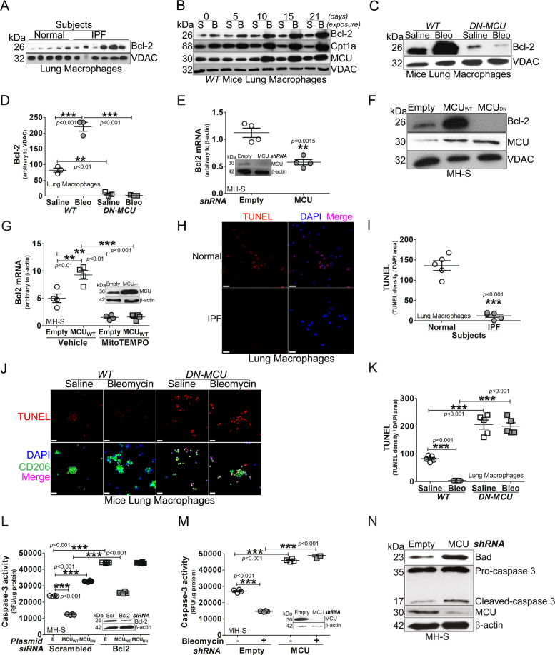
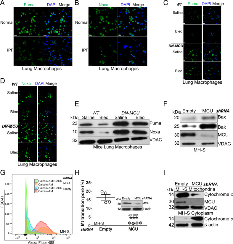
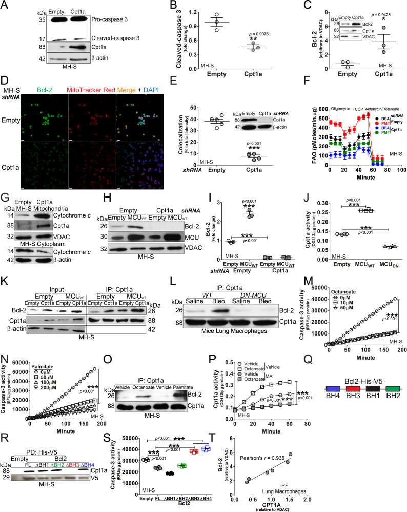
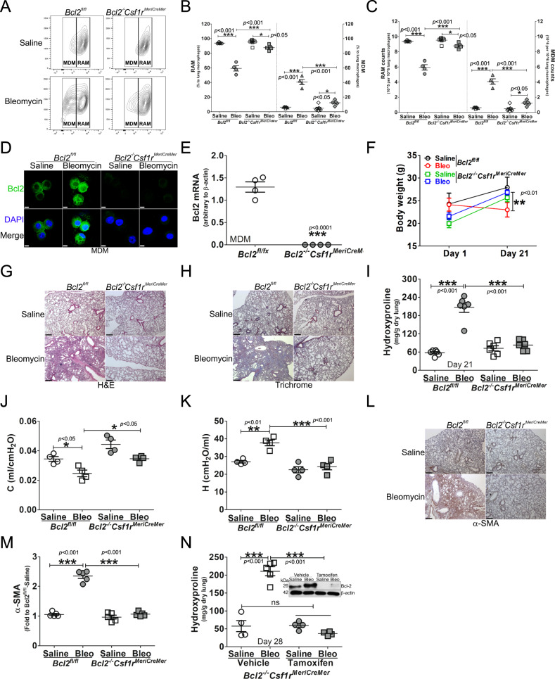
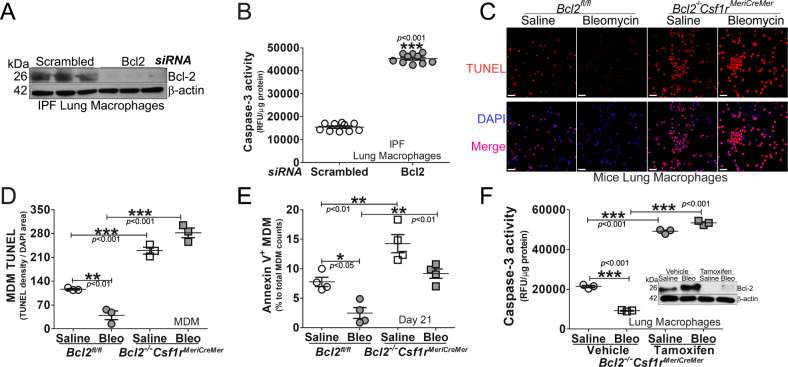
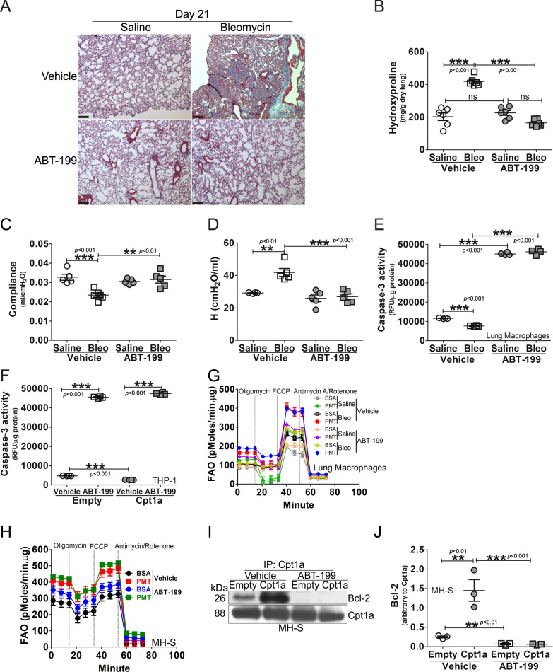
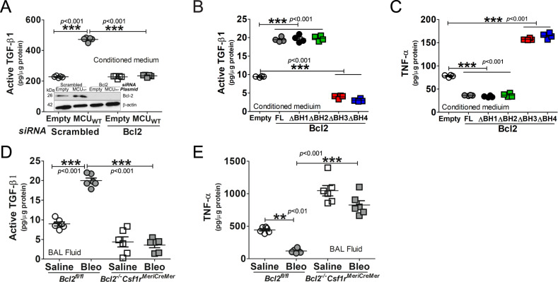
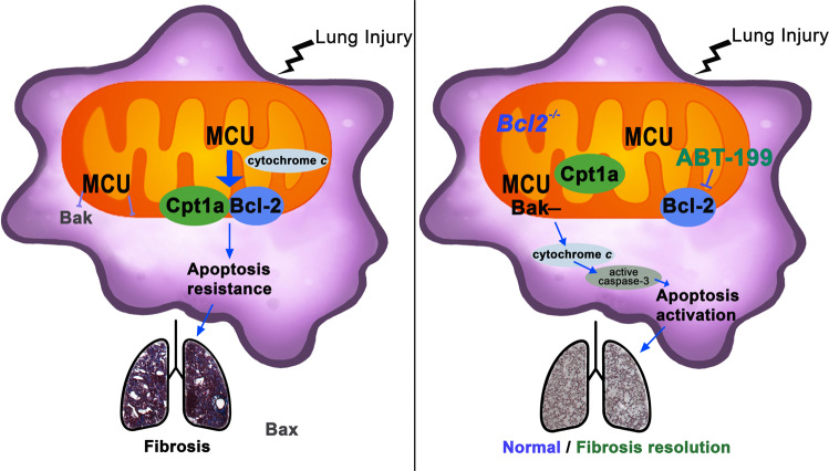
The mitochondrial calcium uniporter (MCU) regulates metabolic reprogramming in lung macrophages and the progression of pulmonary fibrosis. Fibrosis progression is associated with apoptosis resistance in lung macrophages; however, the mechanism(s) by which apoptosis resistance occurs is poorly understood. Here, we found a marked increase in mitochondrial B-cell lymphoma-2 (Bcl-2) in lung macrophages from subjects with idiopathic pulmonary fibrosis (IPF). Similar findings were seen in bleomycin-injured wild-type (WT) mice, whereas Bcl-2 was markedly decreased in mice expressing a dominant-negative mitochondrial calcium uniporter (DN-MCU). Carnitine palmitoyltransferase 1a (Cpt1a), the rate-limiting enzyme for fatty acid β-oxidation, directly interacted with Bcl-2 by binding to its BH3 domain, which anchored Bcl-2 in the mitochondria to attenuate apoptosis. This interaction was dependent on Cpt1a activity. Lung macrophages from IPF subjects had a direct correlation between CPT1A and Bcl-2, whereas the absence of binding induced apoptosis. The deletion of Bcl-2 in macrophages protected mice from developing pulmonary fibrosis. Moreover, mice had resolution when Bcl-2 was deleted or was inhibited with ABT-199 after fibrosis was established. These observations implicate an interplay between macrophage fatty acid β-oxidation, apoptosis resistance, and dysregulated fibrotic remodeling.


Conflict of interest statement

The authors declare no competing interests.

Figures

Fig. 1
Fig. 1. Macrophage MCU regulates apoptosis resistance in lung macrophages by increasing Bcl-2.
A Lung macrophages from subjects (IPF or normal) were subjected to mitochondrial isolation and detection of Bcl-2 by immunoblot analysis. B Lung macrophages from WT mice exposed to bleomycin or saline control were subjected to mitochondrial isolation and detection of Bcl-2, Cpt1a, and MCU by immunoblot analysis. C DN-MCU-Lyz2-cre mice and WT littermates were exposed to saline or bleomycin. Lung macrophages were isolated after 21 days and subjected to mitochondrial isolation and detection of Bcl-2 by immunoblot analysis. DN-MCU (DN-MCU-Lyz2-cre). D Mitochondrial Bcl-2 in C was quantified and normalized to VDAC, n = 3. E MH-S cells were transfected to silence MCU. Total RNA was extracted for the determination of Bcl2 mRNA by quantitative real-time RT-PCR (qRT-PCR), n = 4. F MH-S cells were transfected with MCUWT, MCUDN, or empty control. Mitochondria were isolated for the detection of Bcl-2 and MCU by immunoblot analysis. G MH-S cells were transfected to overexpress MCUWT and treated with MitoTEMPO (50 μM, overnight). Bcl2 mRNA was determined by qRT-PCR, n = 4. BAL cells from normal or IPF subjects were stained with TUNEL. The staining was H imaged by confocal microscopy, scale bars at 20 μm, and I statistically quantitated, n = 5. BAL macrophages from bleomycin- or saline-exposed DN-MCU-Lyz2-cre mice or WT littermates were stained with TUNEL and a macrophage marker, CD206. The staining was J imaged by confocal microscopy, scale bars at 20 μm, and K statistically imaged, n = 5. L MH-S cells were co-transfected with empty, MCUWT, or MCUDN with scrambled or Bcl-2 siRNA. Caspase-3 activity was measured, n = 4. E, empty; Scr, scrambled siRNA. M MH-S cells were transfected to empty or MCU shRNA and exposed to bleomycin (0.0126 U/ml; 1 h). Caspase-3 activity was measured, n = 4. Inset: Immunoblot analysis for MCU. N MH-S cells were transfected with empty or MCU shRNA. Immunoblot analysis for Bad, caspase-3, and MCU was performed. One-way ANOVA with Tukey’s post hoc comparison. Two-tailed student’s t-test (E, I). *p ≤ 0.05, **p ≤ 0.01, and ***p ≤ 0.001. See also Fig. S1.
Fig. 2
Fig. 2. MCU is associated with apoptosis resistance and inhibition of the mitochondrial intrinsic pathway.
Lung macrophages from normal or IPF subjects were stained and imaged for A Puma or B Noxa by confocal microscopy. Scale bars, 20 μm. Lung macrophages from bleomycin- or saline-exposed DN-MCU-Lyz2-cre mice or WT littermates were stained and imaged for C Puma or D Noxa by confocal microscopy. Scale bars, 20 μm. E Lung macrophages from DN-MCU-Lyz2-cre mice or WT littermates were subjected to mitochondrial isolation and immunoblot analysis for Puma and Noxa. MH-S cells were transfected with empty or MCU shRNA. F Immunoblot analysis for Bax and Bak in isolated mitochondria, and G mitochondrial permeability transition pore opening were determined in live cells by flow cytometry, and H quantified, n = 4. I Macrophages were transfected with empty or MCU shRNA. Immunoblot analysis for cytochrome c was performed in isolated mitochondria and cytoplasm. Two-tailed student’s t-test. **p ≤ 0.01. See also Fig. S2.
Fig. 3
Fig. 3. MCU modulated binding of Cpt1a with Bcl-2 to induce apoptosis resistance.
MH-S cells were transfected with empty or Cpt1a. Immunoblot analysis for A caspase-3 with B statistical quantification, n = 3, (C) Bcl-2 in isolated mitochondria with statistical quantification, n = 3. D MH-S was transfected with Cpt1a shRNA plasmid or empty vector. Cells were stained with MitoTracker Red and Bcl-2 24 h later and subjected to confocal imaging. E The colocalization of Bcl-2 to MitoTracker Red in D was quantitated, n = 3. F MH-S was transfected with Cpt1a shRNA plasmid or empty vector, and cultured for 24 h. Cells were subjected to FAO measurement by Seahorse assay, n = 4–6. G MH-S was transfected with Cpt1a plasmid or vehicle. Cytochrome c in isolated mitochondria and cytoplasm was detected by immunoblot analysis. Macrophages were co-transfected with empty or MCUWT with empty or Cpt1a shRNA. Immunoblot analysis for H Bcl-2 with I statistical quantification, n = 3. J MH-S cells were transfected to overexpress MCUWT, MCUDN, or empty vector. Cpt1a activity was measured, n = 4. K MH-S cells were co-transfected empty or MCUWT in combination with empty or Cpt1a. Cpt1a was immunoprecipitated and immunoblot analysis for Bcl-2 and Cpt1a was performed. L DN-MCU-Lyz2-cre mice and WT littermates were exposed to saline or bleomycin. Lung macrophages were isolated at 21 days, subjected to Cpt1a immunoprecipitation, and immunoblot analysis for Bcl-2 and Cpt1a. M MH-S was treated with octanoate at various concentrations for 3 h. Whole lysate was prepared for determining caspase-3 activities, n = 4. N MH-S was treated with palmitate at various concentrations for 3 h. Caspase-3 activities were measured, n = 4. O MH-S was treated with octanoate (10 µM) or palmitate (100 µM) for 3 h. Whole lysate was precipitated with Cpt1a antibody, and elutes were subjected to detection of Bcl-2 and Cpt1a by immunoblot analysis. P MH-S was treated with octanoate (10 µM, 4 h), in combination with malonyl CoA (100 µM, 3 h) or vehicle. Cell lysate was prepared for quantitation of Cpt1a activities, n = 4. Q Schematic of V5-His tagged Bcl-2 with four BH domains. R MH-S cells were transfected with Bcl-2-V5-His full length or truncations of BH1, BH2, BH3, or BH4. Bcl-2-V5-His was purified by pull down and immunoblot analysis for Cpt1a was performed. S MH-S cells were transfected with empty or Bcl-2-V5-His constructs. Caspase-3 activity was performed, n = 4. T Pearson’s correlation of CPT1A and Bcl-2 expression in IPF lung macrophages. One-way ANOVA with Tukey’s post hoc comparison (I, J, M, N, S). Two-tailed student’s t-test (B, C, E). *p ≤ 0.05, **p ≤ 0.01, and ***p ≤ 0.001. See also Fig. S3.
Fig. 4
Fig. 4. Mice harboring a conditional deletion of Bcl2 in monocyte-derived macrophages are protected from pulmonary fibrosis.
Bcl2−/−Csf1rMeriCreMer mice and their Bcl2fl/fl littermates were exposed to saline or bleomycin for 21 days. Lung macrophages from bleomycin- or saline-exposed Bcl2−/−Csf1rMeriCreMer mice and their Bcl2fl/fl littermates were A analyzed by flow cytometry to distinguish MDM and resident alveolar macrophages (RAM), and statistically quantified by B percentage, n = 5, or C cell counts, n = 5. Monocyte-derived macrophages (MDM) from Bcl2−/−Csf1rMeriCreMer mice and their Bcl2fl/fl littermates were D stained and imaged for Bcl-2 protein by confocal analysis, scale bars at 10 μm, or E subjected to total RNA extraction for the detection of Bcl2 mRNA by qRT-PCR, n = 4. F Bcl2−/−Csf1rMeriCreMer mice and their Bcl2fl/fl littermates were exposed to saline or bleomycin. Mice bodyweights were measured at days 1 and 21 post exposure, n = 9–13/group. The lung tissue from bleomycin- or saline-exposed Bcl2−/−Csf1rMeriCreMer mice and their Bcl2fl/fl littermates were subjected to G H&E staining; H Masson’s trichrome staining, represented micrographs from six mice per condition are shown. Scale bars, 200 μm at x5; (I) Hydroxyproline, n = 6. J Compliance (C) and K tissue stiffness (H) were determined by Respiratory mechanics analysis, n = 4. L α-SMA measurement by IHC-P and M corresponding quantitation of α-SMA, n = 5. Bcl2−/−Csf1rMeriCreMer mice and their Bcl2fl/fl littermates were exposed to saline or bleomycin for 21 days. N Bcl2−/−Csf1rMeriCreMer mice were exposed to Bleomycin or saline. Mice were intraperitoneally injected daily with corn oil dissolved tamoxifen or corn oil from day 12 until day 20 (20 mg/kg); break per every two injections, and mice were lavaged at day 28. Mice lungs were processed for determination of hydroxyproline content by hydroxyproline assay, n = 4. Two-way ANOVA (F). One-way ANOVA with Tukey’s post hoc comparison (B, C, I, KN). Two-tailed student’s t-test (E). *p ≤ 0.05, **p ≤ 0.01, and ***p ≤ 0.001. See also Fig. S4.
Fig. 5
Fig. 5. Mice harboring a conditional deletion of Bcl2 potentiates apoptosis of monocyte-derived macrophages.
IPF lung macrophages were transfected with human scrambled or Bcl2 siRNA. A Immunoblot for Bcl-2 and B caspase-3 activity, n = 10. C Lung macrophages from bleomycin- or saline-exposed Bcl2−/−Csf1rMeriCreMer mice and their Bcl2fl/fl littermates were stained with TUNEL and imaged by confocal microscopy. Scale bars, 20 μm. Monocyte-derived macrophages (MDM) from bleomycin- or saline-exposed Bcl2−/−Csf1rMeriCreMer mice and their Bcl2fl/fl littermates were D stained with TUNEL and imaged by confocal microscopy was quantified, n = 3, or E stained for the detection of Annexin V by flow cytometry, n = 4. Bcl2−/−Csf1rMeriCreMer mice were exposed to Bleomycin or saline. Mice were intraperitoneally injected daily with corn oil dissolved tamoxifen or corn oil from day 12 until day 20 (20 mg/kg; break per every two injections), and mice were lavaged at day 28. F BAL macrophages were prepared for detection of caspase-3 activities, n = 3, and Bcl-2 by immunoblot analysis. One-way ANOVA with Tukey’s post hoc comparison. Two-tailed student’s t-test for B. *p ≤ 0.05, **p ≤ 0.01, and ***p ≤ 0.001. See also Fig. S5.
Fig. 6
Fig. 6. Inhibition of Bcl-2 prevents interaction with Cpt1a and protects mice from fibrosis.
Bcl2fl/fl mice were exposed to saline or bleomycin (Bleo). ABT-199, at the concentration of 50 mg/kg, was administered daily to mice at day 12 days post the exposure until day 21. Lung tissue was subjected to A Masson’s trichrome staining, representative micrographs from six mice per condition are shown, with scale bars at 200 μm, x5. B Hydroxyproline, n = 6. Bcl2fl/fl mice were exposed to saline or bleomycin (Bleo). ABT-199, at the concentration of 50 mg/kg, was administered daily to mice at day 12 days post the exposure until day 21. C Compliance and D H stiffness were determined by Respiratory mechanics analysis, n = 5. E WT mice were exposed to saline or bleomycin (Bleo). ABT-199, at the concentration of 50 mg/kg, was administered daily to mice at day 12 days post the exposure, until day 21. Caspase-3 activity was determined in lavaged lung macrophages, n = 4. F THP-1 cells were transfected with empty or Cpt1a and treated with ABT-199 (1 μM, overnight) or vehicle. Cells were subjected to quantification of caspase-3 activity, n = 4. G WT mice were exposed to saline or bleomycin (Bleo). ABT-199, at the concentration of 50 mg/kg, was administered daily to mice at day 12 days post the exposure, until day 21. Fatty acid oxidation of BAL cells was measured by OCR on the Seahorse XF96 bioanalyzer. H MH-S was treated with ABT-199 (1 µM, 3 h), and subjected to FAO measurement, n = 6. MH-S cells were transfected to overexpress Cpt1a and treated with ABT-199 (1 μM, overnight) or vehicle. Cell lysate was I immunoprecipitated with Cpt1a antibody and subjected to immunoblot analysis for Bcl-2, and J Bcl-2 was statistically quantitated, n = 3. One-way ANOVA with Tukey’s post hoc comparison. **p ≤ 0.01, ***p ≤ 0.001. See also Fig. S6.
Fig. 7
Fig. 7. Cpt1a-Bcl-2 binding regulates the macrophage phenotype.
A MH-S cells were co-transfected with empty or MCUWT and scrambled or Bcl-2 siRNA. Conditioned medium was collected for active TGF-β1 ELISA, n = 5. Macrophages were transfected with empty or Bcl-2-V5-His constructs. Conditioned medium was collected for B active TGF-β1 or C TNF-α by ELISA, n = 5. D Active TGF-β1 or E TNF-α was measured in BAL fluid from bleomycin- or saline-exposed Bcl2−/−Csf1rMeriCreMer mice and their Bcl2fl/fl littermates by ELISA, n = 5–6/group. One-way ANOVA with Tukey’s post hoc comparison. **p ≤ 0.01 and ***p ≤ 0.001. See also Fig. S7.
Fig. 8
Fig. 8. Cpt1a-Bcl-2 binding regulates apoptosis and fibrotic remodeling.
Schematic of fibrotic remodeling and apoptosis resistance in monocyte-derived macrophages. MCU increased Cpt1a, which directly binds to Bcl-2 to anchor it in the mitochondria to mediate apoptosis resistance and fibrotic remodeling. Apoptosis activation and resolution of fibrotic remodeling occurred when Bcl-2 was deleted or inhibited with ABT-199 in established fibrosis.

References

    1. Lederer DJ, Martinez FJ. Idiopathic pulmonary fibrosis. N. Engl J Med. 2018;379:797–8. - PubMed
    1. King TE, Jr., Bradford WZ, Castro-Bernardini S, Fagan EA, Glaspole I, Glassberg MK, et al. A phase 3 trial of pirfenidone in patients with idiopathic pulmonary fibrosis. N. Engl J Med. 2014;370:2083–92. - PubMed
    1. Richeldi L, du Bois RM, Raghu G, Azuma A, Brown KK, Costabel U, et al. Efficacy and safety of nintedanib in idiopathic pulmonary fibrosis. N. Engl J Med. 2014;370:2071–82. - PubMed
    1. Diwan A, Wansapura J, Syed FM, Matkovich SJ, Lorenz JN, Dorn GW., 2nd Nix-mediated apoptosis links myocardial fibrosis, cardiac remodeling, and hypertrophy decompensation. Circulation. 2008;117:396–404. - PMC - PubMed
    1. Docherty NG, O’Sullivan OE, Healy DA, Fitzpatrick JM, Watson RW. Evidence that inhibition of tubular cell apoptosis protects against renal damage and development of fibrosis following ureteric obstruction. Am J Physiol Ren Physiol. 2006;290:F4–13. - PubMed

Publication types