Case Reports
. 2021 Aug 13:14:545-550.
doi: 10.2147/IMCRJ.S320678. eCollection 2021.

Multiple Sclerosis Presenting with Sixth Nerve Palsy in a Child

Affiliations
Case Reports

Multiple Sclerosis Presenting with Sixth Nerve Palsy in a Child

Nada Al-Yousuf et al. Int Med Case Rep J. .

Abstract

We report a child with multiple sclerosis who presented with sixth nerve palsy. She is a twelve-year-old Bahraini female presented to the ophthalmology department complaining of double vision. She also had imbalance and paraesthesia. Extraocular muscle examination showed restriction of abduction in the right eye. There was no change in vision. MRI showed right eye optic neuritis (ON) and demyelination which was indicative of multiple sclerosis (MS). Ocular coherence tomography (OCT) showed thinning of nerve fibres of both eyes which was consistent with subclinical optic neuritis. Neurological examination showed brisk knee jerk on the left side and incoordination of movement on the same side. Disability Status Scale (EDSS) showed a score of 3.0. She was given Solu-medrol 500 mg intravenously (IV) in addition to omeprazole 40 mg IV and Vitamin D3 (cholecalciferol) 50,000 IU cap weekly for 8 weeks and Neurorubine forte tablets (vitamin B1, 6, 12) once a day for 2 months. She became asymptomatic in her follow-up visits. Children with MS can present as sixth cranial nerve palsy. Clinicians should have a high index of suspicion for early diagnosis and treatment. In addition to MRI, OCT is a useful diagnostic tool for optic neuritis and MS especially in children.

Keywords: demyelination; diplopia; lateral rectus palsy; ocular coherence tomography.


Conflict of interest statement

The authors report no conflicts of interest in this work.

Figures

Figure 1
Figure 1
The patient’s extraocular eye movements in different gaze positions. (A) The patient is looking straight showing no limitation of extraocular muscles. (B) The patient is looking to the left. There is no limitation of extraocular muscle movements. (C) The patient is looking to the right. There is limitation of abduction of the right eye indicating weakness of the right lateral rectus muscle due to sixth nerve palsy.
Figure 2
Figure 2
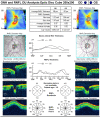
Average RNFL thickness is asymmetrical in both eyes with 58% of asymmetry. RNFL thickness values for different locations of the optic nerve scans showing the average thickness in μm in superior, temporal, inferior and nasal quadrants. The RNFL in the nasal quadrant of the right eye and left eye are 68 μm and 59 μm, respectively. Both readings are significantly thin compared with normal controls (96 μm). The RNFL in the temporal quadrant of the left eye is 55 μm which is severely thin compared with normal controls (73 μm). This thinning of RNFL quadrants is consistent with ON.
Figure 3
Figure 3
MRI of brain. (A) T2 fat sat image right optic nerve hyperintensity indicating optic neuritis. (B) T2 axial image showing left cerebral peduncle plaque. (C) T2 axial showing right optic neuritis. (D) T1 axial post-contrast image showing patchy enhancement of right optic nerve. (E) Periventricular T2 hyperintense focus in right peritrigonal region. (F) Flair sagittal image showing ependymal dot-dash sign sensitive in early MS. (G) Diffusion-weighted image showing left peri ventricular bright focus- T2 shine through effect. (H) Flair axial image showing right parietal juxtacortical and left periventricular foci. (I) Flair axial image showing left parafalcine frontal juxtacortical focus. (J) Sagittal flair image showing juxtacortical and infratentorial plaques. (K) Diffusion-weighted image showing bright right optic nerve. (LN) Open ring enhancement of juxtacortical plaque. (O) Nodular enhancement in periventricular plaque.

References

    1. Charcot M. Histologie de la sclérose en plaques. Gaz Hosp. 1868;41:554–556.
    1. Berer K, Krishnamoorthy G. Microbial view of central nervous system autoimmunity. FEBS Lett. 2014;588(22):4207–4213. doi:10.1016/j.febslet.2014.04.007 - DOI - PubMed
    1. Disease GBD, Injury I, Prevalence C. Global, regional, and national incidence, prevalence, and years lived with disability for 310 diseases and injuries, 1990–2015: a systematic analysis for the Global Burden of Disease Study 2015. Lancet. 2016;388(10053):1545–1602. - PMC - PubMed
    1. Compston A, Coles A. Multiple sclerosis. Lancet. 2008;372(9648):1502–1517. doi:10.1016/S0140-6736(08)61620-7 - DOI - PubMed
    1. Alonso A, Hernan MA. Temporal trends in the incidence of multiple sclerosis: a systematic review. Neurology. 2008;71(2):129–135. doi:10.1212/01.wnl.0000316802.35974.34 - DOI - PMC - PubMed

Publication types