

A Rare Cause of Abdominal and Flank Pain in Children: Nutcracker Syndrome
- PMID: 34414047
- PMCID: PMC8364742
- DOI: 10.7759/cureus.16422
A Rare Cause of Abdominal and Flank Pain in Children: Nutcracker Syndrome
Abstract
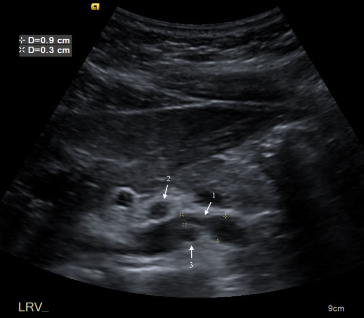
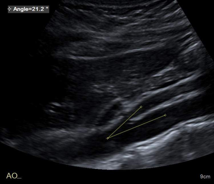
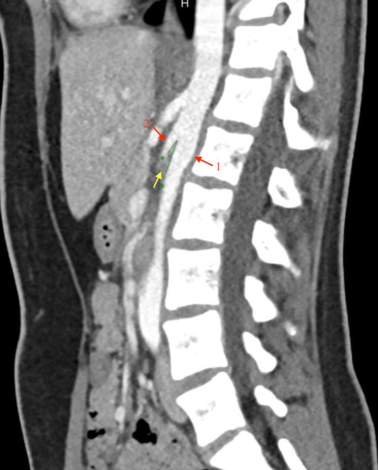
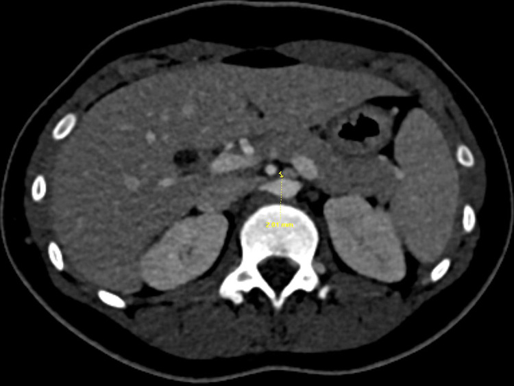
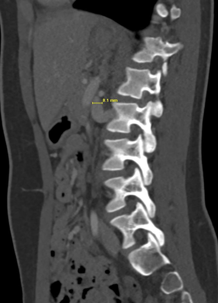
The nutcracker phenomenon is characterized by compression of the left renal vein typically between the abdominal aorta and superior mesenteric artery. It is an uncommon and often undiagnosed condition that has the potential to cause a range of symptoms including hematuria and abdominal or flank pain. The term nutcracker syndrome refers to the clinical manifestations of the nutcracker phenomenon. Diagnosis can be made with Doppler ultrasound, computed tomography angiography, magnetic resonance angiography, or venography. Management can range from conservative treatment in the pediatric population due to high spontaneous remission rate to surgical and endovascular interventions. We discuss the case of a previously healthy young female who presented with abdominal pain. Diagnosis of nutcracker syndrome was made based on imaging. The patient was managed conservatively. This case highlights the importance of considering nutcracker syndrome in the differential diagnosis when evaluating patients with abdominal and flank pain.
Keywords: abdominal pain; flank pain; left renal vein compression; nutcracker phenomenon; nutcracker syndrome.
Copyright © 2021, Agarwal et al.
Conflict of interest statement
The authors have declared that no competing interests exist.
Figures
References
-
- Nutcracker syndrome--how well do we know it? He Y, Wu Z, Chen S, et al. Urology. 2014;83:12–17. - PubMed
-
- Nutcracker syndrome: an update on current diagnostic criteria and management guidelines. Ananthan K, Onida S, Davies AH. Eur J Vasc Endovasc Surg. 2017;53:886–894. - PubMed
-
- Nutcracker syndrome: intravascular stenting approach. Park YB, Lim SH, Ahn JH, Kang E, Myung SC, Shim HJ, Yu SH. Nephrol Dial Transplant. 2000;15:99–101. - PubMed
Publication types
LinkOut - more resources
Full Text Sources