. 2021 Aug 6:2021:6344344.
doi: 10.1155/2021/6344344. eCollection 2021.

Bone Marrow Neutrophils of Multiple Myeloma Patients Exhibit Myeloid-Derived Suppressor Cell Activity

Affiliations

Bone Marrow Neutrophils of Multiple Myeloma Patients Exhibit Myeloid-Derived Suppressor Cell Activity

Julia Petersson et al. J Immunol Res. .

Abstract

Activated normal density granulocytes (NDGs) can suppress T-cell responses in a similar way as myeloid-derived suppressor cells (MDSCs). In this study, we tested the hypothesis that NDGs from blood and bone marrow of multiple myeloma (MM) patients have the ability to suppress T-cells, as MDSC. MM is an incurable plasma cell malignancy of the bone marrow. Like most malignancies, myeloma cells alter its microenvironment to promote tumor growth, including inhibition of the immune system. We found that MM NDG from the bone marrow suppressed proliferation of T-cells, in contrast to healthy donors. The inhibitory effect could not be explained by changed levels of mature or immature NDG in the bone marrow. Moreover, NDG isolated from the blood of both myeloma patients and healthy individuals could inhibit T-cell proliferation and IFN-γ production. On the contrary to previous studies, blood NDGs did not have to be preactivated to mediate suppressive effects. Instead, they became activated during coculture, indicating that contact with activated T-cells is important for their ability to regulate T-cells. The inhibitory effect was dependent on the production of reactive oxygen species and could be reverted by the addition of its inhibitor, catalase. Our findings suggest that blood NDGs from MM patients are suppressive, but no more than NDGs from healthy donors. However, only bone marrow NDG from MM patients exhibited MDSC function. This MDSC-like suppression mediated by bone marrow NDG could be important for the growth of malignant plasma cells in MM patients.


Conflict of interest statement

The authors declare no conflict of interest.

Figures

Figure 1
Figure 1
Inhibition of T-cell proliferation and IFN-γ production by peripheral blood normal density granulocytes. (a) Inhibition of proliferation with healthy donor (HD) and MM NDG. HD NDG inhibits T-cell proliferation (p < 0.0001), and the median proliferation is 54% (95% CI: 38–67). MM NDG also inhibits T-cell proliferation (p = 0.002), and the median proliferation is 48% (95% CI: 23–67). (b) The median of IFN-γ production for each group is 9400 pg/ml (95% CI: 820–13500), 460 pg/ml (95% CI: 0–1900), and 900 pg/ml (95% CI: 0–2000), respectively. Both HD NDG (p = 0.0078) and MM NDG (p = 0.0312) inhibit the production of IFN-γ. (c) The inhibitory effect of HD NDG (p < 0.0001) differs between 5 and 84% and (d) between 13 and 75% for MM NDG (p = 0.0156). (e) The inhibition of T-cell proliferation by NDG is dose dependent (data from representative experiment). (f) NDGs become more inhibitory in the presence of the activator fMLF (n = 9) (p = 0.0078). Error bars indicate median with range. Statistical significance was tested with Wilcoxon matched pairs signed rank test.
Figure 2
Figure 2
Inhibition of T-cell proliferation and IFN-γ production by bone marrow NDG. (a) Median proliferation for each group is 94% (95% CI: 88–95), 91% (95% CI: 82–93), and 71 (95% CI: 50–79), respectively. The proliferation of T-cells was not inhibited by HD NDG (p = 0.1016) but by NDG from MM patients (p = 0.002). (b) The median IFN-γ production for each group is 9700 pg/ml (95% CI: 8200–13500), 3500 pg/ml (95% CI: 1450–7800), and 1600 (95% CI: 600–4500), respectively. Both HD NDG (p = 0.0078) and MM NDG (p = 0.0312) have the ability to inhibit IFN-γ production. (c) The effect of HD NDG on T-cell proliferation (p = 0.1016) differed between +2% and -26% proliferation. (d) Inhibition by NDGs from MM patients (p = 0.002) differed between 1.7 and 60%. Error bars indicate median with range. Statistical significance was tested with Wilcoxon matched pairs signed rank test. Normal density neutrophils become activated by T-cells over time.
Figure 3
Figure 3
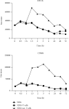
Neutrophil activation markers CD11b and CD66b increase during coculture with CD3/CD28 activated T-cells. (a) CD11b expression over time (72 h) on NDG alone in culture (ring), on NDG in coculture with CD3/CD28 activated T-cells (square), and on NDG in coculture with nonactivated T-cells (rectangle). (b) CD66b expression over time. These are representative plots from two experiments.
Figure 4
Figure 4
Catalase partially recovers the proliferation of T-cells. (a) Addition of the ROS inhibitor catalase to the coculture reverted the inhibitory effect of healthy blood NDG (n = 6), and T-cell proliferation was partially recovered (p = 0.0312). (b) When adding the arginase inhibitor nor-NOHA, the proliferation of T-cells was not recovered (p = 0.780) (n = 10).
Figure 5
Figure 5
Percentage of mature, immature, and immature CD11b neutrophils in blood and bone marrow. (a) Gating strategy for the division of bone marrow neutrophils into mature, immature, and immature CD11b cells. The percentage of (b) mature, (c) immature, and (d) immature CD11b cells in the bone marrow of healthy donors (HDs) and MM patients. (e) The percentage of immature and (f) mature neutrophils in the blood of healthy donors and multiple myeloma patients. Statistical significance was tested with Mann–Whitney, but none was found.

References

    1. Bronte V., Brandau S., Chen S. H., et al. Recommendations for myeloid-derived suppressor cell nomenclature and characterization standards. Nature Communications. 2016;7(1):p. 12150. doi: 10.1038/ncomms12150. - DOI - PMC - PubMed
    1. Millrud C. R., Bergenfelz C., Leandersson K. On the origin of myeloid-derived suppressor cells. Oncotarget. 2017;8(2):3649–3665. doi: 10.18632/oncotarget.12278. - DOI - PMC - PubMed
    1. Sagiv J. Y., Michaeli J., Assi S., et al. Phenotypic diversity and plasticity in circulating neutrophil subpopulations in cancer. Cell Reports. 2015;10(4):562–573. doi: 10.1016/j.celrep.2014.12.039. - DOI - PubMed
    1. Aarts C. E. M., Hiemstra I. H., Béguin E. P., et al. Activated neutrophils exert myeloid-derived suppressor cell activity damaging T cells beyond repair. Blood Advances. 2019;3(22):3562–3574. doi: 10.1182/bloodadvances.2019031609. - DOI - PMC - PubMed
    1. Aarts C. E. M., Hiemstra I. H., Tool A. T. J., et al. Neutrophils as suppressors of T cell proliferation: does age matter? Frontiers in Immunology. 2019;10:p. 2144. doi: 10.3389/fimmu.2019.02144. - DOI - PMC - PubMed

MeSH terms