

Autophagy and Ciliogenesis
- PMID: 34414314
- PMCID: PMC8355725
- DOI: 10.31662/jmaj.2021-0090
Autophagy and Ciliogenesis
Abstract
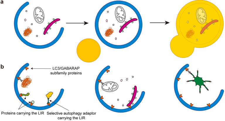
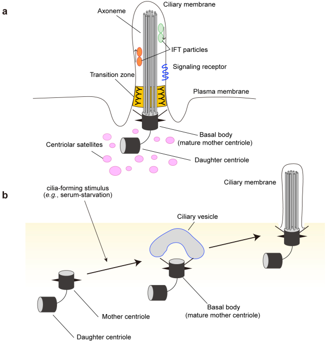
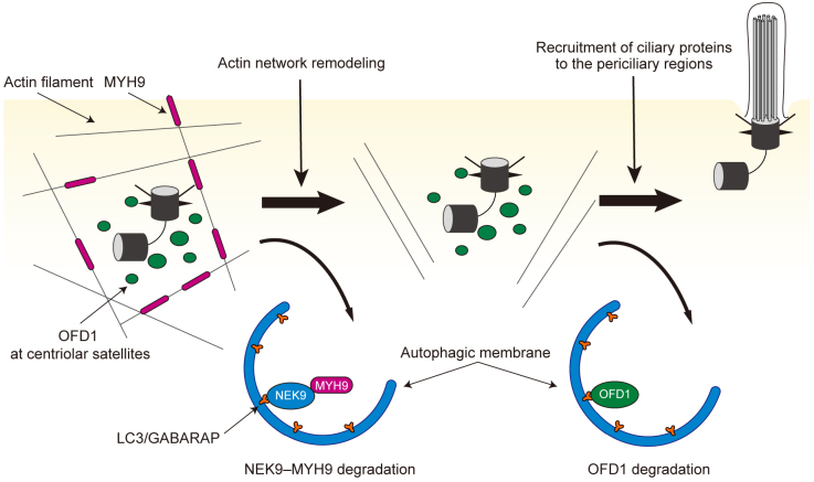
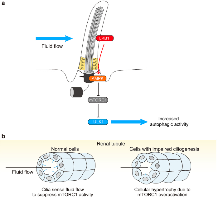
Autophagy is a major intracellular degradation system and plays important roles in various physiological processes such as metabolic adaptation and intracellular homeostasis. It degrades intracellular components both randomly and selectively. Autophagic activity is tightly regulated primarily by nutrient availability, but also by other extracellular and intracellular signals. Growing evidence suggests that there are multiple links between autophagy and the primary cilium. The primary cilium is an organelle present on the cell surface and is important for keeping cellular integrity by transducing extracellular stimuli inside the cell. Recent studies have revealed that autophagy selectively degrades the ciliogenesis inhibitory proteins OFD1 and MYH9, promoting ciliogenesis. Conversely, autophagy also inhibits ciliogenesis under growth conditions. The primary cilium can also regulate autophagic activity. These findings suggest that the relationship between autophagy and the primary cilia is bidirectional, and that both are important for maintaining the normal function of various organs.
Keywords: autophagy; ciliogenesis; ciliopathy.
Copyright © Japan Medical Association.
Conflict of interest statement
None
Figures
Similar articles
-
Self-eating to remove cilia roadblock.Autophagy. 2014 Feb;10(2):379-81. doi: 10.4161/auto.27346. Epub 2013 Dec 12. Autophagy. 2014. PMID: 24343661 Free PMC article.
-
New Roles of the Primary Cilium in Autophagy.Biomed Res Int. 2017;2017:4367019. doi: 10.1155/2017/4367019. Epub 2017 Aug 23. Biomed Res Int. 2017. PMID: 28913352 Free PMC article. Review.
-
The Autophagy-Cilia Axis: An Intricate Relationship.Cells. 2019 Aug 15;8(8):905. doi: 10.3390/cells8080905. Cells. 2019. PMID: 31443299 Free PMC article. Review.
-
Ciliogenesis is reciprocally regulated by PPARA and NR1H4/FXR through controlling autophagy in vitro and in vivo.Autophagy. 2018;14(6):1011-1027. doi: 10.1080/15548627.2018.1448326. Epub 2018 May 17. Autophagy. 2018. PMID: 29771182 Free PMC article.
-
Glucose deprivation induces primary cilium formation through mTORC1 inactivation.J Cell Sci. 2018 Jan 8;131(1):jcs208769. doi: 10.1242/jcs.208769. J Cell Sci. 2018. PMID: 29180513
Cited by
-
The function of the ATG8 in the cilia and cortical microtubule maintenance of Euplotes amieti.Protoplasma. 2024 Nov;261(6):1127-1145. doi: 10.1007/s00709-024-01957-8. Epub 2024 May 20. Protoplasma. 2024. PMID: 38769089
-
HOPS-dependent lysosomal fusion controls Rab19 availability for ciliogenesis in polarized epithelial cells.J Cell Sci. 2024 Mar 1;137(5):jcs261047. doi: 10.1242/jcs.261047. Epub 2023 Sep 4. J Cell Sci. 2024. PMID: 37665101 Free PMC article.
-
Neuronal primary cilia integrate peripheral signals with metabolic drives.Front Physiol. 2023 Mar 29;14:1150232. doi: 10.3389/fphys.2023.1150232. eCollection 2023. Front Physiol. 2023. PMID: 37064917 Free PMC article. Review.
-
Retinal Organoids: Innovative Tools for Understanding Retinal Degeneration.Int J Mol Sci. 2025 Apr 1;26(7):3263. doi: 10.3390/ijms26073263. Int J Mol Sci. 2025. PMID: 40244125 Free PMC article. Review.
-
Human cerebral organoids: cellular composition and subcellular morphological features.Front Cell Neurosci. 2024 Jun 12;18:1406839. doi: 10.3389/fncel.2024.1406839. eCollection 2024. Front Cell Neurosci. 2024. PMID: 38933177 Free PMC article.
References
-
- Mizushima N, Levine B. Autophagy in human diseases. N Engl J Med. 2020;383(16):1564-76. - PubMed
-
- Dikic I, Elazar Z. Mechanism and medical implications of mammalian autophagy. Nat Rev Mol Cell Biol. 2018;19(6):349-64. - PubMed
-
- Nakatogawa H. Mechanisms governing autophagosome biogenesis. Nat Rev Mol Cell Biol. 2020;21(8):439-58. - PubMed
-
- Søreng K, Neufeld TP, Simonsen A. Membrane trafficking in autophagy. Int Rev Cell Mol Biol. 2018;336:1-92. - PubMed
Publication types
LinkOut - more resources
Full Text Sources
Miscellaneous