. 2021 Oct;24(4):729.
doi: 10.3892/mmr.2021.12368. Epub 2021 Aug 20.

Reactive oxygen species induced by uric acid promote NRK‑52E cell apoptosis through the NEK7‑NLRP3 signaling pathway

Affiliations

Reactive oxygen species induced by uric acid promote NRK‑52E cell apoptosis through the NEK7‑NLRP3 signaling pathway

Dongdong Li et al. Mol Med Rep. 2021 Oct.

Abstract

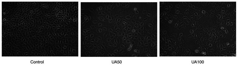
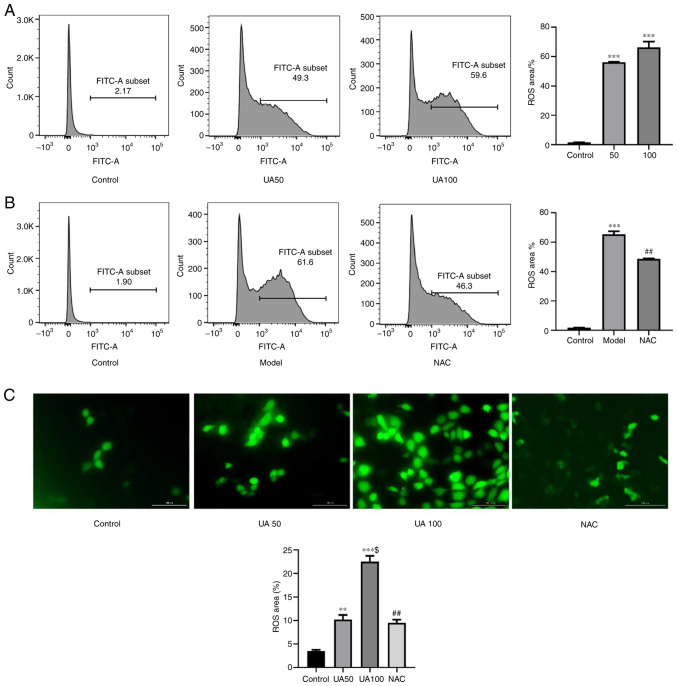
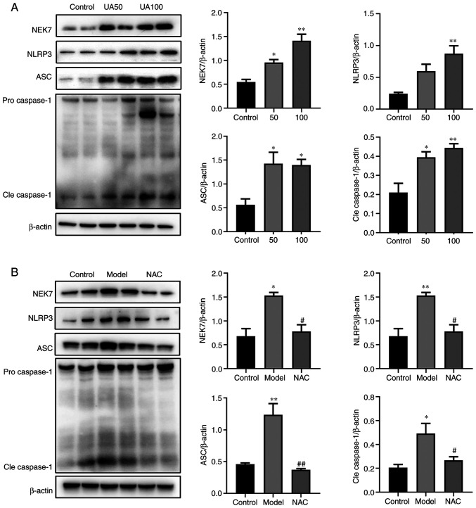
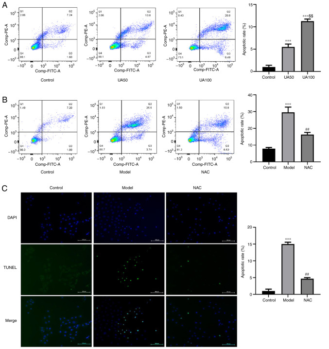
Increasing uric acid (UA) could induce renal tubular epithelial cell (NRK‑52E) injury. However, the specific mechanism by which UA induces renal tubular epithelial cell injury remains unknown. It was hypothesized that UA induces renal tubular epithelial cell injury through reactive oxygen species (ROS) and the Never in mitosis gene A (NIMA)‑related kinase 7 (NEK7)/NLR family pyrin domain containing 3 (NLRP3) signaling pathway. TUNEL assay and flow cytometry were applied to measure apoptosis, and the results of the present study showed that UA treatment induced apoptosis of NRK‑52E cells in a concentration‑dependent manner. Western blotting was performed to determine the expression levels of cleaved caspase‑3, Bax and Bcl‑xl, it was found that levels were significantly increased after UA treatment in NRK‑52E cells. ROS and apoptosis were predominantly induced in NRK‑52E cells and there was an association between ROS and apoptosis. Enhanced expression of NEK7, NLRP3, apoptosis‑associated speck‑like and caspase‑1 were observed in NRK‑52E cells treated with UA. The ROS inhibitor, N‑acetyl‑l‑cysteine, exerted a protective effect on the UA‑induced apoptosis of tubular epithelial cells by reducing excess ROS production, which significantly inhibited NEK7 and NLRP3 inflammasome activation. These results indicated that UA activates ROS and induces apoptosis of NRK‑52E cells. The mechanism might be related to the regulation of the NEK7/NLRP3 signaling pathway.

Keywords: apoptosis; never in mitosis gene A related kinase 7/NLR family pyrin domain containing 3 signaling pathway; reactive oxygen species; uric acid.


Conflict of interest statement

The authors declare that they have no competing interests.

Figures

Figure 1.
Figure 1.
Morphological changes of NRK-52E cells after treatment with UA for 24 h. Magnification, ×400. UA, uric acid.
Figure 2.
Figure 2.
Effect of UA on ROS generation in injured NRK-52E cells. (A and B) Flow cytometry assays showing ROS production in NRK-52E cells treated with 0, 50, 100 µg/ml UA for 24 h and the percentage of ROS-producing cells. NRK-52E cells were pretreated with 20 mM NAC for 1 h and then treated with UA for an additional 24 h and the percentage of ROS-producing cells was examined using flow cytometry. (C) DCFH-DA was used to measure the UA-induced cellular ROS (magnification, ×400). n=4 independent samples; data represent the mean ± standard error of the mean. **P<0.01, ***P<0.001 vs. the control group; $P<0.05 vs. the UA50 group; ##P<0.01 vs. the model group. UA, uric acid; ROS, reactive oxygen species; NAC, N-acetyl-l-cysteine; DCFH-DA, 2′,7′-dichlorofluorescin diacetate.
Figure 3.
Figure 3.
Effects of UA on the apoptotic pathway in injured NRK-52E cells. (A and B) Levels of apoptotic pathway proteins (NEK7, NLRP3, ASC and caspase-1) in response to UA (0, 50 and 100 µg/ml) treatment of NRK-52E cells, as assessed using western blotting. NRK-52E cells were pretreated with 20 mM NAC for 1 h and then treated with 100 µg/ml UA for an additional 24 h and the levels of NEK7, NLRP3, ASC and caspase-1 were determined. Data represent the mean ± standard error of the mean; n=4 independent samples repeated three times. *P<0.05, **P<0.01 vs. the control group. #P<0.05, ##P<0.01 vs. the model group. UA, uric acid; NAC, N-acetyl-l-cysteine; NEK7, Never in mitosis gene A-related kinase 7; NLRP3, NLR family pyrin domain containing 3; ASC, apoptosis-associated speck-like; Cle, cleaved.
Figure 4.
Figure 4.
Effects of UA on the apoptosis of NRK-52E cells. (A) Flow cytometry assays showing apoptosis of NRK-52E cells treated with 0, 50 and 100 µg/ml UA for 24 h and the percentage of apoptotic cells. (B) NRK-52E cells were pretreated with 20 mM NAC for 1 h and then treated with 100 µg/ml UA for an additional 24 h and the percentage of apoptotic cells was examined using flow cytometry. (C) TUNEL assays were used to measure the UA-induced cell apoptosis (magnification, ×200). Data represent the mean ± standard error of the mean. ***P<0.001 vs. the control group; $$P<0.01 vs. the UA50 group; ##P<0.01 vs. the model group. UA, uric acid; NAC, N-acetyl-l-cysteine.
Figure 5.
Figure 5.
Effects of UA on the apoptotic proteins of NRK-52E cells during injury. (A) Levels of apoptotic proteins (Cle caspase-3, Bax and Bcl-xl) in response to UA (0, 50, 100 µg/ml) in NRK-52E cells subjected to injury, as assessed using western blotting. (B) NRK-52E cells were pretreated with 20 mM NAC for 1 h and then treated with 100 µg/ml UA for an additional 24 h and the levels of Cle caspase-3, Bax and Bcl-xl were examined using western blotting. Data represent the mean ± standard error of the mean; n=4 independent samples and repeated three times. *P<0.05, **P<0.01 vs. the control group; #P<0.05, ##P<0.05 vs. the model group. UA, uric acid; NAC, N-acetyl-l-cysteine; Cle, cleaved.

References

    1. He L, Fan Y, Xiao W, Chen T, Wen J, Dong Y, Wang Y, Li S, Xue R, Zheng L, et al. Febuxostat attenuates ER stress mediated kidney injury in a rat model of hyperuricemic nephropathy. Oncotarget. 2017;8:111295–111308. doi: 10.18632/oncotarget.22784. - DOI - PMC - PubMed
    1. Kang DH. Hyperuricemia and progression of chronic kidney disease: Role of phenotype transition of renal tubular and endothelial cells. Contrib Nephrol. 2018;192:48–55. doi: 10.1159/000484278. - DOI - PubMed
    1. Tao M, Shi Y, Tang L, Wang Y, Fang L, Jiang W, Lin T, Qiu A, Zhuang S, Liu N. Blockade of ERK1/2 by U0126 alleviates uric acid-induced EMT and tubular cell injury in rats with hyperuricemic nephropathy. Am J Physiol Renal Physiol. 2019;316:F660–F673. doi: 10.1152/ajprenal.00480.2018. - DOI - PMC - PubMed
    1. Priante G, Gianesello L, Ceol M, Del Prete D, Anglani F. Cell death in the kidney. Int J Mol Sci. 2019;20:3598. doi: 10.3390/ijms20143598. - DOI - PMC - PubMed
    1. Yin W, Zhou QL, OuYang SX, Chen Y, Gong YT, Liang YM. Uric acid regulates NLRP3/IL-1β signaling pathway and further induces vascular endothelial cells injury in early CKD through ROS activation and K+ efflux. BMC Nephrol. 2019;20:319. doi: 10.1186/s12882-019-1506-8. - DOI - PMC - PubMed

MeSH terms