. 2021 Oct;56(10):914-927.
doi: 10.1007/s00535-021-01814-y. Epub 2021 Aug 19.

Mucosal and faecal neutrophil gelatinase-associated lipocalin as potential biomarkers for collagenous colitis

Affiliations

Mucosal and faecal neutrophil gelatinase-associated lipocalin as potential biomarkers for collagenous colitis

Ingunn Bakke et al. J Gastroenterol. 2021 Oct.

Abstract

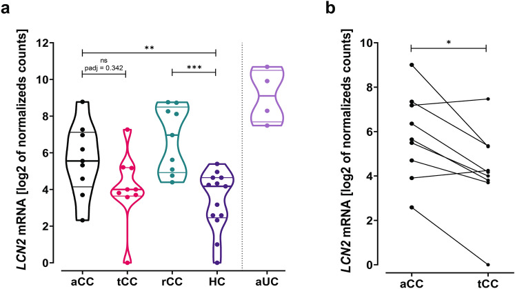
Background: Collagenous colitis (CC) is an inflammatory bowel disease where chronic diarrhoea is the main symptom. Diagnostic markers distinguishing between CC and other causes of chronic diarrhoea remain elusive. This study explores neutrophil gelatinase-associated lipocalin (NGAL) and its mRNA lipocalin2 (LCN2) as histological and faecal disease markers in CC.

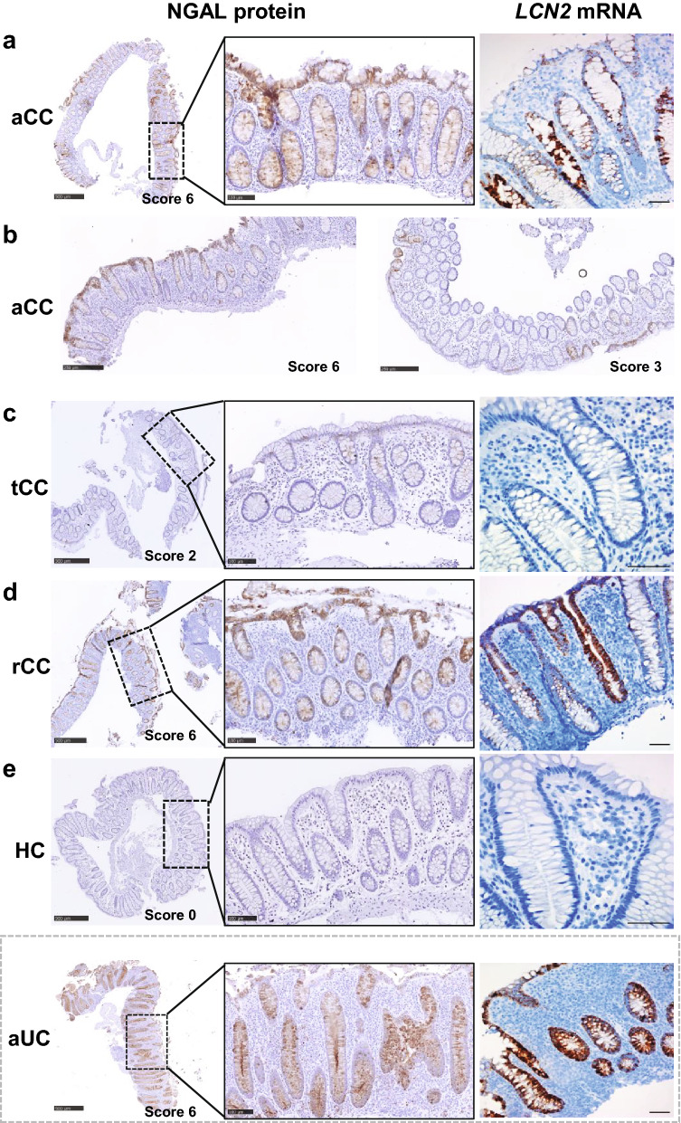
Methods: NGAL/LCN2 were studied in colonic biopsies from CC patients before and during budesonide treatment using RNA sequencing (n = 9/group), in situ hybridization (ISH) (n = 13-22/group) and immunohistochemistry (IHC) (n = 14-25/group). Faecal samples from CC (n = 3-28/group), irritable bowel syndrome diarrhoea (IBS-D) (n = 14) and healthy controls (HC) (n = 15) were assayed for NGAL and calprotectin.

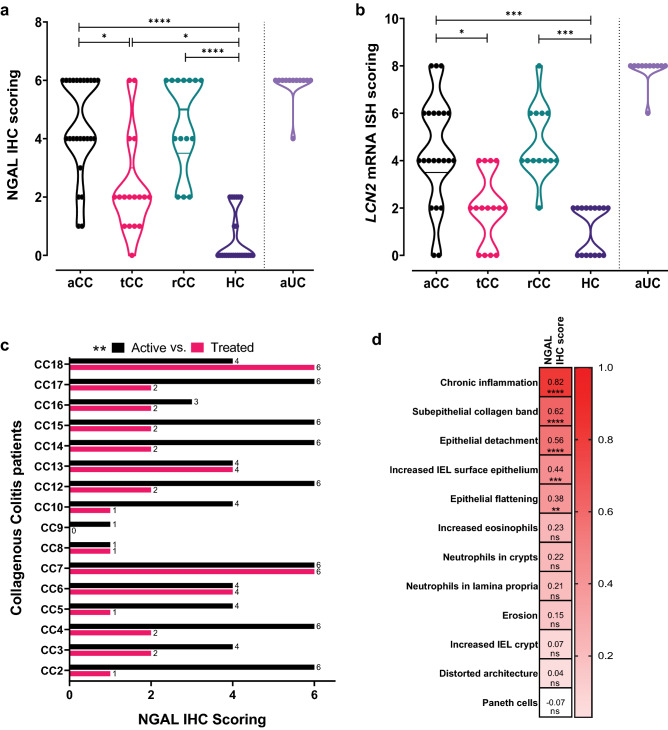
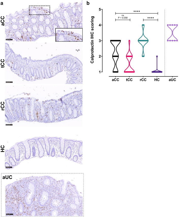
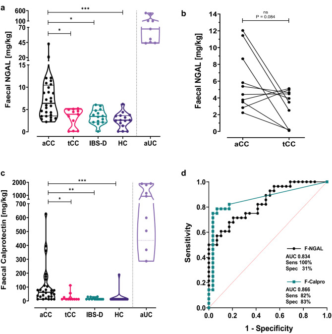
Results: NGAL/LCN2 protein and mRNA expression were upregulated in active CC vs HC, and vs paired samples of treated CC in clinical remission. IHC and ISH localized increased NGAL/LCN2 mainly to epithelium of active CC, compared to almost absence in HC and treated CC. In contrast, calprotectin was solely expressed in immune cells. Despite great individual differences, faecal NGAL was significantly increased in active CC compared to HC, IBS-D and treated CC and had high test sensitivity. Faecal calprotectin levels were variably increased in active CC, but the values remained below usual clinical cut-offs.

Conclusion: NGAL/LCN2 is upregulated in the epithelium of active CC and reduced during budesonide-induced clinical remission to the level of HC and IBD-S. This was reflected in NGAL faecal concentrations. We propose NGAL as an IHC marker for disease activity in CC and a potential faecal biomarker discriminating CC from HC and IBS-D.

Keywords: Calprotectin; Chronic diarrhoea; Inflammatory bowel disease; Irritable bowel syndrome; Microscopic colitis.


Conflict of interest statement

AEØ and AKS received speakers`s honoraria from Takeda, AEØ received support from Tillotts Pharma AGAM, CEH and AM received financial support from Ferring Pharmaceuticals, AM has received salary for consultancies from Tillotts, Ferring, Vifor and Dr Falk Pharma, and speaker’s honoraria from Tillotts and Vifor. All other authors declare that there are no conflicts of interest to disclose.

Figures

Fig. 1
Fig. 1
RNA sequencing of LCN2 mRNA in colonic mucosa of collagenous colitis. a LCN2 mRNA expression in pinch biopsies from colonic mucosa of active collagenous colitis (aCC), budesonide-treated collagenous colitis with clinical remission (tCC), budesonide-refractory collagenous colitis (rCC) and healthy controls (HC). Active ulcerative colitis (aUC) was included for comparison (separated by dotted line). The y-axis shows LCN2 mRNA expression as log2 of normalized counts and the violin plots visualize distribution frequency in addition to individual values (dots), median (thick line), upper and lower quartiles (thin lines). b Paired analysis of LCN2 mRNA expression in colonic mucosa of individual patients with active collagenous colitis before (aCC) and during budesonide treatment with clinical remission (tCC). The y-axis shows LCN2 mRNA expression as log 2 of normalized counts in paired biopsies from nine individual patients. P adj = adjusted P value, ns = non-significant, *P < 0.05, ***P < 0.001, ****P < 0.0001 Analysed as described in “Materials and methods”
Fig. 2
Fig. 2
Representative images of IHC and ISH showing expression of NGAL protein and LCN2 mRNA in colonic epithelium in collagenous colitis. a Overview of NGAL protein expression (left panel), with higher magnification of epithelial staining throughout the crypts (middle panel), and LCN2 mRNA expression (right panel) in colonic mucosa of patients with active collagenous colitis (aCC). b Overview of NGAL expression in aCC mostly in the surface epithelium (left panel) or with a patchy appearance (right panel) ce Overview of NGAL protein expression (left panel), with higher magnification as indicated (middle panel), and LCN2 mRNA expression (right panels) in colonic mucosa of patients with budesonide-treated collagenous colitis in clinical remission (tCC), d budesonide-refractory collagenous colitis (rCC) and e healthy controls (HC). Active ulcerative colitis (aUC) was included for comparison (separated by dotted grey frame). The IHC NGAL scores are given for each image. Scale bars 500 µm (left panel), 100 µm (middle panel), 50 µm (right panel)
Fig. 3
Fig. 3
Semi-quantitative scoring of the colonic epithelial expression levels of NGAL protein and LCN2 mRNA analysed by IHC and ISH staining and correlation to relevant histopathological features. a, b Total score was achieved by multiplying maximum epithelial staining intensities with staining distribution for (a) NGAL IHC and (b) LCN2 mRNA ISH in colonic mucosa of patients with active collagenous colitis (aCC), budesonide-treated collagenous colitis with clinical remission (tCC), budesonide-refractory collagenous colitis (rCC) and healthy controls (HC). Active ulcerative colitis (aUC) was included for comparison (separated by dotted line). The violin plots visualize distribution frequency in addition to individual values (dots), median (thick line), upper and lower quartiles (thin lines). *P < 0.05, ***P < 0.001, ****P < 0.0001 analysed with Kruskal–Wallis with Dunn’s multiple comparisons test. c Paired analysis of the NGAL IHC epithelial scores in individual patients (CC2–CC18) with active collagenous colitis before (aCC) and during budesonide treatment with clinical remission (tCC). **P < 0.01 analysed with Wilcoxon test. d Heatmap showing correlation of the epithelial NGAL IHC scores in the CC groups, HC and IBS-D to different histopathological features relevant to the diagnosis of CC. Spearman correlation coefficient r and *P < 0.05, **P < 0.01, ***P < 0.001, ****P < 0.0001 or ns non-significant are given for all the different features
Fig. 4
Fig. 4
Calprotectin in colonic mucosa of collagenous colitis. a Representative images of IHC showing calprotectin protein expression in the colonic mucosa of patients with active collagenous colitis (aCC), budesonide-treated collagenous colitis with clinical remission (tCC), budesonide-refractory collagenous colitis (rCC) and healthy controls (HC). Higher magnification image of the surface epithelium in inset as indicated. Active ulcerative colitis (aUC) was included for comparison (separated by dotted grey frame). Scale bars 100 µm. b Semi-quantitative scoring of calprotectin-expressing cells in the colonic mucosa of patients in (a). The violin plot visualizes distribution frequency in addition to individual values (dots), median (thick line), upper and lower quartiles (thin lines). Active ulcerative colitis (aUC) was included for comparison (separated by dotted line). ns non-significant, ****P < 0.0001 analysed with Kruskal–Wallis with Dunn’s multiple comparisons test
Fig. 5
Fig. 5
Faecal NGAL and calprotectin in collagenous colitis. a NGAL in faeces measured by ELISA in samples from patients with active collagenous colitis (aCC), budesonide-treated collagenous colitis with clinical remission (tCC), diarrhoea dominant functional irritable bowel-syndrome (IBS-D) and healthy controls (HC). The y-axis shows the concentration of NGAL in mg per kg faeces. *P < 0.05, ***P < 0.001 analysed with Kruskal–Wallis with Dunn’s multiple comparisons test. b Paired analysis of the faecal NGAL concentrations in individual patients with active collagenous colitis before (aCC) and during budesonide treatment with clinical remission (tCC). The y-axis shows the concentration of NGAL in mg per kg faeces in paired samples from 10 individual patients. ns non-significant analysed with Wilcoxon test. c Calprotectin in faeces measured by ELISA in samples from patients in (a). The y-axis shows the concentration of calprotectin in mg per kg faeces. The violin plots visualize distribution frequency in addition to individual values (dots), median (thick line), upper and lower quartiles (thin lines). Active ulcerative colitis (aUC) was included for comparison (separated by dotted line). *P < 0.05, **P < 0.01, ***P < 0.001 analysed with Kruskal–Wallis with Dunn’s multiple comparisons test. d Receiver operating characteristic (ROC) curves of ELISA faecal NGAL (F-NGAL) (black line) and calprotectin (F-calpro) (green line) when comparing aCC against HC and IBS-D. Area under the curve (AUC), and maximal sensitivity (sens) and specificity (spec) when using cut-off values 2.2 mg/kg for F-NGAL and 14.85 mg/kg for F-calpro are given. The red dotted line shows line of identity

References

    1. Langner C, Aust D, Ensari A, et al. Histology of microscopic colitis—review with a practical approach for pathologists. Histopathology. 2015;66:613–626. doi: 10.1111/his.12592. - DOI - PubMed
    1. Lindström CG. 'Collagenous colitis' with watery diarrhoea–a new entity? Pathol Eur. 1976;11:87–89. - PubMed
    1. Pardi DS. Diagnosis and management of microscopic colitis. Am J Gastroenterol. 2017;112:78. doi: 10.1038/ajg.2016.477. - DOI - PubMed
    1. Miehlke S, Guagnozzi D, Zabana Y, et al. European guidelines on microscopic colitis: United European Gastroenterology (UEG) and European Microscopic Colitis Group (EMCG) statements and recommendations. United European Gastroenterol Jo. 2020 doi: 10.1177/2050640620951905. - DOI - PMC - PubMed
    1. Kafil TS, Nguyen TM, Patton PH, et al. Interventions for treating collagenous colitis. Cochrane Database Syst Rev. 2017;11:Cd003575. - PMC - PubMed