Review
. 2021 Nov 2;65(5):813-845.
doi: 10.1042/EBC20200067.

Mammalian lipids: structure, synthesis and function

Affiliations
Review

Mammalian lipids: structure, synthesis and function

Shamshad Cockcroft. Essays Biochem. .

Abstract

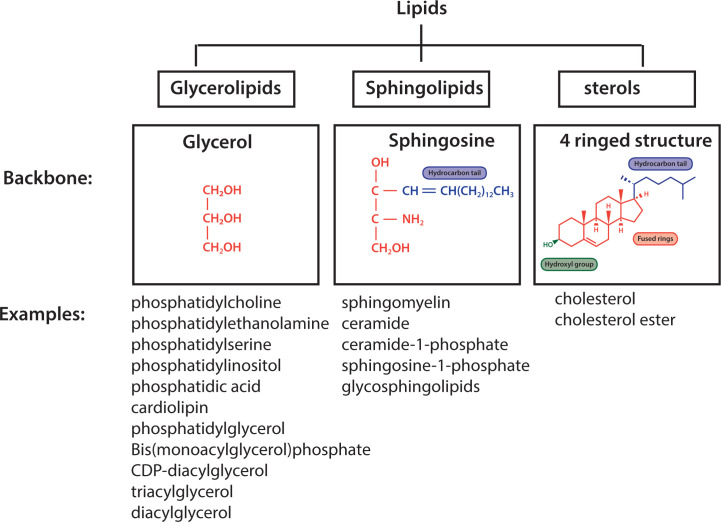
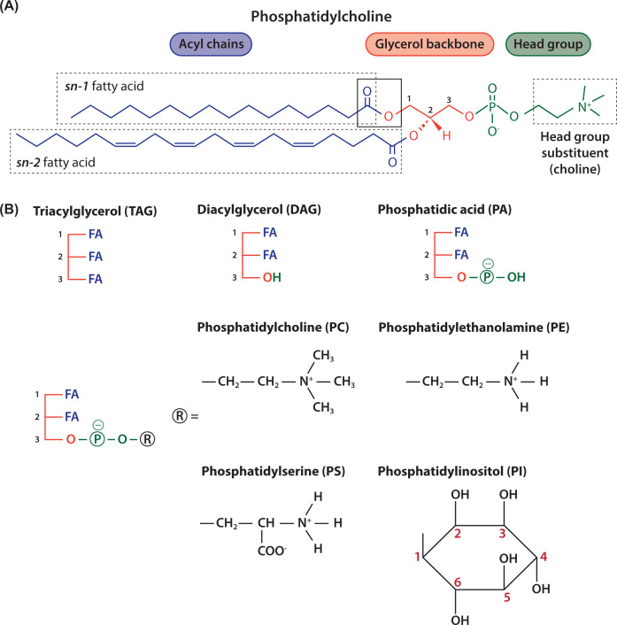
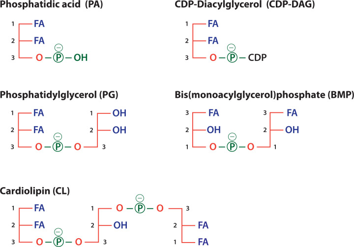
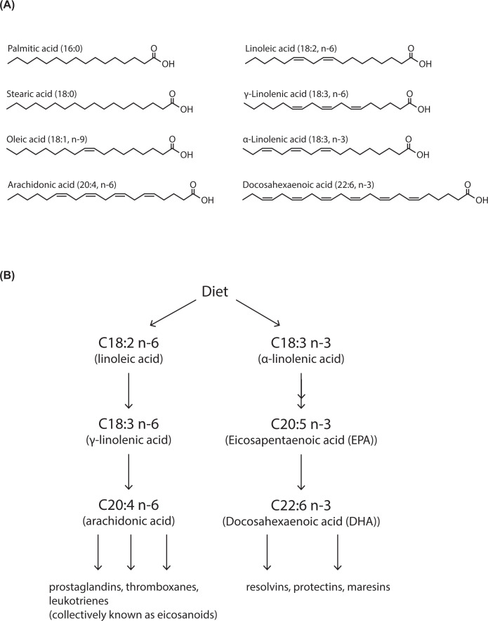
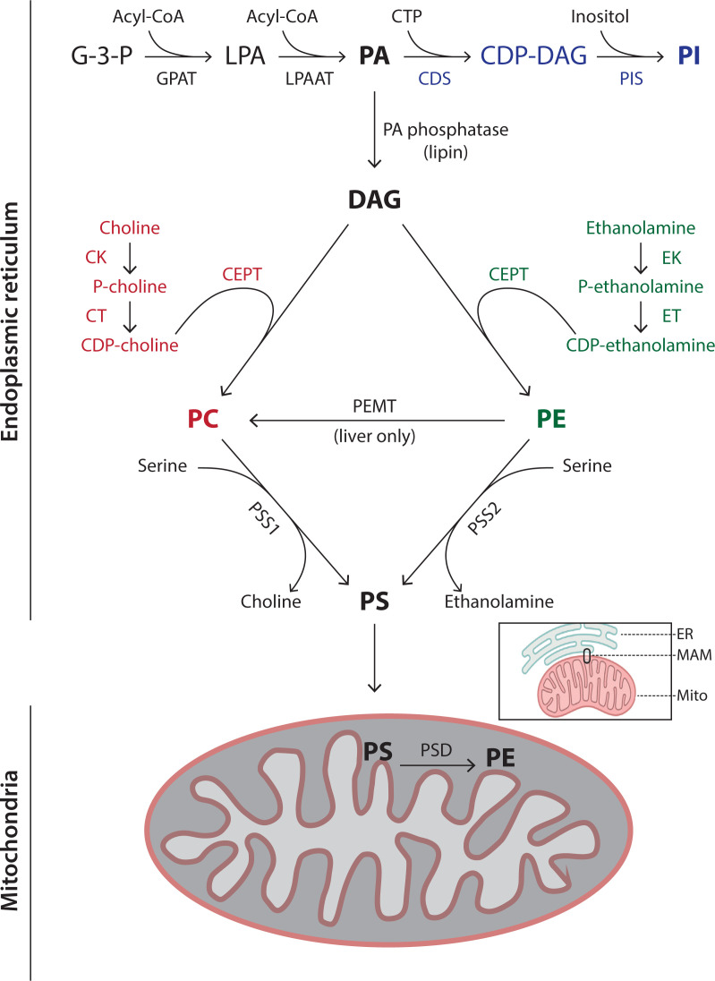
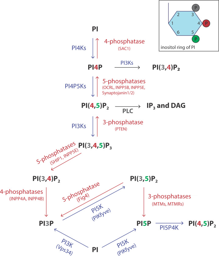
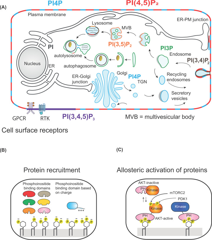
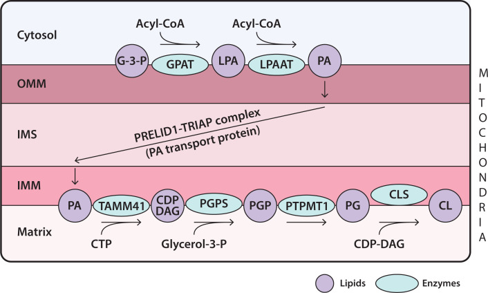
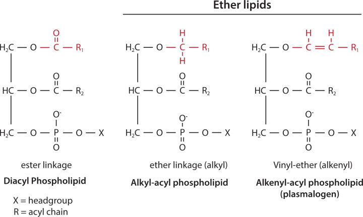
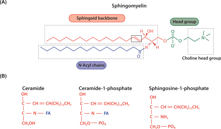
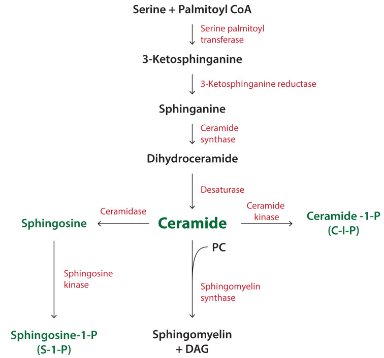
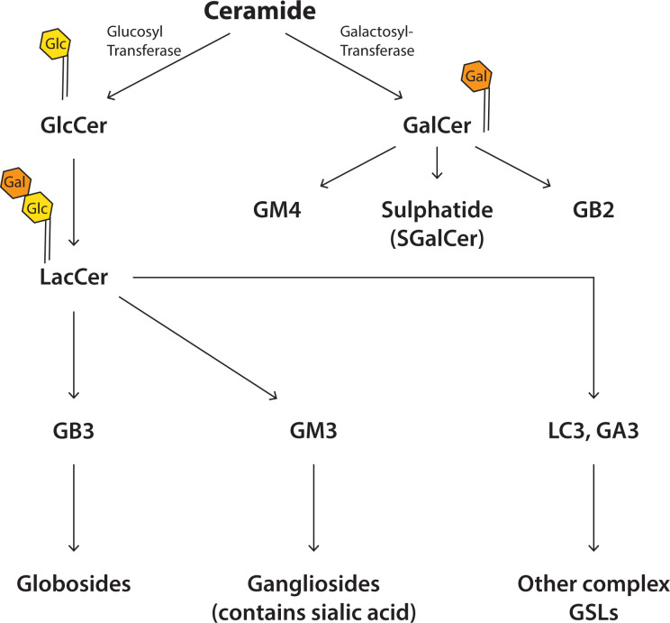
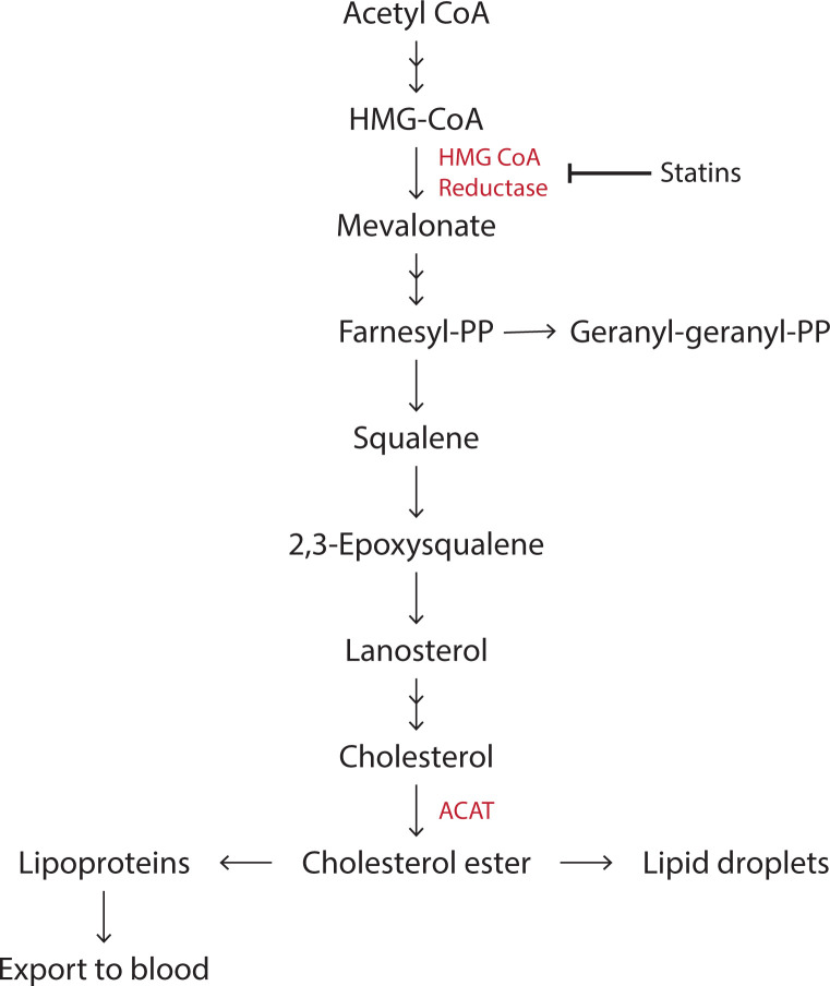
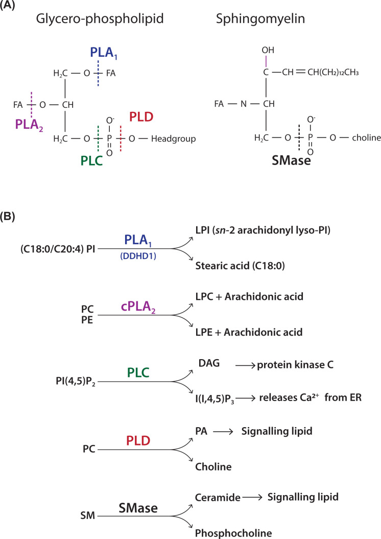
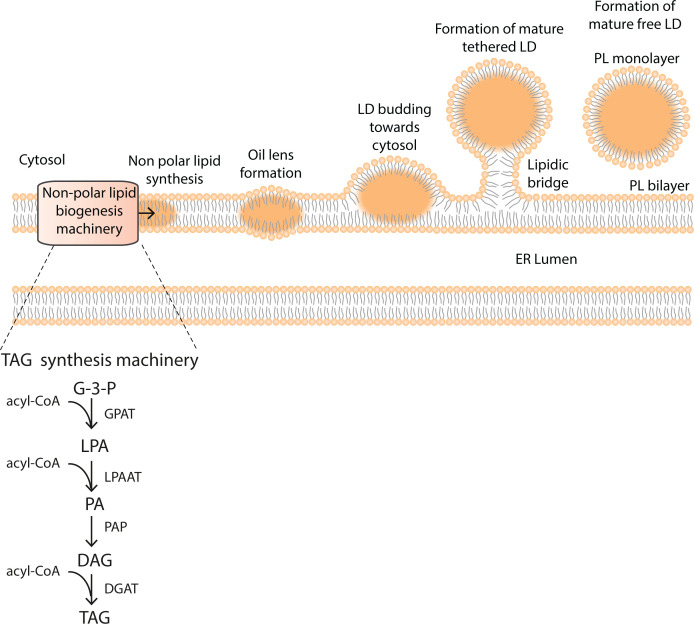
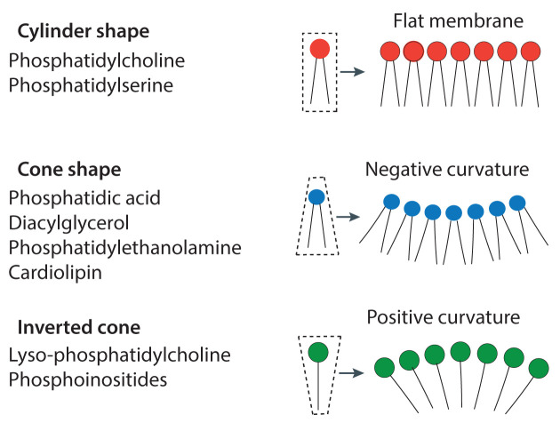
Lipids are essential constituents of cellular membranes. Once regarded merely as structural components, lipids have taken centre stage with the discovery of their roles in cell signalling and in the generation of bioactive metabolites. Lipids regulate many physiological functions of cells and alterations in membrane lipid metabolism are associated with major diseases including cancer, Type II diabetes, cardiovascular disease and immune disorders. Understanding lipid diversity, their synthesis and metabolism to generate signalling molecules will provide insight into the fundamental function of the cell. This review summarises the biosynthesis of the lipids of the mammalian cell; phospholipids, sphingolipids and cholesterol and how lipid diversity is achieved. The fatty acids (FAs) are the main building blocks of lipids and contribute to the diversity. Lipid synthesis is intimately connected to their transport within cells; the contribution by proteins that transport lipids, lipid transport proteins will be described. Cellular lipids are metabolised by phospholipases, lipid kinases and phosphatases to make new bioactive metabolites. These transient bioactive metabolites allow cells to respond to the external environment to maintain cellular health. The function of individual metabolites is also highlighted. Bioactive metabolites can be second messengers, or released to the external medium to regulate other cells. Alternatively, bioactive lipids also provide a platform for reversible recruitment of proteins to membranes using their lipid-binding domains. The wide range of physiological processes in which a specific involvement of lipids has been identified explains the need for lipid diversity present in mammalian cells.

Keywords: Cholesterol; Signalling; phosphatidylinositol; phospholipases; sphingolipids.


Conflict of interest statement

The author declares that there are no competing interests associated with the manuscript.

Figures

Figure 1
Figure 1. Three classes of lipids of mammalian cell
Glycerolipids use glycerol as their backbone, sphingolipids use a sphingoid backbone and sterols are a four-ringed structure with a hydrophobic tail and a hydroxyl group at opposite ends. Examples of lipids from the individual classes present in mammalian cells are provided.
Figure 2
Figure 2. Schematic representation of glycerolipids
(A) Phosphoglycerides have a glycerol backbone (coloured red) with FAs at the sn-1 and sn-2 position (coloured blue) and a phosphate moiety that links to a headgroup at sn-3 position (coloured green). In this example, the headgroup is choline and therefore the phospholipid is phosphatidylcholine (PC) (sn, stereochemical numbering). (B) The glycerol backbone with three FAs is called TAG. The glycerol backbone with two FAs is diacylglycerol (DAG). Addition of a phosphate to DAG makes phosphatidic acid (PA), the simplest phospholipid. Different headgroups (R) can be attached to the phosphate; addition of choline makes PC, ethanolamine makes phosphatidylethanolamine (PE), serine makes phosphatidylserine (PS) and inositol makes phosphatidylinositol (PI).
Figure 3
Figure 3. Schematic representations of PA, PG, CL, CDP-diacylglycerol and bis(monoacylglycero)phosphate
PA is the precursor for the synthesis of CDP-DAG, PG, CL and BMP. PG and BMP are both based on two glycerol molecules attached by a phosphate moiety and two FAs but with different stereochemistry. CL (alternative name: diphosphatidylglygerol) is synthesised from PG and CDP-DAG. Abbreviation: CDP-DAG, CDP-diacylglycerol.
Figure 4
Figure 4. FA diversity due to chain length and degree of unsaturation
(A) FAs comprise a carbon acyl chain with a carboxylic acid at one end. FAs are defined by their acyl chain length and by the number of double bonds. Thus, oleic acid is (C18:1 n-9). C18 denotes the length of the carbon chain and the number of double bonds is indicated after the colon. The position of the first double bond is denoted by n (number) where the carbon chain is counted from the hydrocarbon end. (B) Linoleic acid (C18:2 n-6) and α-linolenic acid C18:3 n−3) are essential FAs and are acquired from the diet. They can be further elongated by addition of acetyl groups by elongase enzymes and double bonds added by desaturase enzymes. n-3 and n-6 FAs are also known as ω3 and ω6 FAs, respectively. See Box 1 for explanation for the nomenclatures used for the position of the double bonds.
Figure 5
Figure 5. Synthesis of the phospholipids, PC, PE, PS and PI
Synthesis of the phospholipids occurs mainly at the ER. PE is additionally synthesised in mitochondria after transfer of PS from the ER through a membrane contact site, called mitochondrial-associated ER membranes (MAMs – see inset) (see text for further details). The names of the enzymes are provided in Table 2.
Figure 6
Figure 6. Synthesis of seven phosphorylated derivatives of PI
Three hydroxyl groups of the inositol ring (positions 3, 4 and 5) of PI are accessible for phosphorylation by lipid kinases. Interconversions between various phosphoinositides occurs through lipid kinases (coloured in blue) and lipid phosphatases (coloured in red). There are multiple kinases and phosphatases with different locations in the cells. Abbreviations: INPP(5), phosphoinositide phosphatase (number at the end identifies the position of the phosphate removed); MTM, myotubularin myopathy; MTMR, MTM related; OCRL, OculoCerebroRenal syndrome of Lowe; PTEN, 3-phosphatase; SHIP, SH2 domain‐containing inositol 5′ phosphatase.
Figure 7
Figure 7. Location of different phosphoinositide species in cells
(A) Different phosphorylated species of PI are enriched in different membrane compartments. PI(3,4,5)P3 (coloured purple) is only made at the plasma membrane when cells are stimulated by either GPCRs or receptor tyrosine kinases (RTKs). PI(4,5)P2 (coloured red) and PI4P (coloured blue) are found at the plasma membranes. PI4P is additionally present at the Golgi. PI3P is present in the endosomal compartment and PI(3,5)P2 is present in multivesicular bodies (MVBs) and lysosomes. (B) Recruitment of proteins by phosphoinositide-binding domains or by patches of positive charge present in proteins by different phosphorylated forms of PI. (C) Activation of AKT by phosphorylation by PDK1 and mTORC2. PIP3 recruits the PH domains of AKT and PDK1 to the plasma membrane bringing the two proteins together. PDK1 phosphorylates AKT. After a second phosphorylation by membrane target of rapamycin complex 2 (mTORC2), AKT is activated and can move away from the membrane to phosphorylate its target proteins (adapted from Figures 1 and 2 from Hammond G.R.V. and Burke J.E. (2020) Current Opinion in Cell Biology, 63:57–67).
Figure 8
Figure 8. Synthesis of CL in mitochondria
Synthesis of CL begins with PA which is synthesised in the OMM. It is transferred to the IMM by the PA transport protein, PRELID1–TRIAP complex. PA is translocated to the matrix side where it is then converted into PG and finally to CL through a series of enzymatic reactions all located in the IMM. See text for abbreviations of enzymes.
Figure 9
Figure 9. Ether-linked phospholipids
Diacyl phospholipids have acyl chains linked to the sn-1 and sn-2 position of the glycerol backbone by ester bonds. Ether phospholipids have an acyl chain attached by an ether bond at the sn-1 position. Ether alkenyl phospholipids have a cis double bond adjacent to the ether linkage at the sn-1 position and are known as plasmalogens. The acyl chain at the sn-2 position in ether lipids is linked via an ester linkage. The polar head group (X) of ether-linked phospholipids is mainly ethanolamine with some choline. The ester, ether (alkyl) and vinyl-ether (alkenyl) linkage at sn-1 is coloured red.
Figure 10
Figure 10. Schematic representation of sphingolipids
(A) Sphingolipids have a sphingoid backbone (coloured red) derived from the condensation of serine with palmitoyl CoA. Attachment of an acyl chain (coloured blue) by an amide link to sphingosine makes ceramide and addition of a headgroup, phosphocholine (coloured green) makes SM, the most abundant sphingolipid of mammalian cells. (B) Sphingosine and ceramide can both be phosphorylated to make bioactive metabolites.
Figure 11
Figure 11. Synthesis of SM and other derivatives of ceramide
Synthesis of ceramide begins with the condensation of serine with palmitoyl CoA. Ceramide can be converted into a variety of metabolites including SM, sphingosine and ceramide-1-phosphate. Sphingosine kinase phosphorylates sphingosine to make sphingosine-1-phosphate. SM can also be converted into ceramide through the action of SMase (see Figure 14). Abbreviation: SMase, sphingomyelinase.
Figure 12
Figure 12. Synthesis of GSLs from ceramide
Addition of the sugar molecules, glucose or galactose to ceramide makes the simple GSLs, GlcCer and GalCer. Addition of further sugar molecules generates a large number of complex GSLs. More than 400 different GSLs have been identified. The most common sugar is galactose, followed by N-acetylglucosamine and by glucose.
Figure 13
Figure 13. Synthesis of cholesterol and isoprenoids
Cholesterol synthesis begins with acetyl CoA and the rate-limiting enzyme is HMG CoA reductase (3-Hydroxy-3-MethylGlutaryl Coenzyme A Reductase) (coloured red). Statins inhibits this enzyme. More than 20 enzymes are involved in cholesterol synthesis and only a few key steps are shown here. Cholesterol can be esterified to cholesterol ester by the enzyme, acyl CoA cholesterol acyl transferase (ACAT). The intermediate farnesyl-PP is a 15-carbon isoprenoid that can be converted into the 20-carbon Geranyl-Geranyl-PP isoprenoid. Both are used for the modification of proteins by prenylation (see Section, ‘Lipid modification of proteins’).
Figure 14
Figure 14. Action of phospholipases on phospholipids
(A) Site of action of various phospholipases (A1, A2, C and D) on a typical glycerol-phospholipid. The FAs at the sn-1 and sn-2 positions are removed by phospholipases A1 and A2 respectively. Phospholipase C and D attack the phosphodiester bond at different positions. Site of action of sphingomyelinase (SMase) on SM. SMase attacks the phosphodiester bond. (B) Examples of well-known phospholipases that cleave specific substrates. Phospholipase A1 removes stearic acid from PI to make sn-2 arachidonyl lyso-PI, a potent signalling molecule. Cytosolic phospholipase A2 (cPLA2) removes arachidonic acid from either PC or PE used for the synthesis of prostaglandins, leukotrienes and thromboxanes. Phospholipase C hydrolyses PI(4,5)P2 that generates two second messengers, I(1,4,5)P3 and DAG. Phospholipase D hydrolyses PC to make PA and choline. SM can be attacked by SMase to make ceramide and phosphocholine.
Figure 15
Figure 15. Lipid droplet formation in the ER membrane
Lipid droplet biogenesis begins with the synthesis of TAG that accumulates between the two leaflets of the ER membrane. An oil lens is formed, which buds towards the cytosol. Finally, the mature lipid droplet, surrounded by a lipid monolayer, is released into the cytosol. The TAG biosynthetic machinery is recruited by a protein called seipin (not shown).
Figure 16
Figure 16. The shape of the lipid contributes to the curvature of the membrane
A lipid can be: cylinder-shaped where the headgroup and the acyl chains occupy a similar-sized space, cone-shaped where the headgroup is smaller than the space occupied by the acyl chains, or inverted cone where the headgroup is larger than the space occupied the acyl chains. When present in a membrane bilayer, the lipids determine the curvature of the membrane.
Figure 17
Figure 17. Analysis of total cellular lipids and localisation of specific lipids in cells
(A) A thin layer chromatographic (TLC) plate showing the separation of total cellular lipids extracted from a human leukaemic cell-line, HL60 cells. HL60 cells were grown for 3 days in radioactive 14C-acetate which will be incorporated into all the lipids. The position of the individual lipids is indicated determined by the use of known lipid standards. Some bands are not identified. The lipids are detected by their radioactivity. The origin indicates where the mixture of lipids was applied on the TLC plate. The solvent used for separation was a mixture of chloroform:methanol:acetic acid:water (75:45:3:1). (B) PI(4,5)P2 is mainly localised at the plasma membrane. The PH domain of phospholipase Cδ1 was fused with GFP and the plasmid containing the DNA was transfected into a mast cell-line, RBL 2H3 cells. After 24 h, the cells were visualised under a fluorescent microscope. This PH domain is highly specific and will only bind to PI(4,5)P2.
Figure 18
Figure 18. Steady-state composition of different membrane compartments of a mammalian cell
The lipid composition of different membranes of the rat liver is variable. The individual graphs shows the lipid composition of the main phospholipids of a specific membrane compartment expressed as a percentage of the total phospholipid. Some minor lipids are not shown and are referred to as ‘others’. Some of the key minor lipids present in a particular compartment are included in the figure. Abbreviation: Chol, cholesterol.

References

    1. Acoba M.G., Senoo N. and Claypool S.M. (2020) Phospholipid ebb and flow makes mitochondria go. J. Cell Biol. 219, e202003131 10.1083/jcb.202003131 - DOI - PMC - PubMed
    1. Arnal-Levron M., Chen Y., Greimel P., Calevro F., Gaget K., Riols F.et al. . (2019) Bis(monoacylglycero)phosphate regulates oxysterol binding protein-related protein 11 dependent sterol trafficking. Biochim. Biophys. Acta. Mol. Cell Biol. Lipids 1864, 1247–1257 10.1016/j.bbalip.2019.05.011 - DOI - PubMed
    1. Balla T. (2013) Phosphoinositides: tiny lipids with giant impact on cell regulation. Physiol. Rev. 93, 1019–1137 10.1152/physrev.00028.2012 - DOI - PMC - PubMed
    1. Braverman N.E. and Moser A.B. (2012) Functions of plasmalogen lipids in health and disease. Biochim. Biophys. Acta. 1822, 1442–1452 10.1016/j.bbadis.2012.05.008 - DOI - PubMed
    1. Brown M.S., Radhakrishnan A. and Goldstein J.L. (2018) Retrospective on cholesterol homeostasis: the central role of Scap. Annu. Rev. Biochem. 87, 783–807 10.1146/annurev-biochem-062917-011852 - DOI - PMC - PubMed

Publication types