. 2021 Dec;41(12):3365-3377.
doi: 10.1177/0271678X211037869. Epub 2021 Aug 20.

Association of autosomal dominant polycystic kidney disease with cerebral small vessel disease

Affiliations

Association of autosomal dominant polycystic kidney disease with cerebral small vessel disease

Woo-Jin Lee et al. J Cereb Blood Flow Metab. 2021 Dec.

Abstract

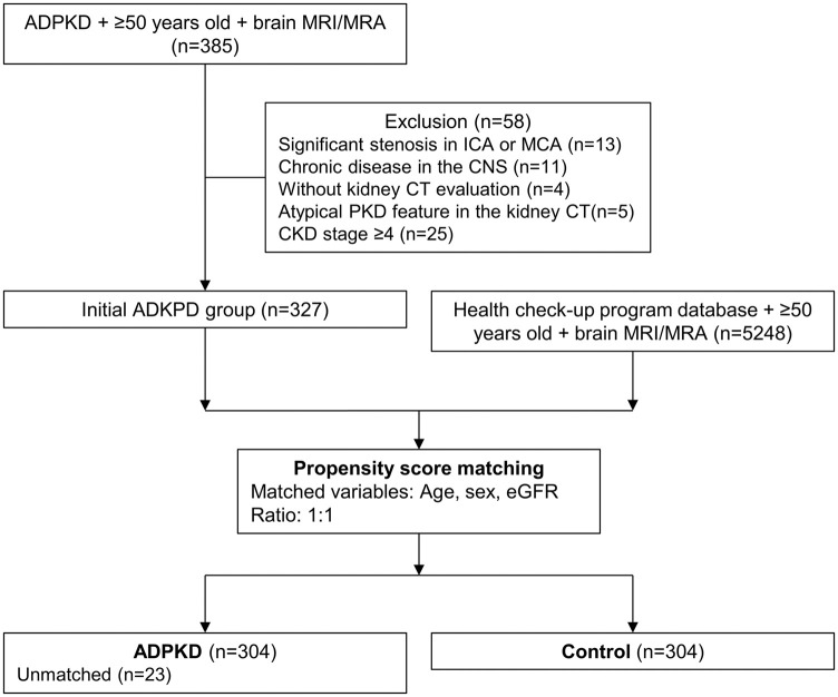
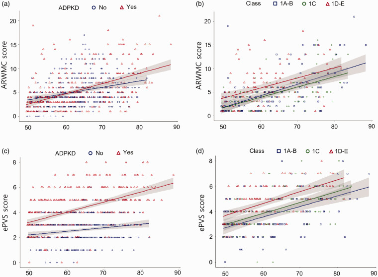
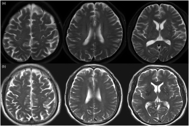
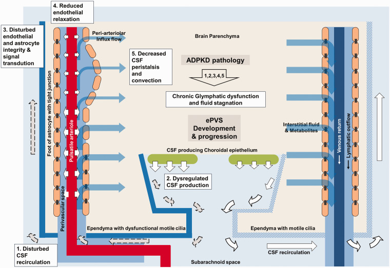
Cilia dysfunction in autosomal-dominant polycystic kidney disease (ADPKD) may impair the integrity of glymphatic system and be implicated in the progression of cerebral small vessel disease (SVD), although the link between the two diseases has not been investigated. We evaluated the association of ADPKD pathology with SVD pattern and severity. Overall, 304 individuals in an ADPKD (chronic kidney disease stage ≤4 and age ≥50 years) cohort and their age, sex, and estimated glomerular filtration rate (eGFR)-matched controls were retrospectively included. ADPKD severity was classified into 1 A-B, 1 C, and 1 D-E, according to age and height-adjusted total kidney volume. SVD parameters included white-matter hyperintensity (WMH) severity scale, enlarged perivascular space (ePVS) score, and degree of lacunes or cerebral microbleeds (CMBs). After adjustments for age, sex, eGFR, and cerebrovascular risk factor parameters, ADPKD was associated with higher ePVS scores (P < 0.001), but not with the WMH severity or degree of lacunes or CMBs. In the ADPKD subgroup, higher ADPKD severity class was associated with higher ePVS scores (P < 0.001), WMH severity (P = 0.003), and degree of lacunes (P = 0.002). ADPKD associated cilia dysfunction may induce chronic cerebral glymphatic system dysfunction, which may contribute to the specific progression of ePVS compared with other SVD markers.

Keywords: Autosomal dominant polycystic kidney disease; cilia dysfunction; enlarged perivascular space; glymphatic system; small vessel disease.


Conflict of interest statement

Declaration of conflicting interests: The author(s) declared no potential conflicts of interest with respect to the research, authorship, and/or publication of this article.

Figures

Figure 1.
Figure 1.
A flow chart illustrating the study participants. MRI: magnetic resonance image; MRA: magnetic resonance angiography; ICA: internal carotid artery; MCA: middle cerebral artery; CNS: central nervous system; PKD: polycystic kidney disease; CKD: chronic kidney disease; eGFR: estimated glomerular filtration rate.
Figure 2.
Figure 2.
Plots for the age and small vessel disease parameters in the whole study population and in the autosomal dominant polycystic kidney disease (ADPKD) group. In panel A, dot plots with linear trend lines with 95% confidence for ADPKD and control groups are depicted for ARWMC scores for the whole study population. ADPKD was not associated with a higher total ARWMC score (P=0.143). In panel B, plots with linear trend lines for each severity subgroups are depicted for ARWMC scores within the ADPKD group. A higher ADPKD severity class was associated with a higher total ARWMC score (P=0.003). In panel C, plots for ePVS scores are depicted for ADPKD and control groups. ADPKD was associated with a higher total ePVS score (P < 0.001). In panel D, plots for ePVS scores are depicted for each severity subgroups within the ADPKD group. A higher ADPKD severity class was associated with a higher total ePVS score (P < 0.001). ARWMC: age-related white matter change, PVS: enlarged perivascular space.
Figure 3.
Figure 3.
Representative cases. Panel A shows magnetic resonance images (MRIs) of a fifty-nine-year-old woman with autosomal dominant polycystic kidney disease (ADPKD) with a severity class of 1C and glomerular filtration rate of 42.6 mL/min. Enlarged periventricular space (ePVS) score was 2 for the centrum semiovale (CS) area, 1 for the basal ganglia (BG), and 3 for the total brain. Panel B shows MRIs of a sixty-three-year-old man with ADPKD severity class of 1D and glomerular filtration rate of 65.3 mL/min. ePVS score was 3 for the CS area, 1 for the BG, and 5 for the total brain.
Figure 4.
Figure 4.
Schematic explanation of the pathophysiologic link between ADPKD and progression of enlarged perivascular space. In the subarachnoid space, white arrows indicate well-regulated CSF flow, and dashed grey arrows indicate disturbed and dysregulated CSF flow. Blue diagonal patterned lines in the right side indicates the ependymal lining with intact motile cilia, and lines filled-up with blue in the left side indicates the ependymal lining with dysfunctional motile cilia. ADPKD: autosomal dominant polycystic kidney disease, CSF: cerebrospinal fluid, ePVS: enlarged perivascular space.

Similar articles

Cited by

References

    1. Yoshida H, Higashihara E, Maruyama K, et al.. Relationship between intracranial aneurysms and the severity of autosomal dominant polycystic kidney disease. Acta Neurochir (Wien) 2017; 159: 2325–2330. - PubMed
    1. Schievink WI, Torres VE, Wiebers DO, et al.. Intracranial arterial dolichoectasia in autosomal dominant polycystic kidney disease. J Am Soc Nephrol 1997; 8: 1298–1303. - PubMed
    1. Wang D, Iversen J, Wilcox CS, et al.. Endothelial dysfunction and reduced nitric oxide in resistance arteries in autosomal-dominant polycystic kidney disease. Kidney Int 2003; 64: 1381–1388. - PubMed
    1. Chapman AB, Johnson A, Gabow PA, et al.. The renin–angiotensin–aldosterone system and autosomal dominant polycystic kidney disease. N Engl J Med 1990; 323: 1091–1096. - PubMed
    1. Wardlaw JM, Smith C, Dichgans M. Mechanisms of sporadic cerebral small vessel disease: insights from neuroimaging. Lancet Neurol 2013; 12: 483–497. - PMC - PubMed

Publication types

MeSH terms