. 2021 Aug 19;184(17):4414-4429.e19.
doi: 10.1016/j.cell.2021.07.006.

Pan-protective anti-alphavirus human antibodies target a conserved E1 protein epitope

Affiliations

Pan-protective anti-alphavirus human antibodies target a conserved E1 protein epitope

Arthur S Kim et al. Cell. .

Abstract

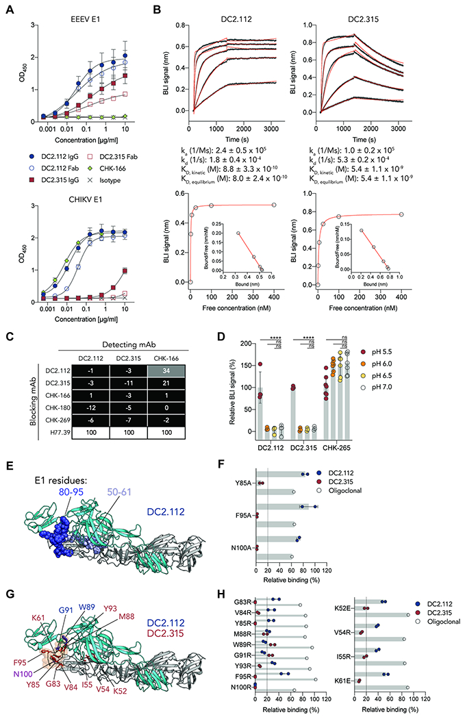
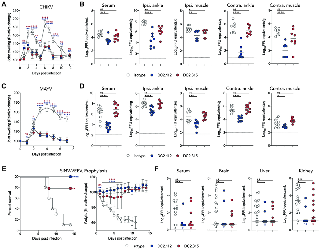
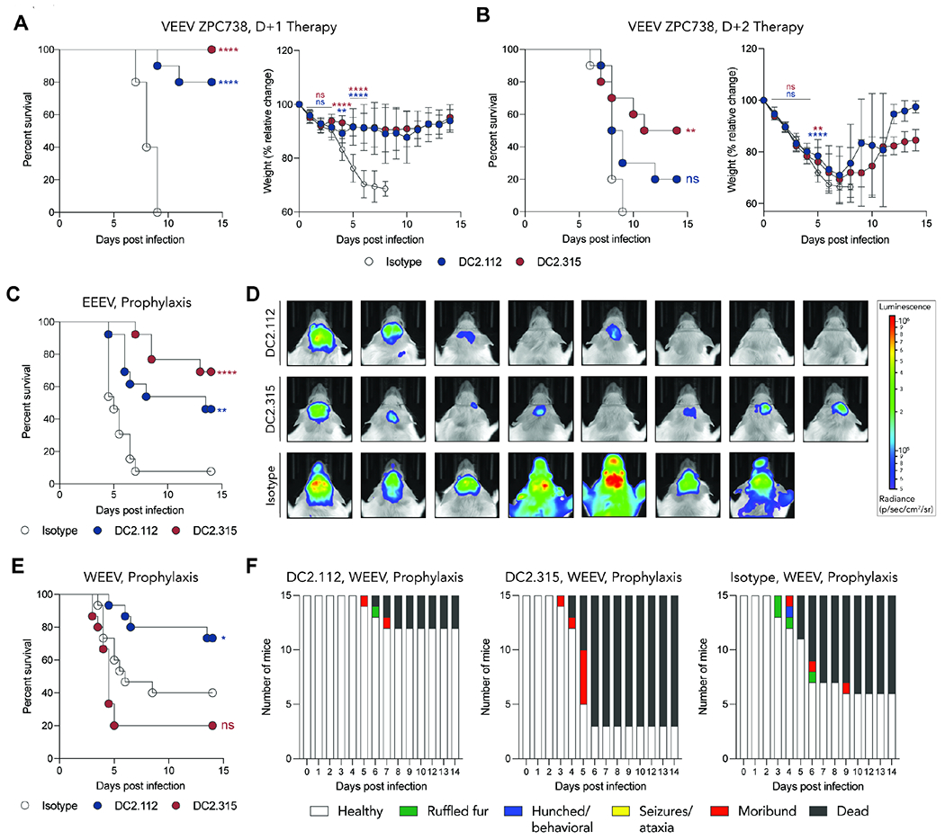
Alphaviruses are emerging, mosquito-transmitted pathogens that cause musculoskeletal and neurological disease in humans. Although neutralizing antibodies that inhibit individual alphaviruses have been described, broadly reactive antibodies that protect against both arthritogenic and encephalitic alphaviruses have not been reported. Here, we identify DC2.112 and DC2.315, two pan-protective yet poorly neutralizing human monoclonal antibodies (mAbs) that avidly bind to viral antigen on the surface of cells infected with arthritogenic and encephalitic alphaviruses. These mAbs engage a conserved epitope in domain II of the E1 protein proximal to and within the fusion peptide. Treatment with DC2.112 or DC2.315 protects mice against infection by both arthritogenic (chikungunya and Mayaro) and encephalitic (Venezuelan, Eastern, and Western equine encephalitis) alphaviruses through multiple mechanisms, including inhibition of viral egress and monocyte-dependent Fc effector functions. These findings define a conserved epitope recognized by weakly neutralizing yet protective antibodies that could be targeted for pan-alphavirus immunotherapy and vaccine design.

Keywords: Fc effector; alphavirus; antibody; arthritis; encephalitis; immunity; inhiibtion; mice; pathogenesis; protection.


Conflict of interest statement

Declaration of interests M.S.D. is a consultant for Inbios, Vir Biotechnology, NGM Biopharmaceuticals, and Carnival Corporation and on the scientific advisory boards of Moderna and Immunome. The Diamond and Fremont laboratories received support from Emergent BioSolutions under a sponsored research agreement. The Diamond laboratory has unrelated research agreements from Moderna and Vir Biotechnology. D.H.F. is a founder of Courier Therapeutics. J.R.L. is a consultant for Celdara Medical. DC2.112 and DC2.315 are the subject of a US patent application with R.J.M., J.A.Q., and J.R.L. as co-inventors. R.H.F., E.D., and B.J.D. are employees of Integral Molecular, and B.J.D. is a shareholder of Integral Molecular. J.E.C. has served as a consultant for Lilly and Luna Biologics, is a member of the Scientific Advisory Boards of CompuVax and Meissa Vaccines, and is founder of IDBiologics. The Crowe laboratory has received unrelated sponsored research agreements from Astra Zeneca and IDBiologics. M.L.G. is an unpaid member of the scientific advisory boards of Protein Metrics and GenNext. G.A. is a founder of SeromYx Systems.

Figures

Figure 1.
Figure 1.. Broadly reactive anti-CHIKV E1 mAbs recognize the structural proteins of arthritogenic and encephalitic alphaviruses.
(A) Anti-CHIKV mAbs were incubated with Vero cells, inoculated with arthritogenic (CHIKV, MAYV, RRV, SINV) or chimeric encephalitic viruses (SINV-VEEV, SINV-EEEV, SINV-WEEV), and anti-alphavirus mAb binding to the cell surface was assessed. Data are representative of 3 experiments. (B) Phylogenetic relationship of alphavirus E1 structural proteins as determined by MUSCLE and neighbor-joining analysis (Edgar, 2004). Alphaviruses are colored by complexes: Semliki Forest, blue; WEE, green; VEE, orange; EEE, purple. (C) Graph (top panel) depicting DC2.112, DC2.315, or CHK-265 mAb binding to alphaviruses: dark green, high binding (EC50 < 100 ng/ml); green, intermediate binding (EC50 100 – 1000 ng/ml); light green, low binding (EC50 > 1000 ng/ml); light blue, no binding; and gray, not tested. EC50 values of DC2.112, DC2.315, or CHK-265 mAb binding to surface of cells infected with alphaviruses (bottom panel). Symbols are colored according to the binding criteria in the graph (top panel). Mean of 2-3 experiments performed in duplicate. See also Figure S1 and S2.
Figure 2.
Figure 2.. Anti-CHIKV E1 mAbs recognize a conserved, cryptic epitope in the E1 protein.
(A) Binding of DC2.112 and DC2.315 mAbs or Fabs, anti-CHIKV E1 mAb CHK-166, or isotype control mAb to immobilized recombinant EEEV E1 (top panel) or CHIKV E1 (bottom panel) proteins. Mean and standard deviations (SD) from 2 experiments performed in duplicate. (B) Representative kinetic sensorgrams (top panels) and steady-state analysis (bottom panels) using BLI by incubating different concentrations of EEEV E1 protein over biosensors immobilized with DC2.112 (left panels) or DC2.315 (right panels). Kinetic affinity (KD, kinetic) values (top panels). Steady-state affinity values (KD, equilibrium) (bottom panels) and Scatchard plots (bottom panel, insets). Mean and SD from 3 experiments. (C) Anti-CHIKV E1 human (DC2.112 or DC2.315), murine (CHK-166, CHK-180, CHK-269), or isotype control (anti-HCV H77.39) mAbs were incubated with CHIKV E1 protein to assess antibody competition binding by BLI. Black boxes: <33% binding; gray boxes: 33 to 67% binding; white boxes: >67% binding of the detecting mAb. (D) CHIKV VLPs captured with CHK-265 were equilibrated in binding buffers of different pH (pH 5.5, 6, 6.5, or 7) and then incubated with mAbs DC2.112, DC2.315, or chimeric CHK-265. The signal at different pH conditions was determined by normalizing to the binding achieved at pH 7.0. Mean and SD of 3-4 experiments (n = 3 to 7; one-way ANOVA with Dunnett’s post-test for each mAb: **** P <0.0001; n.s., not significant). (E) DC2.112 was incubated with EEEV E1 protein and protected regions were identified by HDX-MS. Peptides (residues 50-61 and 80-95) protected by DC2.112 are indicated in blue spheres and mapped onto the CHIKV p62-E1 monomer (PDB 3N42). E2 protein, cyan; E1 protein, gray. (F) Alanine-scanning mutagenesis of key CHIKV binding residues for DC2.112 (blue symbols) and DC2.315 (red symbols) compared to a CHIKV oligoclonal mAb control (open black symbols). The cutoff for binding of key residues (<20%) is indicated by the dotted line. Mean and SD of 2 experiments. (G) CHIKV E1 protein residues necessary for DC2.112 or DC2.315 binding as determined by charged residue mutagenesis and highlighted in blue (DC2.112), red (DC2.315), or purple (DC2.112 and DC2.315) on the CHIKV p62-E1 monomer (PDB 3N42). The fusion loop (residues 84-99) is indicated by the orange outline. (H) Relative binding levels of DC2.112 (blue symbols) or DC2.315 (red symbols) to charged residue mutants are shown and were determined as in panel F. Mean and SD of 2 experiments. See also Figure S3 and S4.
Figure 3.
Figure 3.. Anti-CHIKV E1 mAbs neutralize infection and inhibit viral egress in a cell-type-specific manner.
A-C. Neutralization studies. (A) Anti-CHIKV E1 mAbs DC2.112 and DC2.315 were incubated with indicated alphaviruses before addition to Vero cells. The following mAbs (CHK-265: CHIKV, MAYV, ONNV, RRV; 1A4A-1: SINV-VEEV; EEEV-10: SINV-EEEV; WEEV-209: SINV-WEEV) were included as positive controls. Anti-WNV E16 mAb was included as a negative control. Each graph represents the mean and SD from 2 experiments (n = 4). (B) DC2.112 or DC2.315 IgG were incubated with CHIKV before addition to 3T3 cells. Infected foci were quantitated. Mean and SD of 2 experiments (n = 4). (C) DC2.112 and DC2.315 IgG were incubated with CHIKV before addition to N2a cells. Supernatant was harvested at indicated time points, and viral titers were determined by qRT-PCR. Mean and SD of 5 experiments (n = 6). D-F. Egress inhibition studies. Vero (D-E) or N2a (F) cells were inoculated with CHIKV (D) or SINV-VEEV (E-F) for 2 h, washed extensively to remove unbound virus, and incubated with mAbs or Fabs in the presence of 25 mM NH4Cl to prevent de novo infection. Supernatant was harvested at 1 or 6 hours post infection (hpi), and viral RNA was quantitated by qRT-PCR. Mean and SD of 3 to 5 experiments (n = 6 to 11). C-F: one-way ANOVA with a Bonferroni post-test at each time point and mAb concentration: * P < 0.05, ** P < 0.01, *** P < 0.001, **** p <0.0001; n.s., not significant). See also Figure S4 and S6.
Figure 4.
Figure 4.. DC2.112 and DC2.315 protect in vivo against arthritogenic and encephalitic alphaviruses.
(A-D) Four-week-old male C57BL/6J mice were administered 200 μg of DC2.112, DC2.315, or isotype control mAbs via intraperitoneal (i.p.) route 24 h before subcutaneous inoculation in the foot with 103 FFU of CHIKV AF15561 (A-B) or MAYV BeH407 (C-D). Mice were monitored for joint swelling (A and C). Viral RNA levels in serum were assessed at 2 dpi and in the ipsilateral and contralateral ankles and muscles at 5 (CHIKV) or 7 (MAYV) dpi (B and D). (E-F) Four-week-old male C57BL/6J mice were administered 375 or 750 μg (depending on the commercial preparation) of anti-IFNAR1 mAb (MAR1-5A3) by the i.p. route 24 h before virus inoculation. Anti-E1 or isotype controls mAbs were administered 8 h before i.p. inoculation with 105 FFU of SINV-VEEV-TrD. Mice were monitored for survival and weight change (E). Viral RNA levels in the serum, brain, liver, and kidney were measured at 5 dpi (F). Dotted line indicates limit of detection (LOD), and values indicate the number of animals at the LOD. For A and C (left panels), 2 experiments (n = 9 to 10; two-way ANOVA with Dunnett’s post-test: * P < 0.05; ** P < 0.01, *** P < 0.001, **** P < 0.0001; n.s., not significant). For B (right panel), D (right panel), and F, 2-3 experiments (n = 8 to 15; one-way ANOVA with Dunnett’s post-test: * P < 0.05; ** P < 0.01, *** P < 0.001, **** P < 0.0001; n.s., not significant). For E (left panel), 3 experiments (n = 15; Mantel-Cox log-rank test for survival: ** P < 0.01, **** P < 0.0001). See also Figure S4.
Figure 5.
Figure 5.. DC2.112 and DC2.315 require Fc effector functions for optimal protection in vivo
(A) Four-week-old male C57BL/6J mice were administered 200 μg of intact or aglycosyl (N297Q) DC2.112 or isotype control mAbs via intraperitoneal route (i.p.) route 24 h before subcutaneous inoculation with 103 FFU of CHIKV AF15561. Viral RNA levels were assessed in the ipsilateral and contralateral ankles and muscles at 5 dpi. (B-E) Six-week-old wild-type (B, C, and E) or FcγR−/− (D) C57BL/6J mice (both sexes) were administered 200 μg of intact or N297Q anti-E1 mAbs (DC2.112 or DC2.315) or an isotype control by i.p. injection either 8 h before (B-D) or 24 h after (E) subcutaneous inoculation with 102 FFU of VEEV ZPC738. Mice were monitored for survival and weight change (B, D, and E). In mice treated with mAbs pre-exposure, viral RNA levels were assessed in the serum, brain, liver, and kidney at 6 dpi (C). Values indicate the number of animals with tissue titers at the LOD. For A and C, 2 experiments (n = 8-14; one-way ANOVA with Dunnett’s post-test: * P < 0.05; ** P < 0.01, *** P < 0.001, **** p n.s., not significant). For left panels in B, D, and E, 2-3 experiments (n = 9-15; Mantel-Cox log-rank test for survival: ** or ++ P < 0.01, +++ P < 0.001, **** P < 0.0001, n.s., not significant). For right panels in B, D, and E, data are from 2-3 experiments. Data were analyzed statistically only when all mice were alive in the cohort (n = 9-15; two-way ANOVA with Dunnett’s post-test: * or + P < 0.05; ++ P < 0.01, *** or +++ P < 0.001, **** or +++ P < 0.0001; n.s., not significant). Blue “*”: DC2.112 vs isotype, Blue “+”: DC2.112 N297Q vs isotype, Red “*”: DC2.315 vs isotype, Red “+”: DC2.315 N297Q vs isotype, “ns”: Intact mAbs vs isotype, “ns”: N297Q mAbs vs isotype. See also Figure S4, S5, and S6.
Figure 6.
Figure 6.. Fc effector functions of DC2.112 and monocytes mediate protection against CHIKV infection.
(A) Antibody-dependent cellular phagocytosis (ADCP). THP-1 cells were incubated with DC2.112- or isotype control-bound EEEV E1 immune complex beads, and phagocytosis was measured by flow cytometry. Mean and SD of 2 experiments (n = 4). (B) Antibody-dependent neutrophil activation of ROS release (ADNR). Captured EEEV E1 protein was incubated with DC2.112 or PBS for 2 h at room temperature. Human neutrophils were incubated with luminol and then added. Reactive oxygen species (ROS) release was measured by chemiluminescence. Data are representative of 2 experiments. (C) Antibody-dependent NK cell degranulation (ADNK). Captured EEEV E1 protein was incubated with DC2.112 or isotype control for 2 h. Human NK cells were added and incubated for 5 h at 37°C. Cells were analyzed for CD 107a (left panel) and MIP1-β (right panel) expression by flow cytometry. The upper line indicates a positive control for NK cell degranulation after phorbol myristate acetate and ionomycin stimulation. The lower line indicates the baseline level with a PBS control. Mean and SD of 2 experiments. (D) Antibody-dependent complement deposition (ADCD). DC2.112-bound EEEV E1 immune complexes were incubated with guinea pig complement for 20 min at 37°C, and C3 deposition was analyzed by flow cytometry. Mean and SD of 2 experiments. (E-G) Four-week-old male C57BL/6J mice were administered 25 μg of a depleting anti-CCR2 (MC-21) or an isotype control mAb by intraperitoneal injection 24 h prior to subcutaneous inoculation of 103 FFU of CHIKV AF15561. Mice were administered 200 μg of DC2.112 or isotype control mAb at 1 dpi. Joint swelling (E) and viral RNA levels were measured in the ipsilateral ankle, ipsilateral muscle, and contralateral muscle (at 6 dpi) (F). Flow cytometry plots of peripheral blood leukocytes following administration of anti-CCR2 or isotype control mAb 1 day prior to inoculation with CHIKV (G). For E, 2 experiments (n = 7 to 8; two-way ANOVA with Dunnett’s post-test: **** P < 0.0001; n.s., not significant; blue: isotype vs DC2.112, blue italics: isotype vs DC2.112 + αCCR2, gray: isotype vs isotype + αCCR2). For F, 2 experiments (n = 7 to 8; one-way ANOVA with Dunnett’s post-test: * P < 0.05; ** P < 0.01, *** P < 0.001, **** P < 0.0001; n.s., not significant). See also Figure S4 and S6.
Figure 7.
Figure 7.. DC2.112 and DC2.315 anti-E1 mAbs protect in vivo against encephalitic alphaviruses.
(A-B) Six-week-old male wild-type C57BL/6J mice were administered 200 μg of anti-E1 mAbs or an isotype control by i.p. injection either 24 h (A) or 48 h (B) after subcutaneous inoculation with 102 FFU of VEEV ZPC738. Mice were monitored for survival and weight changes. (C-D) Five-week-old female CD-1 mice were treated with 200 μg of anti-E1 or isotype control mAbs via i.p. injection route before subcutaneous inoculation with 3 x 10³ PFU of EEEV FL93-939 nLuc TAV. Mice were monitored for survival (C). At 4 dpi, IVIS imaging was used to visualize EEEV FL93-939 nLuc TAV infection (D). The total flux (photons s−1) in the dorsal head of each mouse was quantified. IVIS images shown are representative images from 2 experiments (n = 13). (E-F) Four-week-old female CD-1 mice were treated with 200 μg of anti-E1 or isotype control mAbs by i.p. injection before subcutaneous inoculation with 103 PFU of WEEV McMillan. Mice were monitored for survival (E). Clinical disease was assessed (ruffled fur, hunched posture, seizures, ataxia, moribund, or death) (F). Data are from 2 experiments (n = 15; Mantel-Cox log-rank test for survival: * P < 0.05, n.s., not significant). For (A (left panel), B (left panel), C, and E), data are from 2 experiments (n = 10-15; Mantel-Cox log-rank test for survival: * P < 0.05, ** P < 0.01, **** P < 0.0001, n.s., not significant). For (A (right panel) and B (right panel)), statistical analysis of weight change was performed only when all mice were alive in the cohort to avoid survivor bias (n = 10; two-way ANOVA with Dunnett’s post-test: ** P < 0.01, **** P < 0.0001; n.s., not significant).

Similar articles

Cited by

References

    1. Ackerman ME, Moldt B, Wyatt RT, Dugast AS, McAndrew E, Tsoukas S, Jost S, Berger CT, Sciaranghella G, Liu Q, et al. (2011). A robust, high-throughput assay to determine the phagocytic activity of clinical antibody samples. J Immunol Methods 366, 8–19. - PMC - PubMed
    1. Akahata W, Yang ZY, Andersen H, Sun S, Holdaway HA, Kong WP, Lewis MG, Higgs S, Rossmann MG, Rao S, et al. (2010). A virus-like particle vaccine for epidemic Chikungunya virus protects nonhuman primates against infection. Nature medicine 16, 334–338. - PMC - PubMed
    1. Basore K, Kim AS, Nelson CA, Zhang R, Smith BK, Uranga C, Vang L, Cheng M, Gross ML, Smith J, et al. (2019). Cryo-EM Structure of Chikungunya Virus in Complex with the Mxra8 Receptor. Cell 177, 1725–1737.e1716. - PMC - PubMed
    1. Bernard KA, Klimstra WB, and Johnston RE (2000). Mutations in the E2 glycoprotein of Venezuelan equine encephalitis virus confer heparan sulfate interaction, low morbidity, and rapid clearance from blood of mice. Virology 276, 93–103. - PubMed
    1. Boere WA, Harmsen T, Vinje J, Benaissa-Trouw BJ, Kraaijeveld CA, and Snippe H (1984). Identification of distinct antigenic determinants on Semliki Forest virus by using monoclonal antibodies with different antiviral activities. Journal of virology 52, 575–582. - PMC - PubMed

Publication types

MeSH terms

LinkOut - more resources