. 2021 Aug 19;184(17):4430-4446.e22.
doi: 10.1016/j.cell.2021.07.033.

Therapeutic alphavirus cross-reactive E1 human antibodies inhibit viral egress

Affiliations

Therapeutic alphavirus cross-reactive E1 human antibodies inhibit viral egress

Lauren E Williamson et al. Cell. .

Abstract

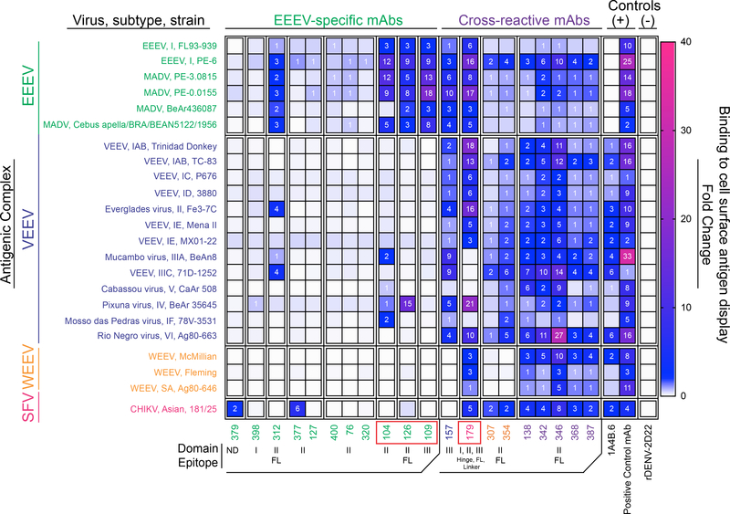
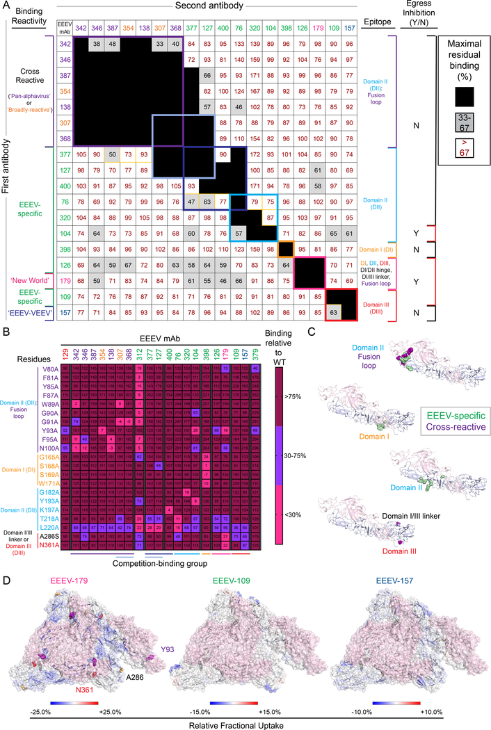
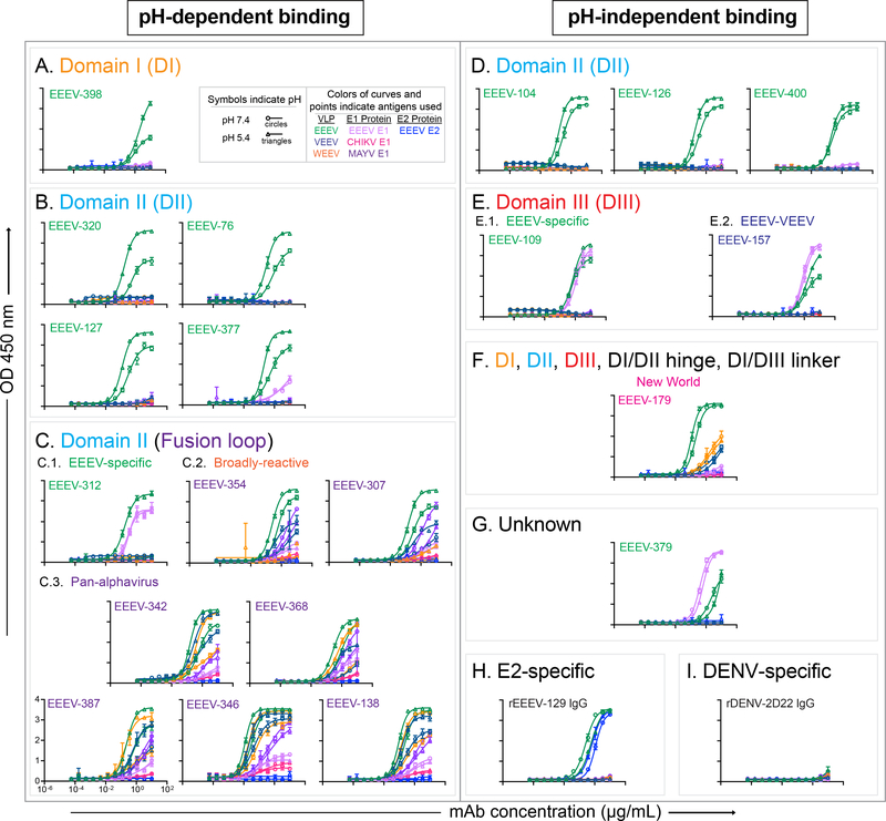
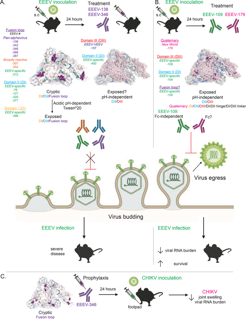
Alphaviruses cause severe arthritogenic or encephalitic disease. The E1 structural glycoprotein is highly conserved in these viruses and mediates viral fusion with host cells. However, the role of antibody responses to the E1 protein in immunity is poorly understood. We isolated E1-specific human monoclonal antibodies (mAbs) with diverse patterns of recognition for alphaviruses (ranging from Eastern equine encephalitis virus [EEEV]-specific to alphavirus cross-reactive) from survivors of natural EEEV infection. Antibody binding patterns and epitope mapping experiments identified differences in E1 reactivity based on exposure of epitopes on the glycoprotein through pH-dependent mechanisms or presentation on the cell surface prior to virus egress. Therapeutic efficacy in vivo of these mAbs corresponded with potency of virus egress inhibition in vitro and did not require Fc-mediated effector functions for treatment against subcutaneous EEEV challenge. These studies reveal the molecular basis for broad and protective antibody responses to alphavirus E1 proteins.

Keywords: alphavirus; cross-reactive antibodies; human monoclonal antibodies; non-neutralizing antibodies; post-exposure prophylaxis.


Conflict of interest statement

Declaration of interests J.E.C. has served as a consultant for Luna Biologics, is a member of the Scientific Advisory Board of Meissa Vaccines, and is Founder of IDBiologics. The Crowe laboratory at Vanderbilt University Medical Center has received unrelated sponsored research agreements from IDBiologics and AstraZeneca. M.S.D. is a consultant for Inbios, Vir Biotechnology, NGM Biopharmaceuticals, and Carnival Corporation and on the Scientific Advisory Boards of Moderna and Immunome. The Diamond laboratory at Washington University School of Medicine has received sponsored research agreements from Emergent BioSolutions, Moderna, and Vir Biotechnology. All other authors report no conflicts. M.E.F., E.D., and B.J.D. are employees of Integral Molecular and B.J.D. is a shareholder. G.A. is a founder and the Chief Scientific Officer (Consulting) of Seromyx.

Figures

Figure 1.
Figure 1.. Binding reactivity of human E1-specific mAbs to virus-like particles (VLPs) or recombinant E1 glycoproteins in the presence of Tween®20 for exposure of potentially cryptic E1 epitopes.
A to E. Representative binding curves for human E1-specific mAbs binding to VLPs for EEEV (green), VEEV (dark blue), or WEEV (orange) VLPs, or recombinant E1 glycoprotein for EEEV (light purple), CHIKV (magenta), or MAYV (purple), or recombinant EEEV E2 glycoprotein (blue) in the presence of Tween®20. MAb concentration (μg/mL) is on the x-axis and optical density at 405 nm on the y-axis. Binding curves are ordered numerically and grouped according to binding reactivity (EEEV-specific [A; green], ‘EEEV-VEEV’ [B; blue], ‘New World’ [C; magenta], ‘broadly-reactive’ [D; orange], or ‘pan-alphavirus’ [E; purple]). F. EC50 (ng/mL) binding ratio of EEEV-specific (green) and cross-reactive (‘pan-alphavirus [purple], ‘broadly-reactive’ [orange], ‘New World’ [magenta], ‘EEEV-VEEV’ [blue]) mAbs to display binding patterns to EEEV VLPs (x-axis) versus recombinant EEEV E1 glycoprotein (y-axis). The dotted line indicates 10 ng/mL EC50 values for binding, and open circles are labeled with antibody clone name [EEEV- #]. G. EC50 values (ng/mL) for mAb binding to EEEV VLPs or recombinant EEEV E1 glycoprotein in the presence of the nonionic detergent Tween®20. The mAbs are grouped according to binding reactivity and are listed in order of increasing EC50 value for binding to EEEV VLPs. EC50 value in ng/mL is indicated by the blue fill color (≤100.00 [dark blue], 101–500 [light blue], and 501 to 5,000 [lightest blue]). Ratio of binding to EEEV VLPs compared to EEEV E1 glycoprotein is indicated as the ratio of EC50 values. Increasing depth of green color (≤1.00 [dark green], 1.01–2.00 [green], 2.01 to 5.00 [light green], 5.01 to 100 [lightest green], and >100 [white]) indicates lower binding ratios and suggests greater dependence on virus particle-specific epitopes. Data in A to G represent mean ± SD of technical triplicates and are representative of 2 independent experiments.
Figure 2.
Figure 2.. Binding breadth of human E1-specific mAbs to diverse alphavirus subtype structural proteins displayed on the surface of cells.
Heatmap of fold-change for mAb binding to alphavirus subtypes (labeled with virus, subtype, and strain; grouped by antigenic complex for either EEEV (green), VEEV (blue), WEEV (orange), or Semliki Forest virus (magenta)). The relative fold-change for mAb binding to each subtype was calculated by background subtraction of median fluorescence to Expi293F cells and normalized relative to the negative control mAb rDENV-2D22. The alphavirus group E1-reactive mouse mAb,1A4B-6 (Roehrig et al., 1990) served as a positive control for E1 mAb binding. The following additional positive control mAbs were used: rEEEV-97 IgG (human mAb; EEEV E2-specific; Williamson et al., 2020), 1A3B-7 (mouse mAb; VEEV E2-specific; Roehrig and Mathews, 1985), 2A3D-5 (mouse mAb; WEEV E1-specific; Hunt and Roehrig, 1985), and mouse anti-CHIKV ascites fluid (CHIKV; ATCC). The mAbs are shown in order based on antigen-specificity (EEEV-specific or cross-reactive) and epitope (as defined in Figure 3). SINV/EEEV egress-inhibiting mAbs are indicated by the red boxes (as defined in Figure 5). Data represents mean ± SD of technical triplicates and are representative values of 2 independent biological replicates. See Figure S1 for multiple sequence alignment and percent E1 amino acid identity of the alphavirus subtypes tested.
Figure 3.
Figure 3.. Human E1-specific mAbs recognize cryptic and exposed epitopes on the EEEV E1 glycoprotein.
A. Competition-binding groups as determined by ELISA using EEEV VLPs. Results for a total of 18 mAbs are shown. EEEV-312 and −379 are not shown due to minimal binding to EEEV VLP under the conditions tested. The first mAb (10 μg/mL) incubated with EEEV VLPs is shown in the left-hand column and the second mAb (biotinylated; 0.5 μg/mL) is shown in the top row. Black boxes indicate competition (reduction in maximal binding to <33%), grey boxes indicate intermediate competition (33 to 67% maximal residual binding), and white boxes indicate no competition (>67% maximal residual binding). Competition-binding groups are indicated by the respective colored boxes. Yellow boxes indicate unidirectional competition. Each mAb is colored according to binding reactivity (EEEV-specific [green], cross-reactive: ‘pan-alphavirus’ [purple], ‘broadly-reactive’ [orange], ‘New World’ [magenta], and ‘EEEV-VEEV’ [blue]). Data are representative values of 3 independent experiments. Epitopes identified for each mAb (as described in B through D) are indicated in the right-hand column. Y = inhibition of SINV/EEEV egress and N = no observed inhibition of SINV/EEEV egress (as described in Figure 5). B. Heatmap of mAb binding to critical interaction residues identified through alanine-scanning mutagenesis library analysis of the EEEV E1 glycoprotein. The average percent binding for each mAb is indicated for the residues identified (<30% binding of mAb relative to wild-type (WT), in which the positive control mAb, rEEEV-129 IgG (Williamson et al., 2020), or at least five mAbs exhibited >75% binding to control for expression. The heatmap indicates >75% (maroon), 30 to 75% (light blue), or <30% (magenta) mAb binding relative to WT. Residues are colored based on E1 domain or region (purple [DII, fusion loop]), cyan [DII], orange [DI], red [DIII], and black [DI/DIII linker]). Each mAb is ordered to correspond with the competition-binding groups (A) as defined by the colored boxes and respective lines at the bottom of the heat map. Data represent mean of at least 2 independent experiments. C. Epitope mapping of critical interaction residues identified by alanine-scanning (B) for mAb binding to the EEEV E1 glycoprotein. Critical interaction residues were mapped onto the cryo-EM reconstruction of SINV/EEEV (PDB: 6MX4). A side view of the EEEV E2-E1 heterodimer (E2 – light pink, E1 – light blue) is shown with critical interaction residues indicated by spheres for the EEEV-specific (green) or cross-reactive (purple) E1-specific mAbs. D. Hydrogen-deuterium exchange mass spectrometry for EEEV-179, −109, and −157 Fab molecules binding to the EEEV E1 glycoprotein. Relative fractional deuterium uptake difference at the 10,000 s time point is mapped onto the cryo-EM reconstruction of EEEV VLP (PDB: 6XO4; E2 – light pink). E1 regions are colored from blue to red based on the relative fractional deuterium uptake difference (%). The color scale corresponds to the highest observed difference and is adjusted to −25% to 25%, −15% to 15%, or −10% to 10% for EEEV-179, −109, or −157 Fab molecules, respectively. Regions with no deuterium uptake difference or with no peptide measurements are shown in white. See Figure S2 for analysis of W89A, F95A, and N100A CHIKV E2/E1 and full panel of EEEV E1 alanine mutants tested. See Figure S3 for HDX-MS analysis summary for binding of EEEV-109, −126, −157, and −179 Fab molecules to the EEEV E1 glycoprotein. See Figure S4 and Table S2 for summary of epitope mapping results.
Figure 4.
Figure 4.. Acidic pH-dependent or -independent binding reactivity of human E1-specific mAbs to virus-like particles (VLPs) or recombinant E1 glycoproteins.
A to C. Representative binding curves of acidic pH-dependent EEEV-specific (green labels) and cross-reactive (‘pan-alphavirus’ [purple labels] and ‘broadly-reactive’ [orange labels]) mAbs to EEEV (green), VEEV (dark blue), or WEEV (orange) VLPs, recombinant EEEV (light purple), CHIKV (magenta), or MAYV (purple) E1 glycoproteins, and recombinant EEEV E2 (blue) glycoprotein at 1× DPBS pH 5.4 (open circles) or 1× DPBS pH 7.4 (open triangles) with mAb concentration (μg/mL) on the x-axis and optical density at 450 nm on the y-axis. Binding curves are ordered by decreasing dependence on pH 5.4 for binding and grouped according to epitope and binding reactivity (DI (A) [EEEV-specific], DII (B) [EEEV-specific], and fusion loop (C) [EEEV-specific (C.1.), ‘broadly-reactive’ (C.2.), and ‘pan-alphavirus’ (C.3.)]. D to G. Representative binding curves of pH-independent EEEV-specific (green labels) and cross-reactive (‘New World’ [magenta labels], ‘EEEV-VEEV’ [blue labels]) mAbs to respective antigens as described in A. Binding curves are ordered numerically and grouped according to epitope and binding reactivity (DII (D) [EEEV-specific], DIII (E) [EEEV-specific (E.1.) and ‘EEEV-VEEV’ (E.2.)], a quaternary epitope including DI, DII, DIII, DI/DII hinge region, and the DI/DIII linker (F) [‘New World’], or an unknown epitope (G). H and I. Representative binding curves of a pH-independent positive control neutralizing, EEEV E2-specific mAb (rEEEV-129 IgG [H]; Williamson et al., 2020) and the negative control DENV-specific mAb (rDENV-2D22 IgG [I]; Fibriansah et al., 2015). Data in A to I represent mean ± SD of technical triplicates and are representative of 2 independent experiments. See Figure 1 for mAb binding by ELISA in the presence of the nonionic detergent Tween®20. See Figures 3 and S4 for epitope summary. See Table S4 for acidic pH-dependent or pH-independent binding summary.
Figure 5.
Figure 5.. Human E1-specific mAbs that bind exposed, pH-independent E1 epitopes inhibit viral egress.
A. Neutralization activity (10 μg/mL) against SINV/EEEV with mAb name on the x-axis and % relative infectivity on the y-axis. The dotted line indicates 30% relative infectivity as an arbitrary cutoff for neutralization activity. Each mAb is colored based on binding reactivity (EEEV-specific [green], ‘EEEV-VEEV’ [blue], ‘New World’ [magenta], ‘broadly-reactive’ [orange], ‘pan-alphavirus’ [purple], or negative control [black]. Data represent mean ± SD of technical triplicates and of at least 2 independent experiments. B. Neutralization curves for SINV/EEEV egress-inhibiting mAbs, EEEV-104, −109, −126, and −179, (C) against SINV/EEEV at 37°C (light orange squares) or 42°C (teal squares) with mAb concentration (μg/mL) on the x-axis and % relative infectivity on the y-axis. Data represent mean ± SD of technical triplicates of a focus reduction neutralization test experiment. C and D. Focus-forming units (FFUs) of supernatant harvested at either 1 hour (C) or 6 hours (D) following mAb addition in an egress inhibition assay. Reduction in SINV/EEEV FFUs were compared to the negative control mAb, rDENV-2D22, using an ordinary one-way ANOVA with Dunnett’s multiple comparisons test (**** p<0.0001). The positive control neutralizing EEEV E2-specific mAb, EEEV-33 (Williamson et al., 2020), was included. Data represent mean ± SD of technical duplicates and of 2 independent experiments. See Figure S6 for neutralization activity of cross-reactive mAbs against several alphaviruses. See Table S5 for neutralization activity summary.
Figure 6.
Figure 6.. SINV/EEEV egress inhibiting mAbs, EEEV-109 (‘EEEV-specific’) and EEEV-179 (‘New World’), treat EEEV-induced disease.
A to H. C57BL/6 mice (5 to 7-weeks old) were inoculated subcutaneously (s.c.) with 103.3 CCID50 of EEEV (strain FL93–939) 24 hours prior to mAb administration intraperitoneally (i.p.) at 20 or 200 μg/mouse (1 mg/kg or 10 mg/kg, respectively; n=10). A mock control was included (n=5; grey). A. EEEV-109 (hybridoma-derived) or rEEEV-109 IgG1 (10 mg/kg; green), rEEEV-109 IgG1 (1 mg/kg; light green), rEEEV-109 LALA-PG (10 mg/kg; blue), or rEEEV-109 LALA-PG (1 mg/kg; light blue) mediated 100, 40, 90, or 60%, respectively, therapeutic survival compared to the negative control mAb rDENV-2D22 (10 mg/kg; black). Survival curves were compared using the log-rank test with Bonferroni multiple comparison correction (**padj<0.01, ***padj<0.001). B and F. Percent body weight change of mAb or mock-treated C57BL/6 mice over the course of 18 days after EEEV inoculation. C and G. Virus titer (log10CCID50/mL; y-axis) in serum collected 3 days post-inoculation was determined by an infectious cell culture assay. mAb or mock-treated controls are indicated on the x-axis. Virus titer in the serum for the treatment groups were compared to rDENV-2D22 using an ordinary one-way ANOVA with Dunnett’s multiple comparisons test (*p<0.05, **p<0.01). D and H. Disease signs (limb weakness [yellow], hunching [light orange], lethargy [orange], paralysis [red], and moribund [dark red]) of mAb or mock-treated C57BL/6 mice over the course of 21 days after EEEV inoculation. E. rEEEV-179 IgG1 (10 mg/kg; magenta), EEEV-138 (10 mg/kg; pink), or rEEEV-346 IgG1 (10 mg/kg; purple) mediated 80, 40, or 30%, respectively, therapeutic survival compared to rDENV-2D22 (10 mg/kg; black). Survival curves were compared using the log-rank test with Bonferroni multiple comparison correction (*padj<0.05). I to M. C57BL/6 mice (4-weeks-old) were administered 200 μg/mouse (10 mg/kg; n=7) of rEEEV-346 IgG1 (purple) or the West Nile virus-specific negative control human mAb hE16 (black), via the i.p. route 24 hours prior to s.c. footpad inoculation with 103 FFU of CHIKV strain LR 2006 OPY1. I. rEEEV-346 IgG1 reduced joint swelling at 2 and 6 dpi. Joint swelling in the rEEEV-346 IgG1 treatment group was compared to hE16 using a two-way ANOVA with Dunnett’s multiple comparisons test (***p<0.001, ****p<0.0001). J to M. Viral RNA levels were assessed in the ipsilateral or contralateral ankles (J and L) and muscles (K and M) 6 dpi. Viral RNA levels present within the ipsilateral or contralateral ankles or muscles of the rEEEV-346 IgG1 treatment group were compared to hE16 using a one-way ANOVA with Dunnett’s multiple comparisons test (**p<0.01, ***p<0.001).
Figure 7.
Figure 7.. Model for mechanism of action of human E1-specific mAbs.
A. Human E1-specific mAbs that target cryptic or partially exposed epitopes do not inhibit SINV/EEEV egress. Exposure for cryptic epitopes depends on pre-treatment conditions, such as acidic pH or addition of the nonionic detergent Tween®20. Treatment efficacy (EEEV-138 and EEEV-346) of EEEV infection following s.c. challenge is minimally significant due to low survival efficacy and presence of viral RNA levels in the serum of treated mice. B. Human E1-specific mAbs that target exposed, pH-independent epitopes can inhibit SINV/EEEV egress. Quaternary epitopes between two adjacent E1 proteins of neighboring trimeric spikes may aid in binding to infected cells for inhibition of virus egress and enable broad alphavirus cross-reactivity (EEEV-179 [‘New World’]). Treatment efficacy (EEEV-109 and −179) of EEEV infection following s.c. challenge corresponds with SINV/EEEV egress inhibition potency due to 100 to 80% survival, respectively, reduction in viral RNA levels in the serum, and disease signs of treated mice. The Fc-mediated effector functions of EEEV-109 did not fully contribute to the treatment efficacy observed. C. A ‘pan-alphavirus’ mAb, EEEV-346, provided cross-protection against CHIKV footpad inoculation through a significant reduction in joint swelling and viral RNA presence. Created with BioRender.com.

References

    1. Ackerman ME, Moldt B, Wyatt RT, Dugast A-S, McAndrew E, Tsoukas S, Jost S, Berger CT, Sciaranghella G, Liu Q, Irvine DJ, Burton DR, and Alter G (2011). A robust, high-throughput assay to determine the phagocytic activity of clinical antibody samples. J Immunol Methods 366, 8–19. - PMC - PubMed
    1. Aguilar PV, Estrada-Franco JG, Navarro-Lopez R, Ferro C, Haddow AD, and Weaver SC (2011). Endemic Venezuelan equine encephalitis in the Americas: hidden under the dengue umbrella. Future Virol 6, 721–740. - PMC - PubMed
    1. Aguilar-Luis MA, Del Valle-Mendoza J, Silva-Caso W, Gil-Ramirez T, Levy-Blitchtein S,Bazan-Mayra J, Zavaleta-Gavidia V, Cornejo-Pacherres D, Palomares-Reyes C, and Del Valle LJ (2020). An emerging public health threat: Mayaro virus increases its distribution in Peru. Int J Infect Dis 92, 253–258. - PubMed
    1. Ahn A, Klimjack MR, Chatterjee PK, Kielian M (1999). An epitope of the Semliki Forest virus fusion protein exposed during virus-membrane fusion. J Virol 73(12): 10029–39. - PMC - PubMed
    1. Armstrong PM, and Andreadis TG (2013). Eastern equine encephalitis virus--old enemy, new threat. N Engl J Med 368, 1670–1673. - PubMed

Publication types

MeSH terms

LinkOut - more resources