. 2021 Nov:563:38-43.
doi: 10.1016/j.virol.2021.08.005. Epub 2021 Aug 15.

Murine BST2/tetherin promotes measles virus infection of neurons

Affiliations

Murine BST2/tetherin promotes measles virus infection of neurons

Katelyn D Miller et al. Virology. 2021 Nov.

Abstract

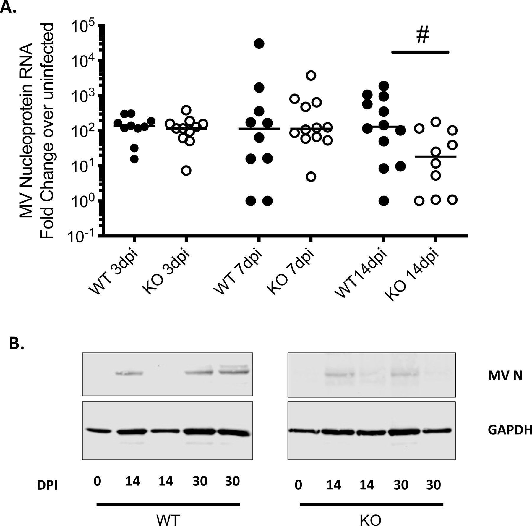
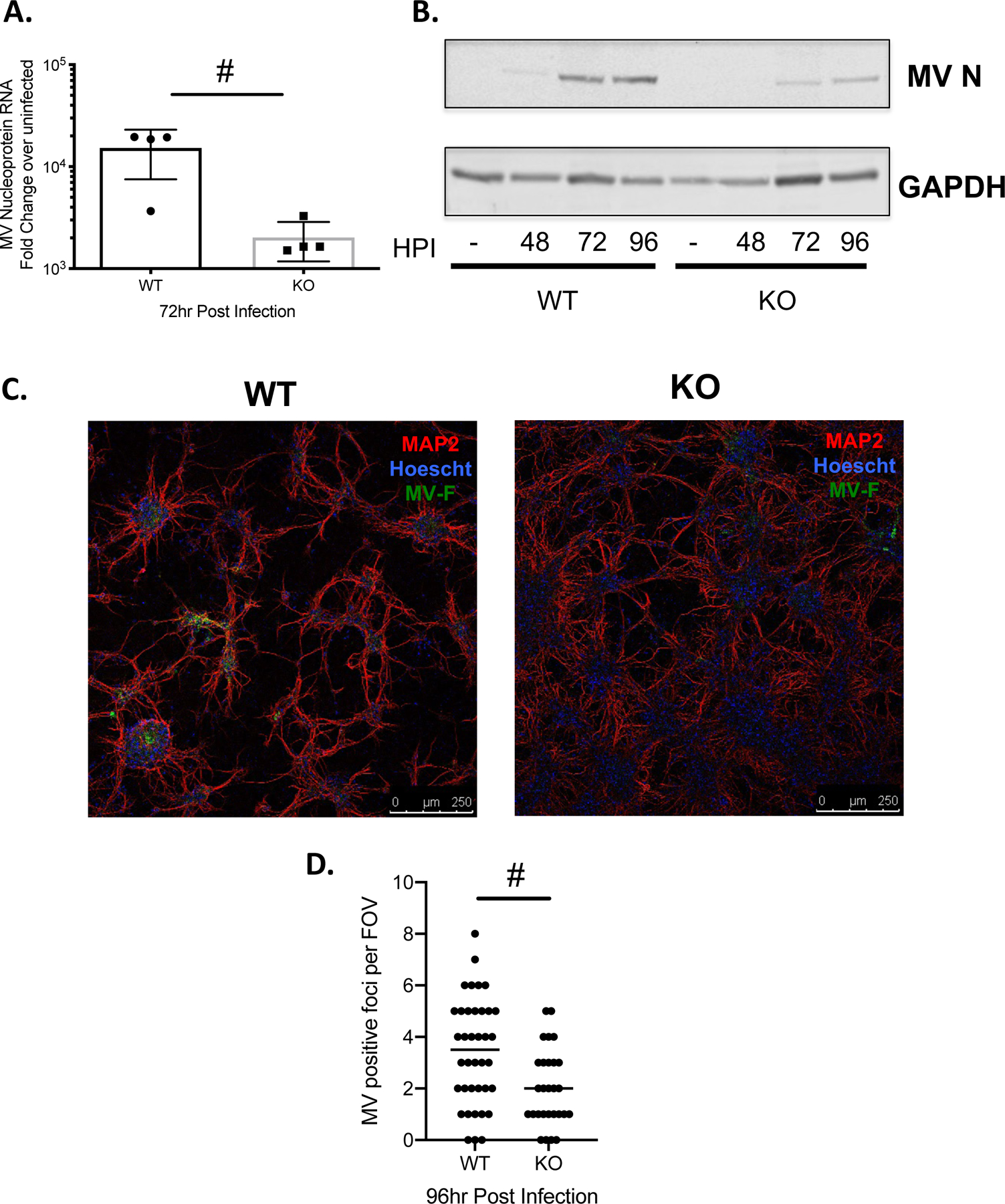
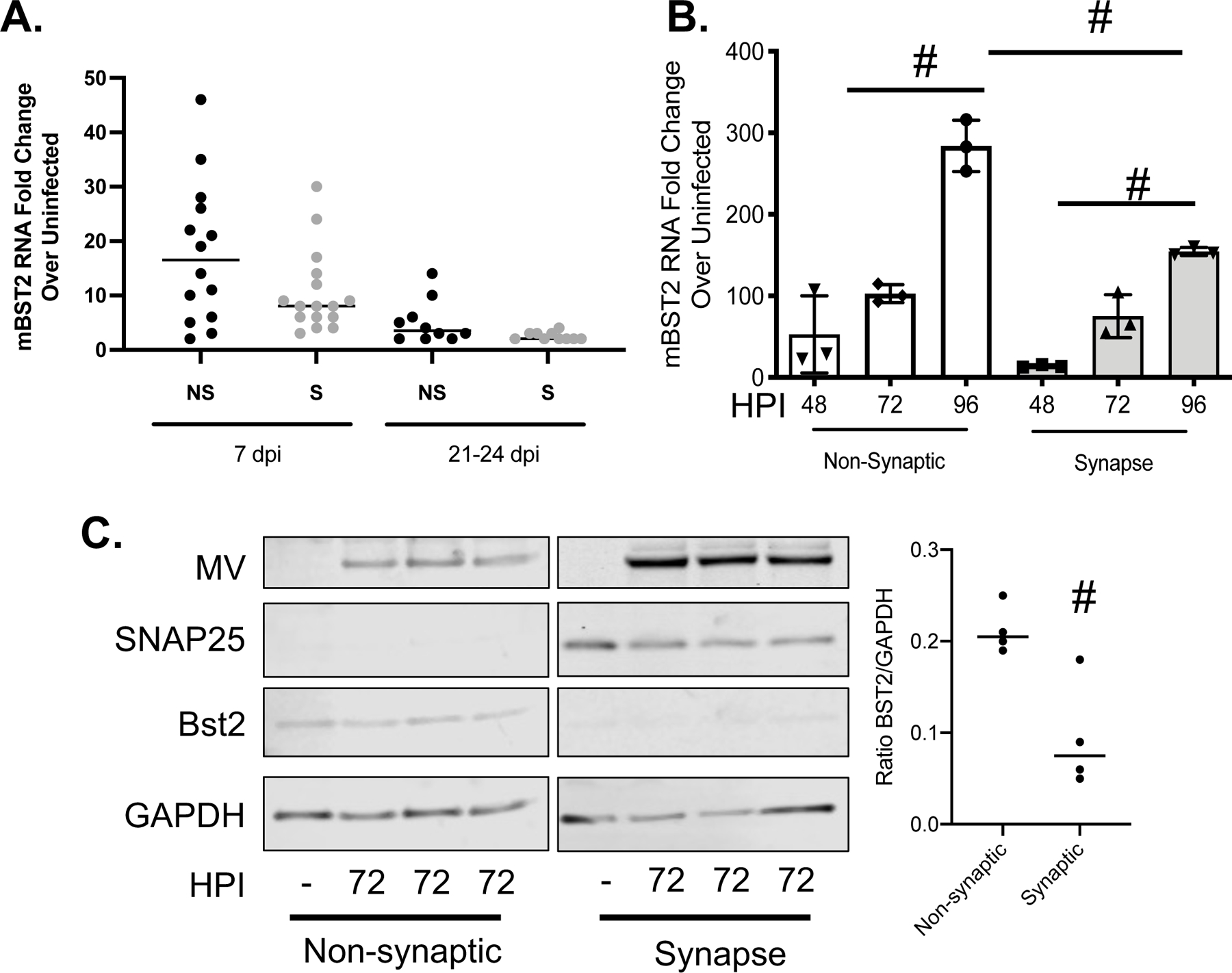
BST2/tetherin is a transmembrane protein with antiviral activity; it is synthesized following exposure to interferons, and restricts the release of budding virus particles by tethering them to the host cell membrane. We previously showed that BST2 is induced in primary neurons following measles virus (MV) infection or type I interferon; however, BST2 was dispensable for protection against challenge with neuron-restricted MV. Here, we define the contribution of BST-2 in neuronal MV infection. Surprisingly, and in contrast to its antiviral role in non-neuronal cells, murine BST2 promotes MV infection in brains of permissive mice and in primary neuron cultures. Moreover, BST2 expression was predominantly observed in the non-synaptic fraction of purified neurons. These studies highlight a cell-type dependent role of a well-characterized antiviral protein in enhancing neuronal infection.

Keywords: BST2; Measles virus; Neuron; Tetherin.


Conflict of interest statement

Conflict of Interest

The authors declare no conflicts of interest.

Declaration of interests

The authors declare that they have no known competing financial interests or personal relationships that could have appeared to influence the work reported in this paper.

Figures

Figure 1:
Figure 1:. Deletion of BST2 leads to decreased MV in vivo.
NSE-CD46+ (WT; black dots) and NSE-CD46+/BST2 KO (KO; white dots) mice were infected intracranially with 1×10⁴ PFU MV-Edmonston. Whole brains were collected, and analyzed for MV nucleoprotein RNA and protein by RT-qPCR (A) and Western blot (B), at the indicated days post infection (dpi). n=10–12/group. Data are represented using the ΔΔCT method. # p <0.05 Mann-Whitney U Test.
Figure 2:
Figure 2:. Deletion of BST2 leads to decreased MV a in primary neurons.
Primary neurons of the indicated genotype (WT: NSE-CD46+; KO: NSE-CD46+/BST2 KO) were infected with MV-Edmonston at an MOI=1. A) RNA was collected at the indicated time point (hours post-infection; hpi) and analyzed by RT-qPCR for MV nucleoprotein RNA. Results from at least 3 independent experiments are represented using the ΔΔCT method. B) Western blot analysis of protein collected at the indicated times post infection. Blots were probed with a polyclonal MV nucleoprotein antibody and an antibody to GAPDH as a loading control. C) Immunofluorescence staining of WT and KO primary neuronal cultures. Red- MAP2; neuronal marker. Blue-Hoescht; nuclei marker. Green- MV fusion protein. Representative images from each genotype are shown. D) MV positive foci were scored across multiple fields of view (FOV). # p <0.05 Unpaired T test.
Figure 3:
Figure 3:. BST2 is not concentrated at the neuronal synapse during MV infection.
A) RNA collected from fractionated whole brain tissue was analyzed by RT-qPCR for murine BST2 (mBST2) RNA and normalized to cyclophilin. B/C) Primary NSE-CD46+ neurons were infected with MV at an MOI=1. Infected cells were collected at the indicated hours post infection followed by synaptosome purification. B) RNA was analyzed by RT-qPCR for murine BST2 (mBST2) RNA and normalized to cyclophilin B. Data represent the results of an experiment performed in triplicate and analyzed using the ΔΔCT method. C) Western blot analysis of protein collected at the indicated times post infection from either the pelleted synaptic or remaining fractions (non-synaptic). Blots were probed with antibodies against BST2, MV, SNAP25 (to indicate synaptic fraction purity) and an antibody to GAPDH (loading control). # p <0.05 Unpaired T test with equal standard deviations. Error bars represent SD.

Similar articles

Cited by

References

    1. Blasius AL, Giurisato E, Cella M, Schreiber RD, Shaw AS, Colonna M. Bone Marrow Stromal Cell Antigen 2 Is a Specific Marker of Type I IFN-Producing Cells in the Naive Mouse, but a Promiscuous Cell Surface Antigen following IFN Stimulation. Journal of Immunology. 2006;177(5):3260–3265. - PubMed
    1. Goto T, Kennel SJ, Abe M, et al.A Novel Membrane Antigen Selectively Expressed on Terminally Differentiated Human B Cells. Blood. 1994;84:1922–1930. - PubMed
    1. Neil SJD, Zang T, Bieniasz PD. Tetherin inhibits retrovirus release and is antagonized by HIV-1 Vpu. Nature. 2008;451(7177):425–430. - PubMed
    1. Fitzpatrick K, Skasko M, Deerinck TJ, Crum J, Ellisman MH, Guatelli J. Direct Restriction of Virus Release and Incorporation of the Interferon-Induced Protein BST-2 into HIV-1 Particles. Hope TJ, ed. PLoS Pathog. 2010;6(3):e1000701–e1000712. - PMC - PubMed
    1. Perez-Caballero D, Zang T, Ebrahimi A, et al.Tetherin Inhibits HIV-1 Release by Directly Tethering Virions to Cells. Cell. 2009;139(3):499–511. - PMC - PubMed

Publication types