. 2021 Nov 18;49(20):e118.
doi: 10.1093/nar/gkab727.

ENDO-Pore: high-throughput linked-end mapping of single DNA cleavage events using nanopore sequencing

Affiliations

ENDO-Pore: high-throughput linked-end mapping of single DNA cleavage events using nanopore sequencing

Oscar E Torres Montaguth et al. Nucleic Acids Res. .

Abstract

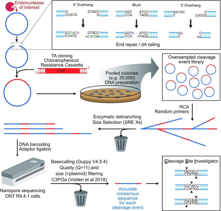
Mapping the precise position of DNA cleavage events plays a key role in determining the mechanism and function of endonucleases. ENDO-Pore is a high-throughput nanopore-based method that allows the time resolved mapping single molecule DNA cleavage events in vitro. Following linearisation of a circular DNA substrate by the endonuclease, a resistance cassette is ligated recording the position of the cleavage event. A library of single cleavage events is constructed and subjected to rolling circle amplification to generate concatemers. These are sequenced and used to produce accurate consensus sequences. To identify the cleavage site(s), we developed CSI (Cleavage Site Investigator). CSI recognizes the ends of the cassette ligated into the cleaved substrate and triangulates the position of the dsDNA break. We firstly benchmarked ENDO-Pore using Type II restriction endonucleases. Secondly, we analysed the effect of crRNA length on the cleavage pattern of CRISPR Cas12a. Finally, we mapped the time-resolved DNA cleavage by the Type ISP restriction endonuclease LlaGI that introduces random double-strand breaks into its DNA substrates.


Figures

Figure 1.
Figure 1.
ENDO-Pore Workflow. A circular DNA substrate (blue circle) is digested with the endonuclease of interest (red Pac-Man) and the linear DNA end-repaired (inset). This will fill-in 5′ overhangs and remove 3′ overhangs, while blunt ends remain intact. dA-tailing allows for TA ligation of a resistance cassette (e.g. chloramphenicol used here) that marks the position of the cleavage site. Each individual DNA molecule represents a single cleavage event. Following transformation of E. coli, DNA preparation from pooled single colonies produces a DNA cleavage library. Rolling circle amplification is used to both linearise the DNA and to produce concatemer copies. The product is debranched by T7 endonuclease and size selection used to favour DNA with multiple copies of the original plasmid substrates (Supplementary Figure S2). DNA barcodes and sequencing adaptors are ligated, and the DNA sequenced using a MinION. Following basecalling, size selection and consensus generation from the concatemers, the cleavage site for each read is identified by the CSI software (Supplementary Figure S3).
Figure 2.
Figure 2.
The consequences of DNA end repair are that cleavage sites identified are always those closest to the 3′ end of each strand. (A) Consequences for strand-specific processing on the site reported by CSI. (B) Consequences for movement and re-cleavage on the site reported by CSI. The solid black lines represent the original dsDNA break and overhangs produced. Black arrows represent movement of the nuclease on one strand (A) or both strands (B). Triangles represent increasing/decreasing apparent spacing between top and bottom stand cleavage loci. Dotted lines represent the cleavage positions and overhangs produced following processing (but not the final cleavage reported). See main text for further details and Supplementary Figure S5 for examples.
Figure 3.
Figure 3.
ENDO-Pore Benchmarking. (A) pUC19 sequencing accuracy at different repeat cut-offs. Read accuracy was determined by aligning consensus sequences to pUC19 sequence using Minimap2 (21) and is represented by a violin plot for the full dataset at each cut-off. Median accuracy is represented by the diamonds and the quartiles are shown by the horizontal lines. (B) Relationship between repeat cut-off and data loss. Average values for the proportion of data retained after different repeat cut-offs were used for the different restriction endonucleases in panel C (points are the average, error bars are the range). (C) Validation of ENDO-Pore using commercial Type II restriction endonucleases. Accuracy is represented as the percentage of sequences with the expected consensus cleavage site at different repeat cut-offs, as indicated by the coloured circles. The vertical dotted line at 95% is the ‘terminal integrity’ guaranteed by New England Biolabs determined by ligation and re-cleavage tests.
Figure 4.
Figure 4.
The effect of crRNA spacer length on the cleavage pattern of CRISPR-Cas12a. (A) R-loop formation between the plasmid target and Cas12a crRNA with 15–20 nt spacer sequences (red). The PAM is shown in green and the bi-lobed Cas12a structure represented by the blue shape. (B) Plasmid DNA cleavage after 2 hours. Supercoiled (SC) substrate, nicked (open circle, OC) intermediate and linear (LIN) product were separated by agarose gel electrophoresis (Supplementary Figure S8) and the tritium-labelled DNA quantified by scintillation counting. Errors bars are SD from three repeat experiments. (C) Strand-linkage plots (SLPs) generated by CSI. Each SLP displays the relative frequency of cleavage events (represented as a connecting line between the NTS and TS according to the heatmap) and the frequency of strand-specific cleavage events above or below the relevant strand (black histograms).
Figure 5.
Figure 5.
Mapping the long-range communication and cleavage by the Type ISP restriction endonuclease LlaGI. (A) Cleavage mechanism of LlaGI. (B) Scatter plot of the 3′-3′ spacing between nicking events reported by CSI. Error bars represent the interquartile distance; thick bars represent the median values; N represents the total number of cleavage events analysed per time point. (C) Tornado plots displaying individual events ranked by spacing between nicking events. Horizonal orange (3′ overhangs) or blue (5′ overhangs) bars represent the spacing between the nicks closest to the 3′ end of each strand. Crosses indicate the midpoint of each event. Positions of the 30 bp and median spacings are shown by grey horizontal lines. (lower panels) The frequency of the cleavage midpoints for each time point are represented as grey vertical bars (bin size 1 bp). The black solid lines represent the theoretical collision distribution at time → ∞ of two motors that initiated at a pair of sites a distance d = 997 steps apart, given by d!/(n!·(dn)!·2d), where n is the position between the sites (32). (D) Dinucleotide cleavage frequency for individual time points calculated by CSI.

References

    1. Tsai S.Q., Zheng Z., Nguyen N.T., Liebers M., Topkar V.V., Thapar V., Wyvekens N., Khayter C., Iafrate A.J., Le L.P.et al. .. GUIDE-seq enables genome-wide profiling of off-target cleavage by CRISPR-Cas nucleases. Nat. Biotechnol. 2015; 33:187–197. - PMC - PubMed
    1. Crosetto N., Mitra A., Silva M.J., Bienko M., Dojer N., Wang Q., Karaca E., Chiarle R., Skrzypczak M., Ginalski K.et al. .. Nucleotide-resolution DNA double-strand break mapping by next-generation sequencing. Nat. Methods. 2013; 10:361–365. - PMC - PubMed
    1. Höijer I., Johansson J., Gudmundsson S., Chin C.-S., Bunikis I., Häggqvist S., Emmanouilidou A., Wilbe M., den Hoed M., Bondeson M.-L.et al. .. Amplification-free long-read sequencing reveals unforeseen CRISPR-Cas9 off-target activity. Genome Biol. 2020; 21:290. - PMC - PubMed
    1. Selkova P., Vasileva A., Pobegalov G., Musharova O., Arseniev A., Kazalov M., Zyubko T., Shcheglova N., Artamonova T., Khodorkovskii M.et al. .. Position of Deltaproteobacteria Cas12e nuclease cleavage sites depends on spacer length of guide RNA. RNA Biology. 2020; 17:1472–1479. - PMC - PubMed
    1. Cameron P., Fuller C.K., Donohoue P.D., Jones B.N., Thompson M.S., Carter M.M., Gradia S., Vidal B., Garner E., Slorach E.M.et al. .. Mapping the genomic landscape of CRISPR–Cas9 cleavage. Nat. Methods. 2017; 14:600–606. - PubMed

Publication types