. 2021 Nov;142(5):841-857.
doi: 10.1007/s00401-021-02354-8. Epub 2021 Aug 21.

PATZ1 fusions define a novel molecularly distinct neuroepithelial tumor entity with a broad histological spectrum

Karam T Alhalabi  1   2   3 Damian Stichel  4   5 Philipp Sievers  4   5 Heike Peterziel  1   3 Alexander C Sommerkamp  1   2 Dominik Sturm  1   2   6 Andrea Wittmann  1   2 Martin Sill  7 Natalie Jäger  1   7 Pengbo Beck  1   7 Kristian W Pajtler  1   7   6 Matija Snuderl  8 George Jour  9 Michael Delorenzo  9 Allison M Martin  10 Adam Levy  10 Nagma Dalvi  11 Jordan R Hansford  12 Nicholas G Gottardo  13 Emmanuelle Uro-Coste  14   15 Claude-Alain Maurage  16   17 Catherine Godfraind  18   19 Fanny Vandenbos  20   21 Torsten Pietsch  22 Christof Kramm  23 Maria Filippidou  1   7   24 Antonis Kattamis  24 Chris Jones  25 Ingrid Øra  26 Torben Stamm Mikkelsen  27 Michal Zapotocky  28 David Sumerauer  28 David Scheie  29 Martin McCabe  30 Pieter Wesseling  31   32   33 Bastiaan B J Tops  32   33 Mariëtte E G Kranendonk  32   33 Matthias A Karajannis  34 Nancy Bouvier  35 Elli Papaemmanuil  36   37 Hildegard Dohmen  38 Till Acker  38 Katja von Hoff  39 Simone Schmid  39 Evelina Miele  40 Katharina Filipski  41   42   43 Lidija Kitanovski  44 Lenka Krskova  45 Johannes Gojo  1   7   46 Christine Haberler  47 Frank Alvaro  48   49 Jonas Ecker  1   6   50 Florian Selt  1   6   50 Till Milde  1   6   50 Olaf Witt  1   6   50 Ina Oehme  1   50 Marcel Kool  1   7   32 Andreas von Deimling  1   4   5 Andrey Korshunov  1   4   5 Stefan M Pfister  1   7   6 Felix Sahm #  1   4   5 David T W Jones  51   52
Affiliations

PATZ1 fusions define a novel molecularly distinct neuroepithelial tumor entity with a broad histological spectrum

Karam T Alhalabi et al. Acta Neuropathol. 2021 Nov.

Abstract

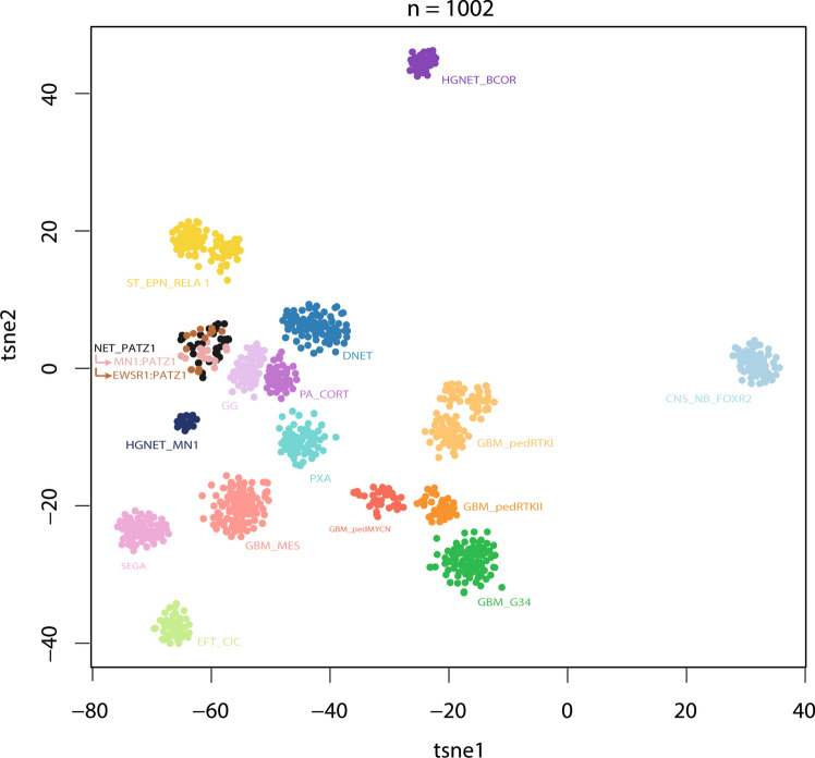
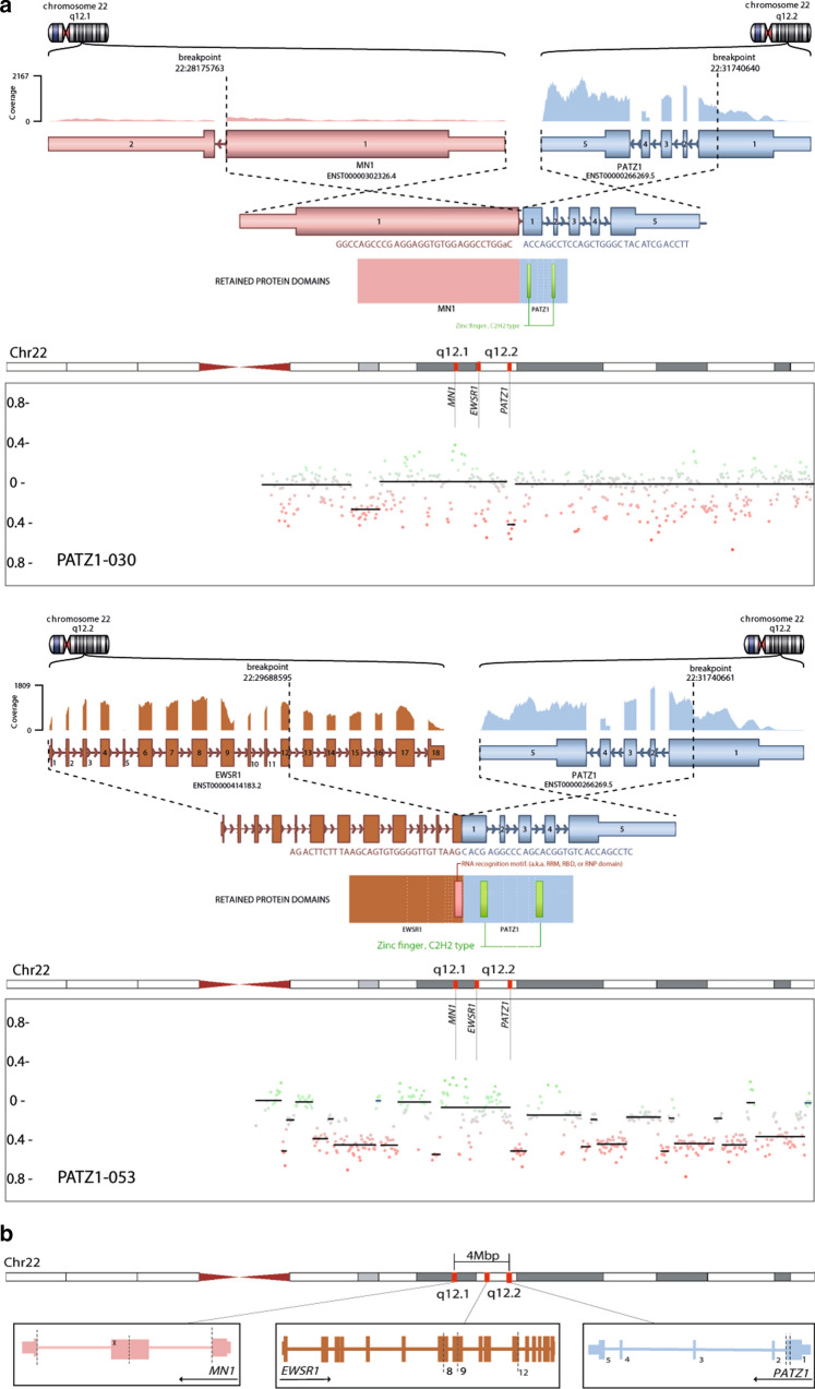
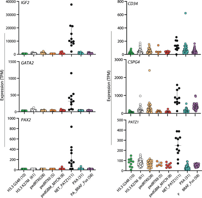
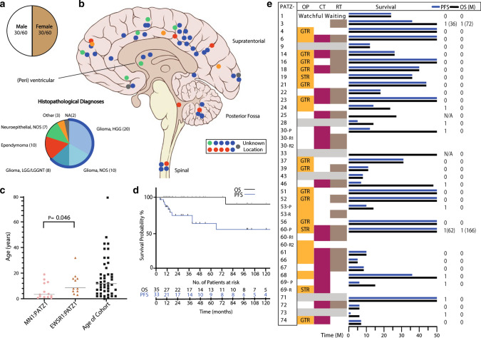
Large-scale molecular profiling studies in recent years have shown that central nervous system (CNS) tumors display a much greater heterogeneity in terms of molecularly distinct entities, cellular origins and genetic drivers than anticipated from histological assessment. DNA methylation profiling has emerged as a useful tool for robust tumor classification, providing new insights into these heterogeneous molecular classes. This is particularly true for rare CNS tumors with a broad morphological spectrum, which are not possible to assign as separate entities based on histological similarity alone. Here, we describe a molecularly distinct subset of predominantly pediatric CNS neoplasms (n = 60) that harbor PATZ1 fusions. The original histological diagnoses of these tumors covered a wide spectrum of tumor types and malignancy grades. While the single most common diagnosis was glioblastoma (GBM), clinical data of the PATZ1-fused tumors showed a better prognosis than typical GBM, despite frequent relapses. RNA sequencing revealed recurrent MN1:PATZ1 or EWSR1:PATZ1 fusions related to (often extensive) copy number variations on chromosome 22, where PATZ1 and the two fusion partners are located. These fusions have individually been reported in a number of glial/glioneuronal tumors, as well as extracranial sarcomas. We show here that they are more common than previously acknowledged, and together define a biologically distinct CNS tumor type with high expression of neural development markers such as PAX2, GATA2 and IGF2. Drug screening performed on the MN1:PATZ1 fusion-bearing KS-1 brain tumor cell line revealed preliminary candidates for further study. In summary, PATZ1 fusions define a molecular class of histologically polyphenotypic neuroepithelial tumors, which show an intermediate prognosis under current treatment regimens.

Keywords: Brain tumor; EWSR1; Gene fusion; MN1; Neuroepithelial; Neurooncology; PATZ1; Pediatric.


Figures

Fig. 1
Fig. 1
t-distributed stochastic neighbor embedding (tSNE) clustering of DNA methylation patterns of 60 NET-PATZ1 tumors alongside 942 in-house reference samples representing 15 other low- and high-grade glial and glioneuronal tumor types, using the 10,000 most variably methylated probes. NET-PATZ1 forms a distinct ‘island’. CNS_NB_FOXR2 CNS neuroblastoma with FOXR2 activation; DNET dysembryoplastic neuroepithelial tumor; EFT_CIC CNS ewing sarcoma family tumor with CIC alteration; GBM_G34 glioblastoma, H3.3 G34 mutant; GBM_MES glioblastoma, subclass mesenchymal; GBM_pedMYCN pediatric-type glioblastoma, subclass MYCN; GBM_pedRTKI pediatric-type glioblastoma, subclass RTKI; GBM_pedRTKII pediatric-type glioblastoma, subclass RTK II; GG ganglioglioma; HGNET_BCOR CNS high-grade neuroepithelial tumor with BCOR alteration; HGNET_MN1 CNS high-grade neuroepithelial tumor with MN1 alteration; NET_PATZ1 neuroepithelial tumor with PATZ1 fusion; PA_CORT hemispheric pilocytic astrocytoma; PXA pleomorphic xanthoastrocytoma; SEGA subependymal giant cell astrocytoma; ST_EPN_RELA supratentorial ependymoma, RELA fused
Fig. 2
Fig. 2
a Illustration of the PATZ1 fusion genes detected by RNA-seq for two selected cases; and corresponding copy number plots of Chromosome 22. The three involved genes MN1, EWSR1 and PATZ1 are marked. PATZ1-030 harbors an in-frame MN1:PATZ1 fusion, retaining an intronic pseudoexon (upper panel). PATZ1-053 demonstrates a variant fusion transcript juxtaposing Exon 12 of EWSR1 onto the usual partner 3′ sequence of PATZ1, in contrast to the more prevalent 5′ breakpoints observed in Exons 8 and 9 of EWSR1 (lower panel). b Schematic view of the loci of the genes involved in the fusions described (MN1, EWSR1, and PATZ1). Note the proximity of the three genes (all lie within approximately 4 Mbp). The dashed lines resemble the genomic breakpoints observed in the gene fusions. An intronic breakpoint observed in a subset of MN1_PATZ1 fusions introduces a novel pseudoexon (marked with x) whilst maintaining the reading frame. Isoforms illustrated: EWSR1 NM_005243; PATZ1 NM_0.13986.4; MN1 NM_002430.3
Fig. 3
Fig. 3
Differential expression analysis between NET-PATZ1 and a reference cohort of other glioma subtypes; IGF2, GATA2, PAX2 and PATZ1 and are more highly expressed in NET-PATZ1 cases when compared with representative pediatric HGG or LGG tumors, while CD34 and NG2 (CSPG4) could represent potential IHC staining markers for NET-PATZ1. Gene expression values are shown as TPM (transcripts per kilobase million). Where relevant, bars indicate median and 1st/3rd Quartile. H3.3 G34R glioblastoma, IDH wildtype, H3.3 G34 mutant; H3.3 K27M diffuse midline glioma H3 K27M mutant: pedGBM_MYCN, glioblastoma, IDH wildtype, subclass MYCN; pedRTKI, glioblastoma, IDH wildtype, subclass RTK I; pedRTKII, glioblastoma, IDH wildtype, subclass RTK II; PXA pleomorphic xanthoastrocytoma; PA_BRAF_Fus pilocytic astrocytoma with BRAF fusion
Fig. 4
Fig. 4
Summary plot of copy number alterations in NET-PATZ1 and categorization of copy number alterations observed on chromosome 22, the most frequent being a dramatic shattering pattern (chromothripsis)
Fig. 5
Fig. 5
Clinical features of NET-PATZ1. a Patient sex distribution. b Distribution of tumor location. The institutional histopathological diagnoses of the series are also shown, representing a broad spectrum of mostly glial diagnoses. c Age distribution with the horizontal line representing median age of our cohort (11 years). MN1:PATZ1-fused tumors appear to be significantly enriched in younger ages (median = 3.5 years) vs EWSR1:PATZ1 (median = 8 years), (p value; 0.046, Student’s t test). d Clinical outcome in terms of OS and PFS of NET-PATZ1. e Overview of the different therapy protocols NET_PATZ1 patients within this cohort received. Where highlighted, the patients received that particular therapy modality, grey bars indicate unknown data. Different management as per primary vs relapse tumor is also shown. Note that some patients benefited from surgery (OP) alone. CT chemotherapy, RT radiation therapy, P primary, R (1/2) relapse 1 or relapse 2, GTR gross total resection, STR subtotal resection, 1 relapse/death, 0 censored. Detailed therapy protocols are listed in Supplementary Table 1, online resource
Fig. 6
Fig. 6
Histology of NET-PATZ1 tumors. a PATZ1-012: perivascular pseudorosettes resembling ependymal morphology are observed. b PATZ1-040 showed an astroblastoma-like morphology. c PATZ1-025: high cellularity was consistently observed across almost all cases. Microcysts are here encircled by monomorphous nuclei. d PATZ1-039: in this tumor monomorphous nuclei with clear cell morphology along with perivascular pseudorosettes were encountered. e, f NG2 staining in PATZ-030 and PATZ-014, respectively, with negative control insets (IDH WT GBM). Thick scale bars represent 100 µm, thin scale bars represent 200 µm

References

    1. Alvarez-Breckenridge C, Miller JJ, Nayyar N, Gill CM, Kaneb A, D’Andrea M, et al. Clinical and radiographic response following targeting of BCAN-NTRK1 fusion in glioneuronal tumor. NPJ Precis Oncol. 2017;1:5. doi: 10.1038/s41698-017-0009-y. - DOI - PMC - PubMed
    1. Benarroch EE. Insulin-like growth factors in the brain and their potential clinical implications. Neurology. 2012;79:2148–2153. doi: 10.1212/WNL.0b013e3182752eef. - DOI - PubMed
    1. Bridge JA, Sumegi J, Druta M, Bui MM, Henderson-Jackson E, Linos K, et al. Clinical, pathological, and genomic features of EWSR1-PATZ1 fusion sarcoma. Mod Pathol. 2019;32:1593–1604. doi: 10.1038/s41379-019-0301-1. - DOI - PubMed
    1. Burel-Vandenbos F, Pierron G, Thomas C, Reynaud S, Gregoire V, Duhil de Benaze G, et al. A polyphenotypic malignant paediatric brain tumour presenting a MN1-PATZ1 fusion, no epigenetic similarities with CNS High-Grade Neuroepithelial Tumour with MN1 Alteration (CNS HGNET-MN1) and related to PATZ1-fused sarcomas. Neuropathol Appl Neurobiol. 2020 doi: 10.1111/nan.12626. - DOI - PubMed
    1. Capper D, Jones DTW, Sill M, Hovestadt V, Schrimpf D, Sturm D, et al. DNA methylation-based classification of central nervous system tumours. Nature. 2018;555:469–474. doi: 10.1038/nature26000. - DOI - PMC - PubMed

Publication types