Review
. 2021 Aug 5:15:650614.
doi: 10.3389/fnsys.2021.650614. eCollection 2021.

The Cerebellar Dopaminergic System

Affiliations
Review

The Cerebellar Dopaminergic System

Paolo Flace et al. Front Syst Neurosci. .

Abstract

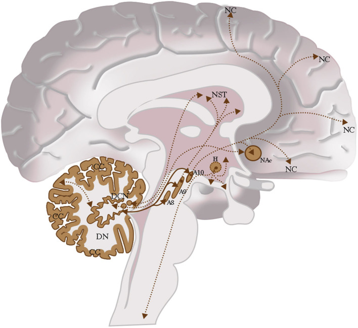
In the central nervous system (CNS), dopamine (DA) is involved in motor and cognitive functions. Although the cerebellum is not been considered an elective dopaminergic region, studies attributed to it a critical role in dopamine deficit-related neurological and psychiatric disorders [e.g., Parkinson's disease (PD) and schizophrenia (SCZ)]. Data on the cerebellar dopaminergic neuronal system are still lacking. Nevertheless, biochemical studies detected in the mammalians cerebellum high dopamine levels, while chemical neuroanatomy studies revealed the presence of midbrain dopaminergic afferents to the cerebellum as well as wide distribution of the dopaminergic receptor subtypes (DRD1-DRD5). The present review summarizes the data on the cerebellar dopaminergic system including its involvement in associative and projective circuits. Furthermore, this study also briefly discusses the role of the cerebellar dopaminergic system in some neurologic and psychiatric disorders and suggests its potential involvement as a target in pharmacologic and non-pharmacologic treatments.

Keywords: Parkinson's disease; autism spectrum disorders; cerebellum; dopamine; dopamine receptors; non-traditional large neurons; schizophrenia.


Conflict of interest statement

The authors declare that the research was conducted in the absence of any commercial or financial relationships that could be construed as a potential conflict of interest.

Figures

Figure 1
Figure 1
Dopamine transporter (DAT) immunoreactivity in the cerebellar cortex. The DAT immunoreactivity is detectable in neuronal bodies and processes of all the layers of the cerebellar cortex. In the molecular layer, DAT immunoreactivity in basket neurons (arrowheads); primary and secondary trunks dendritic and apical dendrites of Purkinje neurons; immunonegative stellate neurons, fine clusters of DAT immunoreactivity in the neuropil of the layer. In the Purkinje neuron layer, DAT immunoreactive Purkinje neuron cell body (single asterisk), DAT immunonegative Purkinje neuron (double asterisk). In the granular layer, DAT immunoreactivity in space of Held, DAT immunoreactivity in the cell body, and axon-like processes of the synarmotic neuron (arrow). (Scale bar: 25 μm).
Figure 2
Figure 2
Dopamine receptor type 2 (DRD2) immunoreactivity is observable in neuronal bodies and processes in the layers of the cerebellar cortex. In the ML:DRD2 immunoreactive basket and stellate neuron cell bodies (arrows), DRD2 immunoreactive primary, secondary, and apical dendrites of Purkinje neurons, DRD2 immunoreactive Purkinje neuron cell bodies (single asterisk). In the granular layer, DRD2 immunoreactivity in space of Held; DRD2 immunoreactive Golgi neuron cell body (arrow) (Scale bar: 20 μm).
Figure 3
Figure 3
Dopamine transporter (DAT) immunoreactivity in the dentate nucleus. The dopamine transporter (DAT) immunoreactivity is detectable in the dentate nucleus gray substance and in the neighboring white substance. DAT immunoreactive small neuron cell bodies (arrowheads); DAT immunonegative small neuron cell body (single asterisks) DAT immunoreactive cell body of projective neuron type, central neuron (arrow); fine clusters of DAT immunoreactivity in the neuropil of the nucleus and in the neighboring withe substance (Scale bar: 15 μm).
Figure 4
Figure 4
Dopamine receptor type 2 (DRD2) immunoreactivity in the dentate nucleus. The (DRD2) immunoreactivity is detectable in the dentate nucleus gray substance and in the neighboring withe substance; DRD2 immunoreactive small neuron cell bodies (arrowheads); DRD2 immunoreactive cell body of projective neuron type, central neuron (arrow); diffuse DAT immunoreactivity in the neuropil of the nucleus (Scale bar: 15 μm).
Figure 5
Figure 5
Structural connectivity of SNpc and VTA, including putative midbrain-cerebellar connectivity. Data were obtained from the 100-unrelated-subjects sample of the HCP repository (see Van Essen et al., 2013) Diffusion datasets were processed using a multi-shell, multi-tissue constrained spherical deconvolution (MSMT-CSD) algorithm (see Jeurissen et al., 2014). A number of 10,000 streamlines passing through the left SNpc (dark red) and VTA (light red) regions of interest (see Pauli et al., 2018) (A) was generated. Streamlines were mapped to structural scans, transformed to MNI152 standard space, binarized, and summed up to obtain tract maximum probability maps (MPMs). A threshold of 50% was applied to show only tracts overlapping in at least half of the sample (B). Tractography was run on 30 high-quality 3T structural and diffusion data from the Human Connectome Project (HCP). Data were downloaded in a minimally pre-processed form and elaborated using the signal processing technique known as Constrained Spherical Deconvolution (CSD). Regions of interest (ROI) were delineated by means of multi-atlas automated segmentation: Substantia nigra (SN) and Ventro Tegmental Area (VTA) were resliced into subject space from Adcock's probabilistic atlas; dentate nucleus (both dorsal and ventral part) using the deep cerebellar nuclei atlas featured in SPM Anatomy Tract colors are attributed according to the spatial orientation of streamlines: superior-inferior (blue), anterior-posterior (green), and latero-lateral (red).
Figure 6
Figure 6
Dentate-nigral interconnections. Coronal view shows the interconnections between the right dentate nucleus and the ipsilateral SN, and the left dentate nucleus and the ipsilateral SN. The fibers exited the cerebellum via the right and left superior cerebellar peduncles.
Figure 7
Figure 7
Dentate-VTA interconnections. Coronal view shows the interconnections between the right dentate nucleus and the ipsilateral and contralateral VTA, and the left dentate nucleus and the ipsilateral and contralateral VTA. The fibers exited the cerebellum via the right and left superior cerebellar peduncles.
Figure 8
Figure 8
Dentate-SN and dentate-VTA interconnections. Coronal view shows the interconnections between the right dentate nucleus and left dentate nucleus to the ipsilateral SN, between the right dentate nucleus and left dentate nucleus to the ipsilateral and contralateral VTA. The fibers exited the cerebellum via the right and left superior cerebellar peduncles.
Figure 9
Figure 9
The dopaminergic CNS and their interconnections. Cerebellum: Cerebellar Cortex (CC), Deep Cerebellar Nuclei (DCN); Midbrain: Retrorubral Nucleus (A8), Substantia Nigra Pars Compacta (A9), Ventral Tegmental Area (A10); Hypothalamus (H); Nucleus Accumbens (NAc); Neostriatum (NST); Neocortex (NC). Intrinsic cerebellar dopaminergic interconnections: Between the dentate nucleus (DN) and the cerebellar cortex (CC); these interconnections are indicated in brown with the double arrow and the bold line. Extrinsic cerebellar dopaminergic interconnections: Between the dentate nucleus (DN) and the nuclei of the midbrain A8, A9, and A10; interconnections are indicated in brown with a double arrow and bold line. Between the dentate nucleus (DN) and the Neostriatum (NST); among others Deep Cerebellar Nuclei Nuclei (DCN) and the NST or the NAc. These interconnections are indicated in brown with the double arrow and the thin dashed line. Other Dopaminergic Interconnections of the CNS: Interconnection between the Ventral Tegmental Area (A10) and the NAc or between the Ventral Tegmental Area (A10) and the Neocortex (NC). These interconnections are indicated in brown with the double arrow and the thin dashed line.

References

    1. Adolfsson R., Gottfries C. G., Roos B. E., Windblad B. (1979). Postmortem distribution of dopamine and homovanillic acid in human brain, variations related to age, and review of the literature. J. Neural. Transm. 45, 81–105. 10.1007/BF01250085 - DOI - PubMed
    1. Alder R., Barbas H. (1995). Complementary distribution of the phosphoproteins DARPP-32 and I-1 in the cerebellar system. Neuroreport 6, 2368–2372. 10.1097/00001756-199511270-00022 - DOI - PubMed
    1. Ambrosi G., Flace P., Lorusso L., Girolamo F., Rizzi A., Bosco L., et al. . (2007). Non-traditional large neuron in the granular layer of the cerebellar cortex. Eur. J. Histochem. 51(Suppl 1), 59–64. - PubMed
    1. Andreasen N. C., Paradiso S., O'Leary D. S. (1998). “Cognitive dysmetria” as an integrative theory of schizophrenia: a cortical-subcortical-cerebellar circuitry? Schizophr. Bull. 24, 203–218. 10.1093/oxfordjournals.schbul.a033321 - DOI - PubMed
    1. Andreasen N. C., Pierson R. (2008). The role of the cerebellum in schizophrenia. Biol. Psychiatry 64, 81–88. 10.1016/j.biopsych.2008.01.003 - DOI - PMC - PubMed

LinkOut - more resources