. 2021 Aug 5:12:690387.
doi: 10.3389/fendo.2021.690387. eCollection 2021.

L-Cell Expression of Melanocortin-4-Receptor Is Marginal in Most of the Small Intestine in Mice and Humans and Direct Stimulation of Small Intestinal Melanocortin-4-Receptors in Mice and Rats Does Not Affect GLP-1 Secretion

Affiliations

L-Cell Expression of Melanocortin-4-Receptor Is Marginal in Most of the Small Intestine in Mice and Humans and Direct Stimulation of Small Intestinal Melanocortin-4-Receptors in Mice and Rats Does Not Affect GLP-1 Secretion

Rune E Kuhre et al. Front Endocrinol (Lausanne). .

Abstract

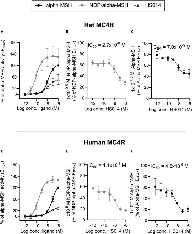
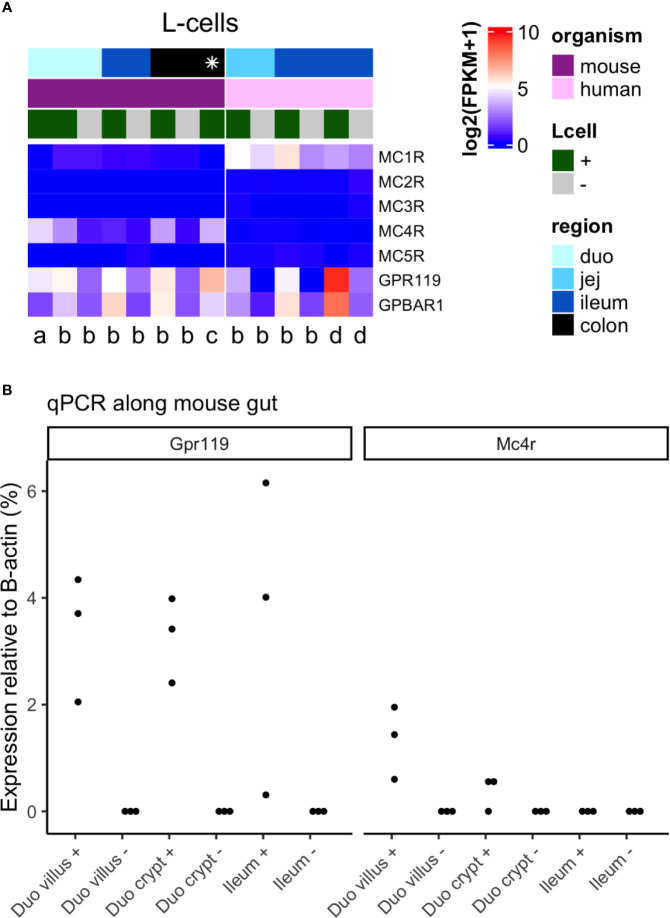
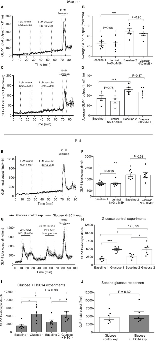
The molecular sensors underlying nutrient-stimulated GLP-1 secretion are currently being investigated. Peripheral administration of melanocortin-4 receptor (MC4R) agonists have been reported to increase GLP-1 plasma concentrations in mice and humans but it is unknown whether this effect results from a direct effect on the GLP-1 secreting L-cells in the intestine, from other effects in the intestine or from extra-intestinal effects. We investigated L-cell expression of MC4R in mouse and human L-cells by reanalyzing publicly available RNA sequencing databases (mouse and human) and by RT-qPCR (mouse), and assessed whether administration of MC4R agonists to a physiologically relevant gut model, isolated perfused mouse and rat small intestine, would stimulate GLP-1 secretion or potentiate glucose-stimulated secretion. L-cell MC4R expression was low in mouse duodenum and hardly detectable in the ileum and MC4R expression was hardly detectable in human L-cells. In isolated perfused mouse and rat intestine, neither intra-luminal nor intra-arterial administration of NDP-alpha-MSH, a potent MC4R agonist, had any effect on GLP-1 secretion (P ≥0.98, n = 5-6) from the upper or lower-half of the small intestine in mice or in the lower half in rats. Furthermore, HS014-an often used MC4R antagonist, which we found to be a partial agonist-did not affect the glucose-induced GLP-1 response in the rat, P = 0.62, n = 6). Studies on transfected COS7-cells confirmed bioactivity of the used compounds and that concentrations employed were well within in the effective range. Our combined data therefore suggest that MC4R-activated GLP-1 secretion in rodents either exclusively occurs in the colon or involves extra-intestinal signaling.

Keywords: L-cells; alpha-MSH; glucagon-like peptide-1 secretion; melanocortin; melanocortin-4-receptor.


Conflict of interest statement

RK is employed by Novo Nordisk (Denmark), but was exclusively employed at University of Copenhagen during the conception and design of experiments forming the basis of this paper. Novo Nordisk was not involved in the conception of study, design and execution of the experiments, interpretation of data or writing of the manuscript. The remaining authors declare that the research was conducted in the absence of any commercial or financial relationships that could be construed as a potential conflict of interest.

Figures

Figure 1
Figure 1
Characterization of agonistic properties of alpha-MSH and NDP-alpha-MSH and antagonizing effects of HS014 on rat and human MC4R in transfected COS-7 cells. Concentration–activation relationship on rat (A) and human (D) MC4R are shown in response to alpha-MSH (black filled circles with black line), NDP-alpha-MSH (gray circles with gray line) and HS014 (open triangle with staged black line). Concentration-dependent antagonizing effects of HS014 (a partial agonist) on rat (B, C) and human (E, F) MC4R activation in response to agonist concentrations that result in sub-maximal activation of the receptors. IC50-values are indicated above respective graphs. EC50 values for (A): alpha-MSH = 1.5 × 10⁻⁸ M, NDP-alpha-MSH = 9.1−11 M, HS014 = 1.5 × 10⁻⁸ M. EC50 values for (×): alpha-MSH = 8.5 × 10⁻⁸ M, NDP-alpha-MSH = 2.8 × 10⁻¹⁰ M, HS014 = 3.5 × 10⁻⁸ M. Data are presented as means ± SEM, n = 3.
Figure 2
Figure 2
Expression of Mc4 receptor in mouse and human L-cells. (A) Heatmap of melanocortin receptor genes in L-cells from multiple RNA sequencing datasets, including corresponding negative samples where available. Each column is a dataset, the left panel being datasets in mouse, and the right in human. Datasets are labeled per publication [a Glass et al. (19); b Roberts et al. (8); c Billing et al. (20); d Goldspink et al. (21)], per region of the gut (shades of blue, where the distal gut is darker blue; * colon and rectum), and for L-cell positive or negative population (+ = green, − = gray). Data is plotted as log2 (mean fragments per kilobase million, FPKM), increasing from blue → white → red. Data from Billing et al. was calculated as the average across both L-cell clusters (Insl5 and Nts), while data from Glass et al. was calculated as the average across all cells. (B) Relative expression of Gpr119 and Mc4r in L-cells from duodenum (duo) villus and crypt, and ileum (n = 3 mice). Samples include both L-cells (+) and their corresponding negatives (−). Values are 2ΔCT relative to β-actin.
Figure 3
Figure 3
MC4 receptor activity neither controls GLP-1 secretion from isolated perfused mouse and rat small intestine per se nor inhibits glucose-stimulated GLP-1 secretion. GLP-1 (total) outputs are shown in response to luminal or vascular NDP-α-MSH administration in mice (A–D) or rats (E, F) (1 µM in both cases) or in response to luminal glucose (20%, w/v) with or without co-administration of the MCR-4 partial agonist HS014 (rats, 30 nM) (G, H). Outputs are presented as min–min concentrations (A–D, fmol/min) or as total outputs during respective experimental periods (each 15 min of duration) (E–J, pmol). Dots in (B, D, F, H, I, J) represent outputs from different experiments. Data are shown as means ± SEM. Statistical significance was tested by Student t-test (H) or by One-way ANOVA for repeated measurements followed by Tukey post hoc test (remaining). P <0.05 was considered significant. *P <0.05, ***P <0.001. n (A–F) = 6. Black color in (G) are the control glucose experiment, gray line is the glucose + HS014 experiment.

References

    1. Baggio LL, Drucker DJ. Biology of Incretins: GLP-1 and GIP. Gastroenterology (2007) 132(6):2131–57. 10.1053/j.gastro.2007.03.054 - DOI - PubMed
    1. Holst JJ. The Physiology of Glucagon-Like Peptide 1. Physiol Rev (2007) 87(4):1409–39. 10.1152/physrev.00034.2006 - DOI - PubMed
    1. Müller TD, Finan B, Bloom SR, D’Alessio D, Drucker DJ, Flatt PR, et al. . Glucagon-Like Peptide 1 (GLP-1). Mol Metab (2019) 30:72–130. 10.1016/j.molmet.2019.09.010 - DOI - PMC - PubMed
    1. Wewer Albrechtsen NJ, Kuhre RE, Deacon CF, Holst JJ. Targeting the Intestinal L-Cell for Obesity and Type 2 Diabetes Treatment. Exp Rev Endocrinol Metab (2014) 9(01):61–72. 10.1586/17446651.2014.862152 - DOI - PubMed
    1. Gribble FM, Reimann F. Enteroendocrine Cells: Chemosensors in the Intestinal Epithelium. Annu Rev Physiol (2016) 78:277–99. 10.1146/annurev-physiol-021115-105439 - DOI - PubMed

Publication types

LinkOut - more resources