

MiR-130a-3p Alleviates Liver Fibrosis by Suppressing HSCs Activation and Skewing Macrophage to Ly6Clo Phenotype
- PMID: 34421906
- PMCID: PMC8375151
- DOI: 10.3389/fimmu.2021.696069
MiR-130a-3p Alleviates Liver Fibrosis by Suppressing HSCs Activation and Skewing Macrophage to Ly6Clo Phenotype
Abstract
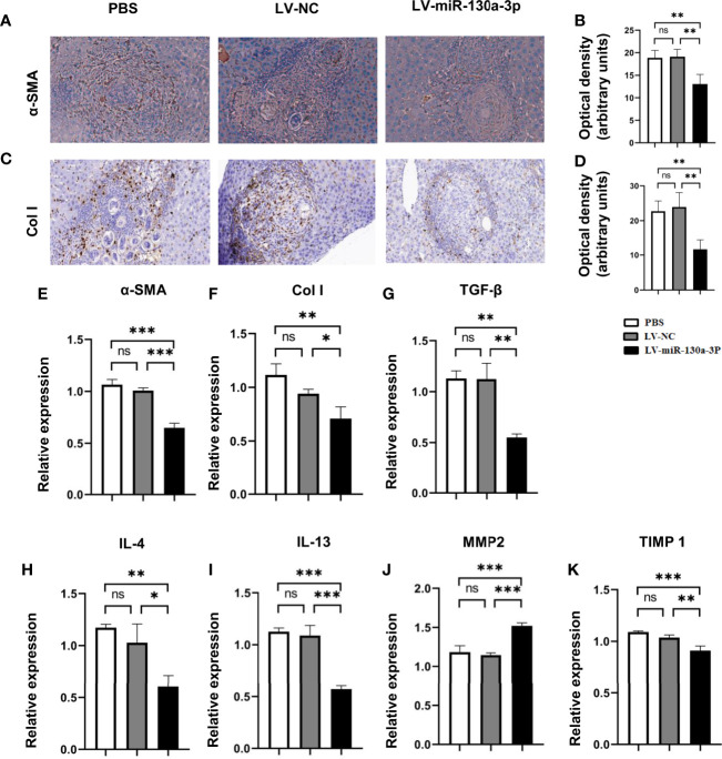
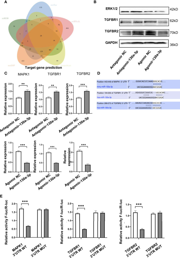
Emerging evidences have highlighted the crucial role of microRNAs (miRNAs) in the liver cirrhosis, but the relationship between miR-130a-3p and liver cirrhosis is not entirely clear. As we all know, schistosomiasis, as one of the zoonoses, can lead to liver cirrhosis when it advances. In this study, we investigated the biological functions of miR-130a-3p on the liver fibrosis of schistosomiasis in vivo and in vitro. The mice infected with Schistosoma japonicum (S. japonicum) were treated with lentivirus vector (LV)-miR-130a-3p by hydrodynamic injection through the tail vein. Our findings showed significantly decreased expression of miR-130a-3p both in the serum of patients with cirrhosis and in the liver of mice infected with S. japonicum. The results showed that LV-miR-130a-3p could effectively enter into the liver and alleviate liver granulomatous inflammation and collagen deposition. Simultaneously, LV-miR-130a-3p-promoted macrophages presented the Ly6Clo phenotype, concomitant with the decreased expression of the tissue inhibitor of metalloproteinases (TIMP) 1, and increased the expression of matrix metalloproteinase (MMP) 2, which contributed to the dissolution of collagen. Furthermore, overexpression of miR-130a-3p not only inhibited the activation and proliferation of hepatic stellate cells (HSCs) but also induced the apoptosis of HSCs. In addition, we also confirmed that miR-130a-3p enables to bind with mitogen-activated protein kinase (MAPK) 1 and transforming growth factor-beta receptors (TGFBR) 1 and TGFBR2 genes and inhibit the expressions of these genes. Our findings suggested that miR-130a-3p might represent as the potential candidate biomarker and therapeutic target for the prognosis identification and treatment of schistosomiasis liver fibrosis.
Keywords: HSCs; Ly6Clo; liver fibrosis; miR-130a-3p; schistosomiasis.
Copyright © 2021 Liu, Wang, Wang, Zhang, Li, Yang, Liu, Zhan, Xu and Xia.
Conflict of interest statement
The authors declare that the research was conducted in the absence of any commercial or financial relationships that could be construed as a potential conflict of interest.
Figures


Similar articles
-
Schistosoma japonicum egg antigen up-regulates fibrogenesis and inhibits proliferation in primary hepatic stellate cells in a concentration-dependent manner.World J Gastroenterol. 2013 Feb 28;19(8):1230-8. doi: 10.3748/wjg.v19.i8.1230. World J Gastroenterol. 2013. PMID: 23482848 Free PMC article.
-
MiR-130a-3p attenuates activation and induces apoptosis of hepatic stellate cells in nonalcoholic fibrosing steatohepatitis by directly targeting TGFBR1 and TGFBR2.Cell Death Dis. 2017 May 18;8(5):e2792. doi: 10.1038/cddis.2017.10. Cell Death Dis. 2017. PMID: 28518142 Free PMC article.
-
ATF3 is involved in rSjP40-mediated inhibition of HSCs activation in Schistosoma japonicum-infected mice.J Cell Mol Med. 2024 Jun;28(12):e18458. doi: 10.1111/jcmm.18458. J Cell Mol Med. 2024. PMID: 39031798 Free PMC article.
-
Galectin-3, histone deacetylases, and Hedgehog signaling: Possible convergent targets in schistosomiasis-induced liver fibrosis.PLoS Negl Trop Dis. 2017 Feb 23;11(2):e0005137. doi: 10.1371/journal.pntd.0005137. eCollection 2017 Feb. PLoS Negl Trop Dis. 2017. PMID: 28231240 Free PMC article. Review.
-
Macrophages: master regulators of inflammation and fibrosis.Semin Liver Dis. 2010 Aug;30(3):245-57. doi: 10.1055/s-0030-1255354. Epub 2010 Jul 21. Semin Liver Dis. 2010. PMID: 20665377 Free PMC article. Review.
Cited by
-
MiR-4465-modified mesenchymal stem cell-derived small extracellular vesicles inhibit liver fibrosis development via targeting LOXL2 expression.J Zhejiang Univ Sci B. 2024 May 17;25(7):594-604. doi: 10.1631/jzus.B2300305. J Zhejiang Univ Sci B. 2024. PMID: 39011679 Free PMC article.
-
Regulatory Functions and Mechanisms of Circular RNAs in Hepatic Stellate Cell Activation and Liver Fibrosis.Cells. 2023 Jan 19;12(3):378. doi: 10.3390/cells12030378. Cells. 2023. PMID: 36766720 Free PMC article. Review.
-
METTL3 inhibits liver fibrosis via RBX1 stability and TXNIP-mediated ferroptosis.Hepatol Commun. 2025 Jul 14;9(8):e0752. doi: 10.1097/HC9.0000000000000752. eCollection 2025 Aug 1. Hepatol Commun. 2025. PMID: 40658786 Free PMC article.
-
Signaling pathways that activate hepatic stellate cells during liver fibrosis.Front Med (Lausanne). 2024 Sep 18;11:1454980. doi: 10.3389/fmed.2024.1454980. eCollection 2024. Front Med (Lausanne). 2024. PMID: 39359922 Free PMC article. Review.
-
Pathology and molecular mechanisms of Schistosoma japonicum-associated liver fibrosis.Front Cell Infect Microbiol. 2022 Oct 28;12:1035765. doi: 10.3389/fcimb.2022.1035765. eCollection 2022. Front Cell Infect Microbiol. 2022. PMID: 36389166 Free PMC article. Review.
References
Publication types
MeSH terms
Substances
LinkOut - more resources
Full Text Sources
Medical
Research Materials
Miscellaneous