Review
. 2021 Aug 4:12:716499.
doi: 10.3389/fimmu.2021.716499. eCollection 2021.

Intestinal Regulatory T Cells as Specialized Tissue-Restricted Immune Cells in Intestinal Immune Homeostasis and Disease

Affiliations
Review

Intestinal Regulatory T Cells as Specialized Tissue-Restricted Immune Cells in Intestinal Immune Homeostasis and Disease

Justin Jacobse et al. Front Immunol. .

Abstract

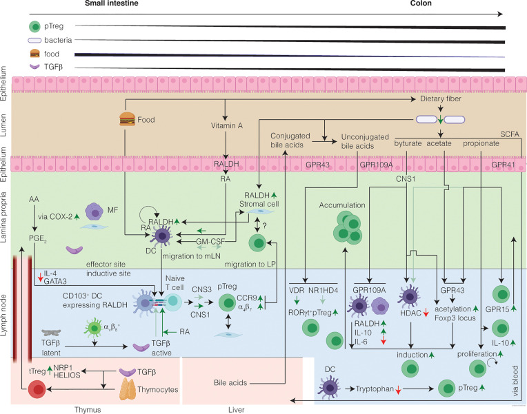
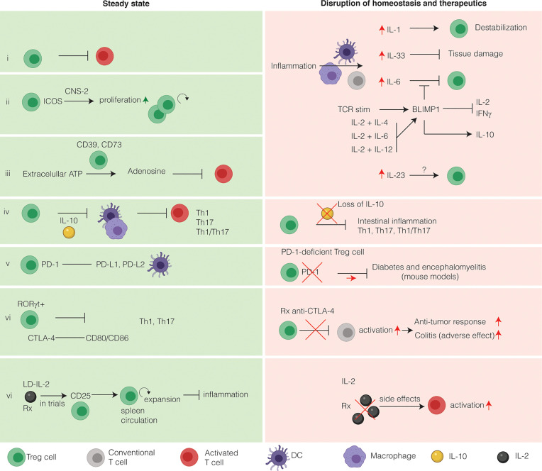
FOXP3+ regulatory T cells (Treg cells) are a specialized population of CD4+ T cells that restrict immune activation and are essential to prevent systemic autoimmunity. In the intestine, the major function of Treg cells is to regulate inflammation as shown by a wide array of mechanistic studies in mice. While Treg cells originating from the thymus can home to the intestine, the majority of Treg cells residing in the intestine are induced from FOXP3neg conventional CD4+ T cells to elicit tolerogenic responses to microbiota and food antigens. This process largely takes place in the gut draining lymph nodes via interaction with antigen-presenting cells that convert circulating naïve T cells into Treg cells. Notably, dysregulation of Treg cells leads to a number of chronic inflammatory disorders, including inflammatory bowel disease. Thus, understanding intestinal Treg cell biology in settings of inflammation and homeostasis has the potential to improve therapeutic options for patients with inflammatory bowel disease. Here, the induction, maintenance, trafficking, and function of intestinal Treg cells is reviewed in the context of intestinal inflammation and inflammatory bowel disease. In this review we propose intestinal Treg cells do not compose fixed Treg cell subsets, but rather (like T helper cells), are plastic and can adopt different programs depending on microenvironmental cues.

Keywords: IBD; homeostasis; intestinal inflammation; intestine; regulatory T (Treg) cells.


Conflict of interest statement

The authors declare that the research was conducted in the absence of any commercial or financial relationships that could be construed as a potential conflict of interest.

Figures

Figure 1
Figure 1
The healthy intestine favors Treg-cell induction in response to harmless antigen and local microenvironmental conditioning maintains the Treg cell phenotype. The mechanisms of intestinal Treg cell induction depend on the structure of the encountered antigen and on the inductive site: in the SI encounter of harmless food antigens predominates eliciting DC-mediated Treg cell induction from naïve T cells. In the colon, commensal microbiota produce SCFA which in presence of TGFβ induce RORγt+ Treg cells. In the SI inductive sites, DC-derived TGFβ induction converts circulating naïve T cells into pTreg cells, which is further potentiated by diet-derived RA. Treg cell induction in the SI is CNS1-dependent with a minor contribution from CNS3. In the colon inductive sites, the SCFA induce Treg cells. SCFA induce acetylation of the Foxp3 locus and alter the phenotype of the pTreg cells by increasing RORγt. Moreover, bile acids unconjugated by the microbiome contribute to the induction of colonic RORγt+ Treg cells. After induction, pTreg cells migrate from the inductive sites to the LP, where they accumulate and maintain immune homeostasis. In the LP, Treg cells might interact with stromal cells, which receive signals from the microbiome, through a cell-contact dependent mechanism. A portion of the intestinal Treg cells is tTreg cells, induced in the thymus in presence of TGFβ, and that have migrated to the intestine and are highly expressing putative markers for tTreg cells. Finally, a gradient of Treg cells exists increasing from SI to colon. Question marks indicate research areas that remain relatively unexplored.
Figure 2
Figure 2
Intestinal Treg cells tightly maintain intestinal immune homeostasis and relevance for immunotherapy. Intestinal Treg cells have several markers that discriminate them from other Treg cells. Under homeostatic conditions, Treg cells suppress effector T cells to prevent inflammation (i). During inflammation, several inflammatory cytokines alter the phenotype of intestinal Treg cells. (ii) Inducible co-stimulator (ICOS) stabilizes intestinal Treg cells in a CNS2-dependent manner. (iii) Extracellular ATP is converted to the immunosuppressive adenosine by Treg cells expressing CD39 and CD73. CD73 is induced by TGFβ. (iv) The main immunosuppressive cytokine secreted by intestinal Treg cells is IL-10. IL-10 functions to inhibit T-helper 1 (Th1), Th17 and Th1/Th17 inflammation in a STAT3-dependent manner via IL-10R signaling on APCs, limiting inflammasome activation. On the other hand, loss of IL-10 both in mice and in humans leads to intestinal inflammation and IBD, respectively. The main co-stimulatory receptors expressed on Treg cells are programmed death receptor (PD-1) (v) and cytotoxic T-lymphocyte associated protein 4 (CTLA-4) (vi). Both these co-stimulatory receptors are exploited therapeutically to increase the anti-cancer immune response. Anti-CTLA-4 therapy has colitis as a frequent side-effect, whereas colitis is not associated with blockade of the PD-1 pathway. (vi) Because Treg cells express the high affinity receptor CD25, they respond to low doses of IL-2 by expansion, thus limiting inflammation. LD-IL-2 is investigated in a clinical trial for its use in patients with IBD. In contract to LD-IL-2, a high dose of IL-2 will activate T cells and lead to deleterious side effects and therefore is not used in clinical practice.

References

    1. Miller JFAP. Immunological Function of the Thymus. Lancet (1961) 278:748–9. 10.1016/S0140-6736(61)90693-6 - DOI - PubMed
    1. Miller JFAP. Role of the Thymus in Transplantation Immunity*. Ann N Y Acad Sci (1962) 99:340–54. 10.1111/j.1749-6632.1962.tb45319.x - DOI
    1. Gershon RK, Kondo K. Cell Interactions in the Induction of Tolerance: The Role of Thymic Lymphocytes. Immunology (1970) 18:723–37. - PMC - PubMed
    1. Kojima A, Tanaka-Kojima Y, Sakakura T, Nishizuka Y. Prevention of Postthymectomy Autoimmune Thyroiditis in Mice. Lab Invest (1976) 34:601–5. - PubMed
    1. Nishizuka Y, Sakakura T. Thymus and Reproduction: Sex-Linked Dysgenesia of the Gonad After Neonatal Thymectomy in Mice. Science (1969) 166:753–5. 10.1126/science.166.3906.753 - DOI - PubMed

Publication types

MeSH terms