

doi: 10.1107/S2056989021007167.
eCollection 2021 Aug 1.
Crystal structures of two polymorphs of tixocortol pivalate
Affiliations
- PMID: 34422306
- PMCID: PMC8340970
- DOI: 10.1107/S2056989021007167
Item in Clipboard
Crystal structures of two polymorphs of tixocortol pivalate
Acta Crystallogr E Crystallogr Commun.
.
Abstract
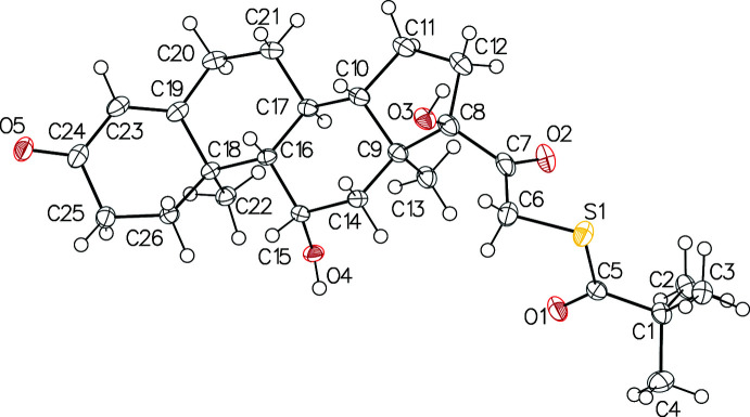
Two polymorphs, (I) and (II), of (S)-{2-[(8S,9S,10R,11S,13S,14S,17R)-11,17-dihy-droxy-10,13-dimethyl-3-oxo-2,6,7,8,9,11,12,14,15,16-deca-hydro-1H-cyclo-penta-[a]phenanthren-17-yl]-2-oxoeth-yl} 2,2-di-methyl-propane-thio-ate, C26H38O5S, have been identified. They are ortho-rhom-bic, non-centrosymmetric (P212121). The structures display layers of mol-ecules conected via O-H⋯O hydrogen bonds along the b-axis direction in polymorph (I) and along the c-axis direction in polymorph (II). The structure of (II) exhibits disorder of the main mol-ecule.
Keywords: crystal structure; pivalone; polymorphs; tixocortol.
© Rousselin et al. 2021.
Figures

ORTEP view of polymorph (I). Displacement ellipsoids are drawn at the 50% probability level.
ORTEP view of polymorph (II). Displacement ellipsoids are drawn at the 30% probability level. The minor component of the disorder is omitted for clarity.

View of the hydrogen bond-network in polymorph (I).

View of the hydrogen-bond network in polymorph (II).
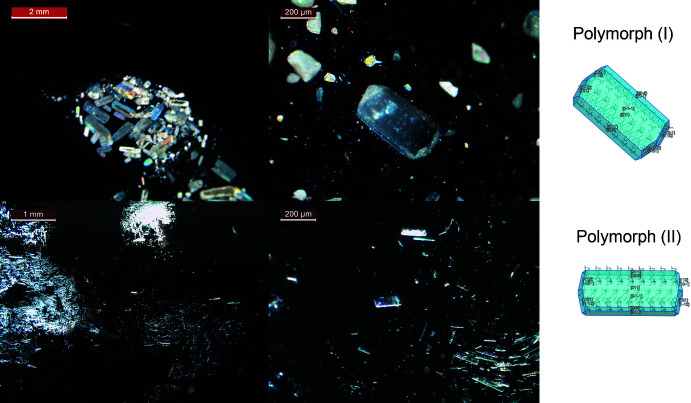
View of the crystal morphology of polymorph (I) (top) and (II) (bottom).

Reaction scheme for the synthesis of tixocortol pivalate.

PXRD patterns of polymorphs (I) and (II) and their simulated patterns from the SCXRD study at room temperature.
Similar articles
-
Crystal structure of (S)-2-[(3S,8S,9S,10R,13S,14S,17R)-3-hy-droxy-10,13-dimethyl-2,3,4,7,8,9,10,11,12,13,14,15,16,17-tetra-deca-hydro-1H-cyclo-penta[a]phenanthren-17-yl]-N-meth-oxy-N-methyl-pro-pan-amide (Fernholz Weinreb amide).Acta Crystallogr E Crystallogr Commun. 2015 Feb 18;71(Pt 3):275-7. doi: 10.1107/S2056989015001747. eCollection 2015 Mar 1. Acta Crystallogr E Crystallogr Commun. 2015. PMID: 25844186 Free PMC article.
-
Crystal structure of (20S)-21-[4-(2-hy-droxy-propan-2-yl)-1H-1,2,3-triazol-4-yl]-20-(4-methyl-pent-yl)-5-pregnen-3β-ol with an unknown solvate.Acta Crystallogr E Crystallogr Commun. 2018 Mar 6;74(Pt 4):465-468. doi: 10.1107/S2056989018003286. eCollection 2018 Apr 1. Acta Crystallogr E Crystallogr Commun. 2018. PMID: 29765747 Free PMC article.
-
3β-Acet-oxy-6-hy-droxy-imino-cholestane.Acta Crystallogr Sect E Struct Rep Online. 2011 Apr 1;67(Pt 4):o773-4. doi: 10.1107/S1600536811007306. Epub 2011 Mar 5. Acta Crystallogr Sect E Struct Rep Online. 2011. PMID: 21754066 Free PMC article.
-
Loteprednol.2024 Dec 15. Drugs and Lactation Database (LactMed®) [Internet]. Bethesda (MD): National Institute of Child Health and Human Development; 2006–. 2024 Dec 15. Drugs and Lactation Database (LactMed®) [Internet]. Bethesda (MD): National Institute of Child Health and Human Development; 2006–. PMID: 29999696 Free Books & Documents. Review.
-
Segesterone Acetate.2024 Aug 15. Drugs and Lactation Database (LactMed®) [Internet]. Bethesda (MD): National Institute of Child Health and Human Development; 2006–. 2024 Aug 15. Drugs and Lactation Database (LactMed®) [Internet]. Bethesda (MD): National Institute of Child Health and Human Development; 2006–. PMID: 30371999 Free Books & Documents. Review.
References
-
- Bircher, A. J., Thürlimann, W., Hunziker, T., Pasche-Koo, F., Hunziker, N., Perrenoud, D., Elsner, P. & Schultheiss, R. (1995). Dermatology, 191, 109–114. - PubMed
-
- Bouley, E. (2013). Patent EP 2853528.
-
- Boultif, A. & Louër, D. (2004). J. Appl. Cryst. 37, 724–731.
-
- Bruker (2016). APEX3 and SAINT. Bruker AXS, Inc., Madison, Wisconsin, USA.
-
- Burden, A. D. & Beck, M. H. (1992). Br. J. Dermatol. 127, 497–500. - PubMed
LinkOut - more resources
Full Text Sources
Research Materials