Review
. 2021 Jul 28;9(7):e3673.
doi: 10.1097/GOX.0000000000003673. eCollection 2021 Jul.

Tips on Raising Reliable Local Perforator Flaps

Affiliations
Review

Tips on Raising Reliable Local Perforator Flaps

Mark W Ashton. Plast Reconstr Surg Glob Open. .

Abstract

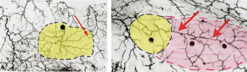
From early on in the development of plastic surgery, it was quickly realized that utilizing locally adjacent tissue, or "matching like with like," yielded superior aesthetic reconstructions to those in which the tissue was derived from a distant location. In many cases, the use of a local perforator flap is a simpler procedure with less patient morbidity and a quicker recovery from surgery. The difficulty with local perforator flaps has been locating the supplying perforators, ensuring that the flap has a robust and reliable blood supply, and that sufficient tissue is able to be transferred. The recent reappraisal of our understanding of the blood supply of the integument has allowed, for the first time, the capacity to accurately and inexpensively, without the need for "high tech equipment," locate perforators, as they emerge from the deep fascia into the overlying integument, and through a better understanding of the interconnecting anastomotic vessels between perforators reliably predict how much tissue can be safely raised on a single perforator, before surgery. Further, through the use of strategic "delay," it is possible to manipulate the interconnecting vessels between the selected perforator and its surrounding neighbors to design a flap of tissue of any dimension, composed of whatever tissue we require, and safely transfer that tissue locally, or if required, distantly, as a free flap. This article will highlight these advances, explain their relevance in raising reliable local perforator flaps, and will, where possible, call attention to any pearls and pitfalls, and how to avoid complications.


Figures

Fig. 1.
Fig. 1.
The distance of perfusion of disulphine blue in Figures 3 and 5 above is the same and is not related to the width of the individual flaps. This distance corresponds to the eventual line of tissue necrosis and the length of flap survival. The length of tissue survival is not dependent on the width of the flap, but rather is determined by its internal vascular anatomy. Reprinted with permission from Br J Surg. 1970;57(7):502–508.
Fig. 2.
Fig. 2.
The angiosomes of the head and neck study of a 20-week-old fetus, a 28-week-old fetus, and an adult. The vascular pattern of the vessels of the scalp is constant and the blueprint is established very early on in fetal development. In particular, note that the superficial temporal artery is unchanged from the 28-week fetus to the adult. Reprinted with permission from Taylor GI, Pan WR, The Angiosome Concept and Tissue Transfer. Volume 2:656–657. Chapter 4, Figures 4.31 and 4.32.
Fig. 3.
Fig. 3.
Stages of vascular development of the ventral limb bud of a quail. Reprinted with permission from Taylor GI and Pan WR, The Angiosome Concept. Volume 1:183. Chapter 2, Figures 2–5.
Fig. 4.
Fig. 4.
Layers 1, 2, and 3 are rigidly fixed together. They are attached to the underlying deep fascia or periosteum by ligaments binding them to the deep fascia below (Layer 5). These ligaments comprise layer 4.
Fig. 5.
Fig. 5.
Where layers 1, 2, and 3 must move over the deep fascia, layer 4 becomes a space. Original concept and diagram: Mr BC Mendelson, FRACS, FACS. Slide used in his lecture series 2008. With permission from Mr. Bryan Mendelson, MD, MBBS, FRCS, FRACS, FACS. For more information, see Melbourne Advanced Facial Anatomy Course.
Fig. 6.
Fig. 6.
Blood vessels, nerves, and lymphatics all use the ligaments of layer 4 to cross between the deep fascia and the superficial integument.
Fig. 7.
Fig. 7.
The illustration above shows two different patterns of attachment of the superficial integument to the deep fascia. In the top diagram, the superficial integument (layers 1, 2, and 3) is rigidly fixed to the deep fascia by multiple ligaments in layer 4. In the bottom illustration, the layers 1, 2, and 3 must glide over the deep fascia and hence, layer 4 is composed of loose areolar tissue and the ligaments are spaced widely apart. Because the blood vessels use the ligaments to cross layer 4, the orientation of the ligaments predicts the geography of the perforating blood vessels.
Fig. 8.
Fig. 8.
Where the superficial integument must glide over the deep fascia, layer 4 becomes a space. Ligaments attaching the two components are located at the boundaries of the space. Nerves and blood vessels use the ligaments to cross layer 4 and are therefore also located at the boundaries of the space. The spaces are avascular.
Fig. 9.
Fig. 9.
The top photograph shows the premasseter space viewed from just in front of the ear, looking forward toward the mouth and nose. The facial nerve can be seen coursing over the masseter in the floor of the space. The ligaments fixing the superficial fascia to the deep fascia are at the periphery of the space have been marked with blue ink. The branches of the facial nerve pass directly on top of the masseter muscle below the deep fascia, and use the massenteric ligaments to enter the platysma and other muscles of the SMAS. Because the Platysma in layer 3 must move over the masseteric deep fascia in layer 5, there is a space in layer 4. This space (the lower premasseter) is avascular, and contains no branches of the facial nerve. This knowledge provides for safe and rapid dissection of the middle lower face. Concept and original drawing: Mr BC Mendelson, MD, MBBS, FRCS, FRACS, FACS. Arterial overlay by the author. Image Courtesy: Mr BC Mendelson, MD, MBBS, FRCS, FRACS, FACS.
Fig. 10.
Fig. 10.
Seminal study by Taylor. In a T-shaped flap on the abdomen of a dog, three flaps of equal width are raised based on the central green perforator. The geographic location of the surrounding red perforators determines the length of the flap survival. In the inferior flap, the red perforator is located at the flap’s tip, and the entire flap survives. Reprinted with permission from Plast Reconst Surg. 2017;140(4):721–733. Figure 5.
Fig. 11.
Fig. 11.
Perforators are interconnected by either choke or true anastomoses. Choke anastomoses, as their name suggests, restrict flow between perforators, whilst true anastomoses allow unrestricted flow between perforators effectively joining them together as one. The top diagram shows choke anastomoses above, and a true anastomosis below. The bottom photographs of the side of the nose show pink perspex filled anastomoses between the dorsal nasal branches of the facial artery and supra-trochlear and supra-orbital branches of the ophthalmic artery. In the left photograph these are choke anastomoses. In the right these same anastomoses are true anastomoses. Reprinted with permission from Plast Reconst Surg. 2013;132(6):1447–1456. Figure 1.
Fig. 12.
Fig. 12.
This seminal study from Taylor and Dahr shows the functional influence of choke anastomoses between perforators—in this case the thoracodorsal and the deep circumflex iliac arteries of a rabbit. When the deep circumflex artery is ligated, fluorescein traveling from the thoracodorsal perforator is held up at the choke anastomoses (arrows), and restricted from entering the distal territory, before eventually being able to enter the deep circumflex artery angiosome at 30 minutes. Reprinted with permission from Plast Reconstr Surg 1999;104:2079–2091. Note: Descriptors of the part labels A-D were not included in the original publication.
Fig. 13.
Fig. 13.
The dimensions of tissue that can be safely captured on a perforator are not just dependent upon the distance between perforators, but also upon the interconnection between those perforators. In the two images above, the perforator on the left is surrounded by choke anastomoses (red arrow) and, hence, only the area in yellow can be reliably captured. In the image on the right, the three perforators are interconnected by true anastomoses and, hence, they will act as one, and the additional pink area can also be captured, on the same chosen perforator, without the risk of necrosis. (The two true anastomoses are identified with the two red arrows in the diagram on the right.)
Fig. 14.
Fig. 14.
CT angiogram of the abdomen. This technology allows clear identification of perforators as they emerge through the deep fascia. In this CT angiogram a large perforator (central blue arrow) can be seen emerging through the left rectus muscle. This CTA also shows the “law of equilibrium;” that is, where a perforator is unusually large, the contralateral or adjacent perforators will be correspondingly small. In this case there are no large perforators on the right hemiabdomen, and the right SIEA (left blue arrow above) is dominant. On the patient's left, the perforator (central blue arrow) is the main blood supply to the abdomen, and the left SIEA (right blue arrow above) is small. Concept introduced in Rozen WM, Grinsell D, Koshima I, et al. Dominance between angiosome and perforator territories: a new anatomical model for the design of perforator flaps. J Reconstr Micro. 2010 ;26:539-45.
Fig. 15.
Fig. 15.
This figure shows the rate of rewarming of tissue between perforators. There is a statistical difference in the rate at which true and choke anastomoses re-heat tissue after that tissue has been cooled. This difference can be used clinically to identify the interconnections between perforators and whether they are true or choke anastomoses. Reprinted with permission from Plastic Reconst Surg. 2013;132(6):1457–1464.
Fig. 16.
Fig. 16.
The above sequence shows the posterior aspect of the left calf that has initially been precooled with an icepack to 20 degrees (A). (The arrows show the uniform blue/green color). Upon removal of the icepack, warm blood below the deep fascia emerges through perforators, to heat the cooled integument and highlights their position; seen as orange spots in (B). As the tissue is warmed, the color of the perforator location changes from orange to red. Critically, image C also identifies the nature of the interconnections between perforators; the lower most perforators are connected by true anastomoses in red (identified with arrows), whilst the top perforator is connected with the others by a choke anastomosis. This corresponds with the angiogram in image D. The clinical significance of this study is that, counterintuitively, this flap is more reliable when raised on the distal (rather than on the proximal) perforator. Reprinted with permission from Plast Reconstr Surgery. 2013; 132(6):1457-1464.
Fig. 17.
Fig. 17.
CT angiography and Thermal imaging of the same abdomen. A, The CT Angiogram provides detail on the exact location of the perforators as they emerge through the deep fascia, and their caliber. The largest perforator is seen emerging through the left rectus muscle (blue arrows). This perforator has been identified with thermography and confirmed with Doppler Ultrasound. B, A pen marks the location in the middle diagram. C, Importantly, in addition to being able to locate the perforators (in yellow), the thermography also shows the interconnections between perforators—the true anastomoses are also yellow.
Fig. 18.
Fig. 18.
A summary of key points to be learned from this article.
Fig. 19.
Fig. 19.
A series of photographs to show the method used by the author to transfer a local perforator flap. This patient had a sarcoma excised from the shin of her left leg. The defect was initially closed with a skin graft to allow monitoring for recurrence. After 2 years, she requested replacement with more robust tissue. A, A local perforator is identified preoperatively using either CTA, Doppler, or thermography. Before any skin is removed, an exploratory incision is made at the edge of the scar to confirm the perforator is adequate. This is done by carefully dissecting beneath the deep fascia. B, A flap is marked and then incised. The direction of the flap is based upon skin laxity to preselect for longer vessels and to allow direct closure of the secondary defect. C, The flap is raised, protecting the perforator, and preserving the underlying saphenous nerve. D, Until it is completely islanded on the perforator. E, The flap is then rotated 180 degrees to sit upon the proposed defect. F, Once confirmed that the flap is adequate, the skin graft is excised, and the donor defect approximated.
Fig. 20.
Fig. 20.
Postoperative results. The final healed result at 2 weeks (A) and at 3 months postoperative (B).

References

    1. Gillies HD, Pilcher LS. Plastic Surgery of the Face. 1920. New York: Thieme-Stratton Corp
    1. Gillies H, Millard DR. The Principles and Art of Plastic Surgery. Boston, Toronto: Butterworth; 1957.
    1. Milton SH. Pedicled skin-flaps: the fallacy of the length: width ratio. Br J Surg. 1970;57:502–508. - PubMed
    1. Taylor GI, Pan WR. The Angiosome Concept and Tissue Transfer. New York: Thieme Medical Publishers. Vol 2:656–657.
    1. Bates D, Taylor GI, Newgreen DF. The pattern of neurovascular development in the forelimb of the quail embryo. Dev Biol. 2002;249:300–320. - PubMed