Editorial
. 2021 Jul;10(4):527-537.
doi: 10.21037/acs-2021-rp-31.

The Ross-Konno procedure for congenital aortic stenosis

Affiliations
Editorial

The Ross-Konno procedure for congenital aortic stenosis

Sameh M Said. Ann Cardiothorac Surg. 2021 Jul.

Abstract

Left ventricular outflow tract (LVOT) obstruction can result from supravalvular, valvular and/or subvalvular etiologies. Congenital aortic valvular stenosis is frequently associated with aortic annular hypoplasia. Aortoventriculoplasty with pulmonary autograft, "The Ross-Konno" operation, provides more or less a radical solution to multilevel LVOT obstruction by enlarging the aortic annulus and the subvalvular area, thus relieving both valvular and subvalvular obstructions. In addition to this, the procedure carries the major advantage of having a competent autograft in the LVOT. An autograft that has the potential for growth and provides excellent quality of life without the need for anticoagulation. The procedure is most commonly performed as a complete root implantation, harvesting the coronary arteries as buttons, and harvesting the autograft with a muscle skirt to allow a single unit reconstruction of the LVOT. The procedure has been modified over time to minimize the risk of conduction tissue injury and the development of complete heart block by modifying the interventricular septal incision. The Ross-Konno procedure has changed the approach to patients with complex multilevel LVOT obstruction. However, while it can be performed early in life, one should be aware that this procedure is technically demanding and can be associated with a higher surgical risk. Nevertheless, it is considered "the ultimate solution" for those with complex multilevel LVOT obstruction.

Keywords: Ross; Ross-Konno; aortoventriculoplasty; autograft; hypoplastic aortic annulus.


Conflict of interest statement

Conflicts of Interest: SMS is a consultant for Cryolife, and Stryker.

Figures
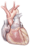
Figure 1
Figure 1
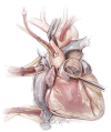
The proposed incisions for the pulmonary autograft harvest and ascending aorta transection. A skirt of right ventricular free wall muscle is harvested with the autograft for the planned anterior aortic root enlargement part (Konno) of the procedure.
Figure 2
Figure 2
The distal ascending aorta and both cavae are cannulated. Once cardiopulmonary bypass is initiated, the main pulmonary artery is transected just proximal to its bifurcation and the pulmonary valve is inspected to determine its suitability as an autograft. This part can also be completed after arresting the heart and determining the non-suitability of the native aortic valve for repair.
Figure 3
Figure 3
Once the pulmonary valve is determined to be suitable, the main pulmonary artery is completely transected and mobilized using electrocautery and the autograft is harvested. A right-angled clamp is passed through the pulmonary valve 5 mm below the nadir of the non-facing cusp and used as a guide to incise the right ventricular free wall safely below the pulmonary valve cusps.
Figure 4
Figure 4
Lifting the pulmonary artery vertically from the plane of the heart during mobilization creates a gap from the coronary arteries. It is important to be aware of the close proximity of the left main and left anterior descending coronary arteries, especially in reoperative settings where landmarks are obscured. LMCA, left main coronary artery; LAD, left anterior descending.
Figure 5
Figure 5
The autograft is harvested by a combination of sharp and electrocautery dissection. The autograft is carefully removed from its posterior attachment through “enucleation”, thus preserving the first septal perforator branch of the left anterior descending coronary artery. A useful landmark for the location of the first septal perforator is the medial papillary muscle of the tricuspid valve. Administration of cardioplegia can help hemostasis at this stage by visualizing any bleeding point from the autograft harvest site. RCA, right coronary artery; LAD, left anterior descending.
Figure 6
Figure 6
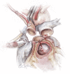
Preparation of the aortic root can be done prior to harvesting the autograft. The aortic valve leaflets are excised and the coronary buttons are harvested and mobilized off the epicardial surface of the heart. The left ventricular outflow tract is evaluated and a septal myectomy, and/or resection of endocardial fibroelastosis can be performed at this stage. Notice the proposed interventricular septal incision (Konno incision) (dotted line).
Figure 7
Figure 7
A Konno incision is performed to the left of the right coronary artery button or along the commissure between the left and right coronary sinuses of Valsalva. The extent of this incision depends on the degree of enlargement of the left ventricular outflow tract required and it should be done in an oblique or transverse fashion to avoid the conduction tissue.
Figure 8
Figure 8
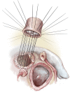
The pulmonary autograft with its infundibular extension is then implanted in the left ventricular outflow tract (A). This can be performed using running or interrupted sutures. It is important to trim the infundibular skirt to fit the septal incision and minimize the risk of outflow tract obstruction by leaving a bulk of infundibular muscle below the neoaortic valve. (B) An alternative option, especially when the length of the septal incision is longer than the infundibular skirt, is to close the created ventricular septal defect with a prosthetic patch and reimplant the autograft as a full root technique.
Figure 9
Figure 9
The left followed by the right coronary arteries are implanted in the middle of the corresponding sinuses of Valsalva of the autograft. The completed autograft and the reconstructed neoaortic root are shown. The distal pulmonary homograft anastomosis can be done at this stage prior to reconstructing the ascending aorta to facilitate exposure and ensure hemostasis.
Figure 10
Figure 10
Implantation of the pulmonary homograft. The distal anastomosis is constructed to the pulmonary artery bifurcation, followed by suturing the proximal homograft to the right ventricular outflow tract. It is important to take the endocardium and part of the myocardium only along the posterior suture line to avoid compromising the first septal perforator branch of the left anterior descending coronary artery which is in close proximity. This can be supported with a strip of pericardium to ensure hemostasis. We prefer to use the largest possible pulmonary homograft in this position.
Figure 11
Figure 11
The completed Ross-Konno operation.
Figure 12
Figure 12
The mini Ross-Konno procedure. The initial steps of the procedure are similar to the standard Ross-Konno. However, a smaller Konno incision combined with an extended left ventricular septal myectomy is performed to eliminate all potential subaortic obstruction without the creation of a ventricular septal defect. The aortic annulus is then enlarged to accommodate the autograft.
Figure 13
Figure 13
The modified Ross-Konno procedure is performed by amending the ventricular septal incision. The left ventricular outflow tract is enlarged posterior to the course of the left main coronary artery (dotted line), thus avoiding the interventricular septum and the conduction tissue.
Figure 14
Figure 14
The left ventricular outflow tract is enlarged posteriorly by incising the free wall of the left ventricle posterior to the course of the left main coronary artery. The autograft is then implanted using multiple pledgeted mattress sutures to ensure stability and hemostasis.
Video
Video
The Ross-Konno procedure for congenital aortic stenosis.

References

    1. Konno S, Imai Y, Iida Y, et al. A new method for prosthetic valve replacement in congenital aortic stenosis associated with hypoplasia of the aortic valve ring. J Thorac Cardiovasc Surg 1975;70:909-17. 10.1016/S0022-5223(19)39673-4 - DOI - PubMed
    1. Rastan H, Koncz J. Aortoventriculoplasty: a new technique for the treatment of left ventricular outflow tract obstruction. J Thorac Cardiovasc Surg 1976;71:920-7. 10.1016/S0022-5223(19)40132-3 - DOI - PubMed
    1. Ross DN. Replacement of aortic and mitral valves with a pulmonary autograft. Lancet 1967;2:956-8. 10.1016/S0140-6736(67)90794-5 - DOI - PubMed
    1. Reddy VM, Rajasinghe HA, Teitel DF, et al. Aortoventriculoplasty with the pulmonary autograft: the "Ross-Konno" procedure. J Thorac Cardiovasc Surg 1996;111:158-65; discussion 165-7. 10.1016/S0022-5223(96)70412-9 - DOI - PubMed
    1. Al Halees Z. The Mini-Ross-Konno procedure. Eur J Cardiothorac Surg 2011;39:1067-9. 10.1016/j.ejcts.2010.10.008 - DOI - PubMed

Publication types