Review
. 2021 Aug 5:9:691644.
doi: 10.3389/fcell.2021.691644. eCollection 2021.

When Viruses Cross Developmental Pathways

Affiliations
Review

When Viruses Cross Developmental Pathways

Pankaj Trivedi et al. Front Cell Dev Biol. .

Abstract

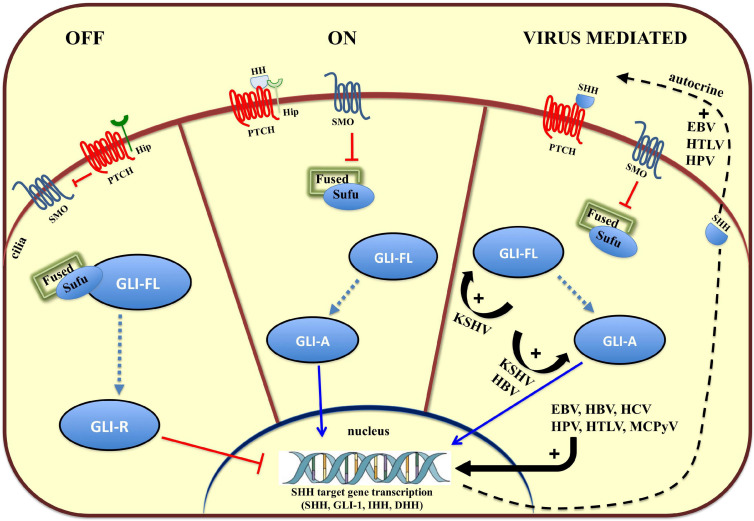
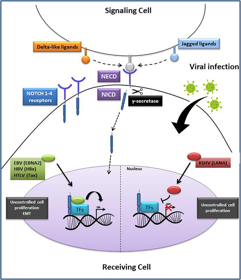
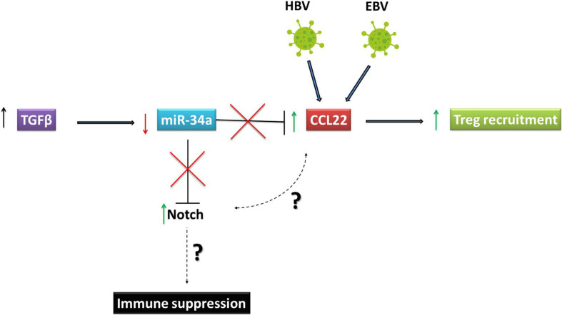
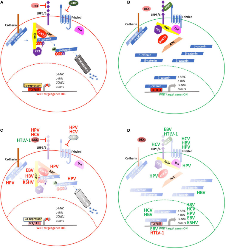
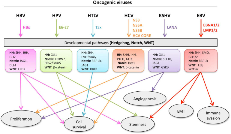
Aberrant regulation of developmental pathways plays a key role in tumorigenesis. Tumor cells differ from normal cells in their sustained proliferation, replicative immortality, resistance to cell death and growth inhibition, angiogenesis, and metastatic behavior. Often they acquire these features as a consequence of dysregulated Hedgehog, Notch, or WNT signaling pathways. Human tumor viruses affect the cancer cell hallmarks by encoding oncogenic proteins, and/or by modifying the microenvironment, as well as by conveying genomic instability to accelerate cancer development. In addition, viral immune evasion mechanisms may compromise developmental pathways to accelerate tumor growth. Viruses achieve this by influencing both coding and non-coding gene regulatory pathways. Elucidating how oncogenic viruses intersect with and modulate developmental pathways is crucial to understanding viral tumorigenesis. Many currently available antiviral therapies target viral lytic cycle replication but with low efficacy and severe side effects. A greater understanding of the cross-signaling between oncogenic viruses and developmental pathways will improve the efficacy of next-generation inhibitors and pave the way to more targeted antiviral therapies.

Keywords: Hedgehog; Notch; WNT; immune evasion; microRNA; oncogenic viruses; targeted therapies.


Conflict of interest statement

The authors declare that the research was conducted in the absence of any commercial or financial relationships that could be construed as a potential conflict of interest.

Figures

FIGURE 1
FIGURE 1
Overview of the Hedgehog (HH) signaling pathway and modulation by oncoviruses. On the left, in the absence of HH ligands (SHH, IHH, and DHH), the HH receptor Patched (PTCH) keeps the pathway off (OFF) by inhibiting Smoothened (SMO), and keeping the glioma-associated oncogene (GLI) transcription factors in an inactive form (GLI-R). In the middle, binding of HH ligand to HH receptor, PTCH, turns the pathway on (ON), and inhibits its activity, relieving the repression of SMO, which converts full-length GLI (GLI-FL) into a transcriptional activator (GLI-A). In vertebrates, cilia are required for production of GLI-repressor (GLI-R) and/or GLI-activator (GLI-A). On the left, viruses hyperactivate HH signaling with multiple mechanisms (VIRUS MEDIATED). EBV, Epstein Barr Virus; HBV, Hepatitis B Virus; HCV, Hepatitis C Virus; HPV, Human Papilloma Virus, KSHV, Kaposi’s Sarcoma-associated Herpes Virus; MCPyV, Merkel Cell Polyoma Virus.
FIGURE 2
FIGURE 2
Oncogenic viruses exploit the Notch signaling pathway. Notch ligands (Delta-like or Jagged) on the signaling cell bind to Notch 1–4 receptors on the receiving cell, generating the Notch extracellular domain (NECD) and the Notch intracellular domain (NICD). Subsequently, the proteolytic cleavage of NECD by γ-secretase, generates NICD translocation into the nucleus where it binds with the transcription factors (TFs): recombining binding protein Jk (RBP–Jκ), cAMP response element-binding protein (CREB), forming an active complex that regulates the transcription of Notch target genes. On the (Left side): EBNA2, HBx and Tax proteins encoded by EBV, HBV, and HTLV, respectively, promote cell proliferation or epithelial-mesenchymal transition (EMT) through a direct interaction with the transcription complex, leading to tumorigenesis. On the (Right side): LANA protein, encoded by KSHV, prevents the activation of the transcription complex, resulting in an accumulation of NICD and increased cancer cells proliferation. EBV, Epstein Barr Virus; HBV, Hepatitis B Virus; HTLV-1, Human T Lymphocyte Virus-1; KSHV, Kaposi’s Sarcoma-associated Herpesvirus.
FIGURE 3
FIGURE 3
Oncoviruses exploit Notch signaling to escape immune responses. Reed Sternberg cells, which represent the Hodgkin’s Lymphoma tumor component are high expressers of Notch 1 and 2 and produce high levels of CCL22. This chemokine is important for recruiting regulatory T (Treg) cells to the tumor microenvironment. In HBV associated HCC, PVTT symptoms are positively correlated with TGFβ. The increase in TGFβ led to downregulation of miR-34a, which targets CCL22. Downregulation of miR-34a induced by TGFβ in HCC led to an increase in CCL22 and consequently in regulatory T cell recruitment to create an immunosuppressive tumor environment. HBV, Hepatitis B virusVirus; EBV, Epstein Barr Virus; HCC, Hepatocellular Carcinoma.
FIGURE 4
FIGURE 4
WNT signaling is targeted by oncogenic viruses during tumorigenesis. (A) WNT signaling is maintained in an “OFF” state by active processes that result in proteosomal degradation of cytosolic β-catenin and prevent transcription of WNT target genes. (B) Secretion of WNT ligands activate signaling though FZD and LRP5/6 to recruit the AxinAXIN-APC-GSK3αGSK3b complex away from β-catenin, resulting in its cytosolic stabilization and eventual translocation to the nucleus where it activates the TCF/LEF transcriptional complex to induce expression of WNT target genes. The oncogenic viruses, HBV, HCV, HPV, EBV, HTLV-1, and KSHV target various aspects of WNT signaling either to (C) repress inhibitors of or (D) enhance positive regulators of WNT signaling. (red) Negative regulation; (green) positive regulation. See text for details.
FIGURE 5
FIGURE 5
Developmental pathways co-opted during viral oncogenesis. Viruses deregulate developmental signaling to support malignant transformation and disease progression, via a variety of mechanisms, including (but not limited) to, increased proliferation and cell survival, induced stemness to improve fitness of the cancer cells, accrued neo-vascularization, activation of the epithelial-to-mesenchymal transition (EMT), evasion of tumor-targeting immune response. Inhibitors of developmental pathways may prevent the acquisition of new properties by the cancer cells, such as virus-induced transcriptional, metabolic, and functional reprogramming. Each color is associated with a specific virus and the cell processes controlled the developmental pathway.

References

    1. Aicher S., Kakkanas A., Cohen L., Blumen B., Oprisan G., Njouom R., et al. (2018). Differential regulation of the Wnt/β-catenin pathway by hepatitis C virus recombinants expressing core from various genotypes. Sci. Rep. 8:11185. 10.1038/s41598-018-29078-2 - DOI - PMC - PubMed
    1. Al-Shabanah O. A., Hafez M. M., Hassan Z. K., Sayed-Ahmed M. M., Abozeed W. N., Alsheikh A., et al. (2014). Methylation of SFRPs and APC genes in ovarian cancer infected with high risk human papillomavirus. Asian Pac. J. Cancer Prev. 15 2719–2725. 10.7314/apjcp.2014.15.6.2719 - DOI - PubMed
    1. Anastasiadou E., Stroopinsky D., Alimperti S., Jiao A. L., Pyzer A. R., Cippitelli C., et al. (2019). Epstein-Barr virus-encoded EBNA2 alters immune checkpoint PD-L1 expression by downregulating miR-34a in B-cell lymphomas. Leukemia 33 132–147. 10.1038/s41375-018-0178-x - DOI - PMC - PubMed
    1. Andersen P., Uosaki H., Shenje L. T., Kwon C. (2012). Non-canonical Notch signaling: emerging role and mechanism. Trends Cell Biol. 22 257–265. 10.1016/j.tcb.2012.02.003 - DOI - PMC - PubMed
    1. Arensdorf A. M., Marada S., Ogden S. K. (2016). Smoothened regulation: a tale of two signals. Trends Pharmacol. Sci. 37 62–72. 10.1016/j.tips.2015.09.001 - DOI - PMC - PubMed

LinkOut - more resources