. 2021 Aug 6:8:680775.
doi: 10.3389/fcvm.2021.680775. eCollection 2021.

Inhibition of Rac1 GTPase Decreases Vascular Oxidative Stress, Improves Endothelial Function, and Attenuates Atherosclerosis Development in Mice

Affiliations

Inhibition of Rac1 GTPase Decreases Vascular Oxidative Stress, Improves Endothelial Function, and Attenuates Atherosclerosis Development in Mice

Sebastian Zimmer et al. Front Cardiovasc Med. .

Abstract

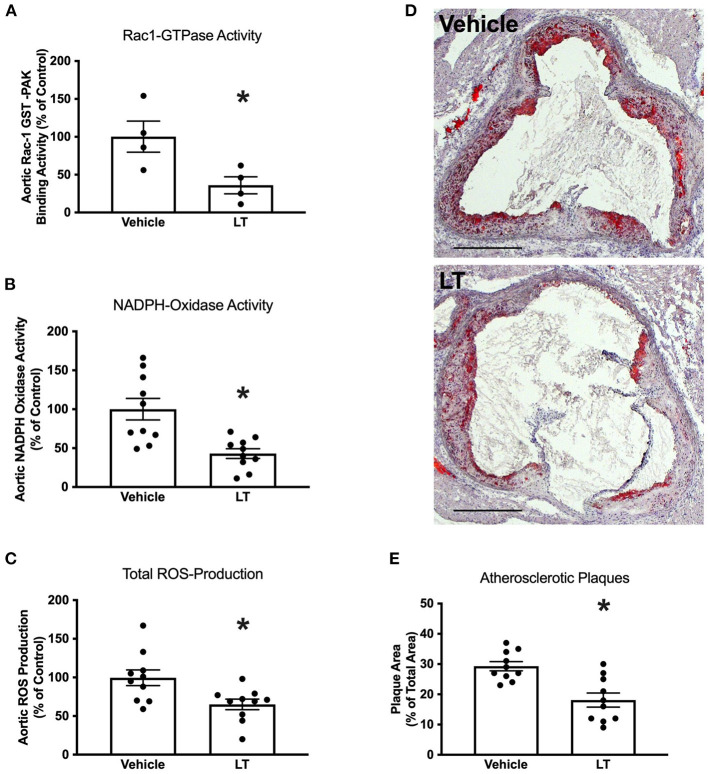
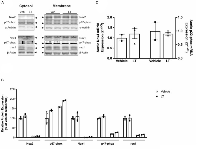
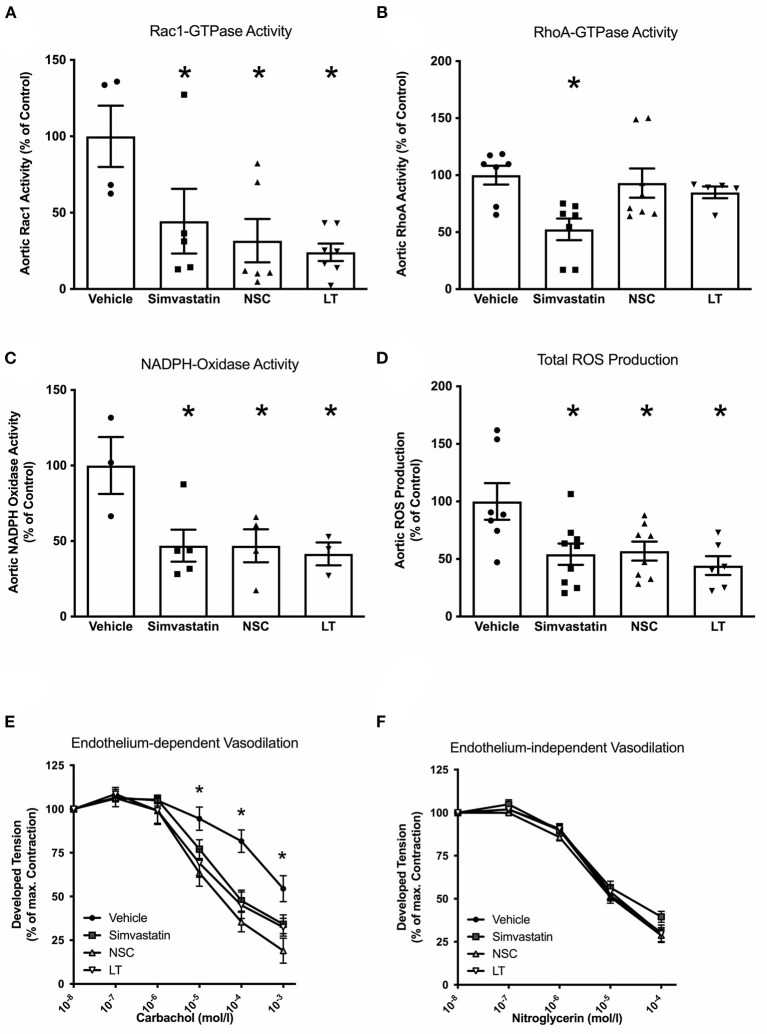
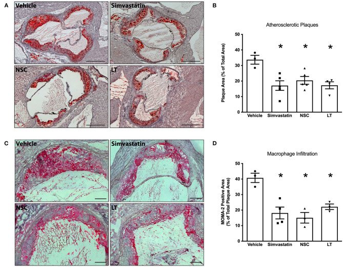
Aims: Oxidative stress and inflammation contribute to atherogenesis. Rac1 GTPase regulates pro-oxidant NADPH oxidase activity, reactive oxygen species (ROS) formation, actin cytoskeleton organization and monocyte adhesion. We investigated the vascular effects of pharmacological inhibition of Rac1 GTPase in mice. Methods and Results: We treated wild-type and apolipoprotein E-deficient (ApoE-/-) mice with Clostridium sordellii lethal toxin (LT), a Rac1 inhibitor, and assessed vascular oxidative stress, expression and activity of involved proteins, endothelial function, macrophage infiltration, and atherosclerosis development. LT-treated wild-type mice displayed decreased vascular NADPH oxidase activity and ROS production. Therapeutic LT doses had no impact on behavior, food intake, body weight, heart rate, blood pressure, vascular and myocardial function, differential blood count, and vascular permeability. ApoE-/- mice were fed a cholesterol-rich diet and were treated with LT or vehicle. LT treatment led to decreased aortic Rac1 GTPase activity, NADPH oxidase activity and ROS production, but had no impact on expression and membrane translocation of NADPH oxidase subunits and RhoA GTPase activity. LT-treated mice showed improved aortic endothelium-dependent vasodilation, attenuated atherosclerotic lesion formation and reduced macrophage infiltration of atherosclerotic plaques. Concomitant treatment of cholesterol-fed ApoE-/- mice with LT, the specific synthetic Rac1 inhibitor NSC 23766 or simvastatin comparably reduced aortic Rac1 activity, NADPH oxidase activity, oxidative stress, endothelial dysfunction, atherosclerosis development, and macrophage infiltration. Conclusions: These findings identify an important role of the small GTPase Rac1 in atherogenesis and provide a potential target for anti-atherosclerotic therapy.

Keywords: GTPases; Rac1; atherosclerosis; endothelial function; free radicals; oxidative stress.


Conflict of interest statement

The authors declare that the research was conducted in the absence of any commercial or financial relationships that could be construed as a potential conflict of interest.

Figures

Figure 1
Figure 1
Assessment of Clostridium sordellii lethal toxin (LT) toxicity. The main characteristic of LT toxicity is increased vascular permeability with perivascular edema. Representative HE stainings of heart and lung tissue from mice treated with vehicle or a therapeutic dose of LT for 4 weeks show no signs of tissue edema. In contrast, mice that received a lethal dose of LT display edema in the connective tissue surrounding larger arteries (arrow heads).
Figure 2
Figure 2
Long-term Rac1 inhibition, oxidative stress and atherosclerosis. (A) Aortic homogenates of ApoE−/− mice that were fed a cholesterol-rich diet for 7 weeks and were concomitantly treated with 0.1 μg LT/week via osmotic mini-pumps showed reduced Rac1 GTPase activity compared to vehicle-treated mice in a Rac1 GST-PAK pull-down assay (*p < 0.05, n = 4 per group). (B,C) LT-treated ApoE−/− mice displayed reduced aortic NADPH oxidase activity (B, *p < 0.05, n = 10 per group) and aortic ROS production (C, *p < 0.05, n = 10 per group), as measured by lucigenin-enhanced chemiluminescence and L-012 chemiluminescence, respectively. (D) Representative histological cross-sections of the aortic root stained with oil red O to display atherosclerotic plaques. LT-treated ApoE−/− mice showed decreased atherosclerotic plaque formation. (E) Quantification of atherosclerotic plaque formation, expressed as plaque area in percent of total area (*p < 0.05, n = 10 per group). Scale bars indicate 0.5 mm.
Figure 3
Figure 3
NADPH oxidase subunit expression. (A) Western blot analysis of NADPH oxidase subunit protein expression in cytosolic and membrane fractions of aortic homogenates of cholesterol-fed ApoE−/− mice treated with vehicle or LT. There was no difference in Rac1, Nox1, Nox2, p47-phox, and p67-phox expression. (B) Densitometric quantification of the Western analyses. Expression of genes-of-interest was normalized to housekeeping gene expression. These values were normalized to membrane protein expression of vehicle-treated mice (n = 2 per group). For each subunit, membrane expression is displayed on the left, cytosolic expression on the right. (C) Real-time PCR analysis of Nox4 and p22-phox subunit mRNA expression in aortic homogenates of cholesterol-fed ApoE−/− mice treated with vehicle or LT. Neither aortic Nox4 nor p22-phox expression was altered by LT treatment in these mice (n = 2–3 independent measurements (pooled from n=2-3 mice per sample) per group).
Figure 4
Figure 4
Vascular effects of different Rac1 inhibitors: GTPase activity, oxidative stress and endothelial function. To investigate the vascular effects of different Rac1 GTPase inhibitors, ApoE−/− mice were fed a cholesterol-rich diet for 7 weeks and were concomitantly treated with vehicle, LT (0.1 μg LT/week), NSC 23766 (10 mg/kg/d), or simvastatin (20 mg activated simvastatin/kg/d). All three compounds significantly inhibited aortic Rac1 GTPase activity (A, Rac1 GTPase activity G-LISA, *p < 0.05 vs. vehicle, n = 4–7 per group), but only simvastatin significantly decreased aortic rhoA GTPase activity (B, rhoA GTPase activity G-LISA, *p < 0.05 vs. vehicle, n = 5–8 per group). All three compounds significantly reduced aortic NADPH oxidase activity (C, lucigenin-enhanced chemiluminescence, *p < 0.05 vs. vehicle, n = 3–5 per group) and aortic ROS production (D, L-012 chemiluminescence, *p < 0.05 vs. vehicle, n = 6–9 per group) and significantly improved endothelium-dependent vasodilation (E, organ chamber experiments with isolated aortic segments, *p < 0.05 vs. vehicle, n = 5 per group) compared to vehicle-controls. Endothelium-independent vasodilation was not affected by the Rac1 inhibitors (F, n = 5 per group).
Figure 5
Figure 5
Vascular effects of different Rac1 inhibitors: atherosclerosis and macrophage infiltration. ApoE−/− mice were fed a cholesterol-rich diet for 7 weeks and were concomitantly treated with vehicle, LT (0.1 μg LT/week), NSC 23766 (10 mg/kg/d), or simvastatin (20 mg activated simvastatin/kg/d). All three compounds significantly reduced atherosclerotic plaque formation in the aortic root compared to vehicle-control. (A) Representative histological cross-sections. Scale bars indicate 0.5 mm. (B) Quantification of atherosclerotic plaque formation, expressed as plaque area in percent of total area (*p < 0.05 vs. vehicle, n = 3–5 per group). Furthermore, all three compounds significantly reduced macrophage infiltration of atherosclerotic plaques in these mice. (C) Representative MOMA-2 immunohistochemical staining using a monoclonal rat anti-mouse MOMA-2 antibody. Scale bars indicate 0.15 mm. (D) Quantification of macrophages infiltrating aortic atherosclerotic plaques (*p < 0.05 vs. vehicle, n = 3–4 per group).

References

    1. Darley-Usmar V, Halliwell B. Blood radicals: reactive nitrogen species, reactive oxygen species, transition metal ions, and the vascular system. Pharmaceut Res. (1996) 13:649–62. 10.1023/A:1016079012214 - DOI - PubMed
    1. Laursen JB, Rajagopalan S, Galis Z, Tarpey M, Freeman BA, Harrison DG. Role of superoxide in angiotensin II-induced but not catecholamine-induced hypertension. Circulation. (1997) 95:588–93. 10.1161/01.CIR.95.3.588 - DOI - PubMed
    1. Li P-F, Dietz R, von Harsdorf R. Reactive oxygen species induce apoptosis of vascular smooth muscle cell. Febs Lett. (1997) 404:249–52. 10.1016/S0014-5793(97)00093-8 - DOI - PubMed
    1. Burtenshaw D, Kitching M, Redmond EM, Megson IL, Cahill PA. Reactive Oxygen Species (ROS), intimal thickening, and subclinical atherosclerotic disease. Front Cardiovasc Med. (2019) 6:89. 10.3389/fcvm.2019.00089 - DOI - PMC - PubMed
    1. Nowak WN, Deng J, Ruan XZ, Xu Q. Reactive oxygen species generation and atherosclerosis. Arterioscler Thromb Vasc Biol. (2017) 37:e41–52. 10.1161/ATVBAHA.117.309228 - DOI - PubMed