. 2020 Nov:2020:10.1109/ismr48331.2020.9312927.
doi: 10.1109/ismr48331.2020.9312927. Epub 2021 Jan 11.

Improved Integrated Robotic Intraocular Snake

Affiliations

Improved Integrated Robotic Intraocular Snake

Makoto Jinno et al. Int Symp Med Robot. 2020 Nov.

Abstract

Retinal surgery can be performed only by surgeons possessing advanced surgical skills because of the small, confined intraocular space, and the restricted free motion of instruments in contact with the sclera. Snake-like robots could be essential for use in retinal surgery to overcome this problem. Such robots can approach from suitable directions and operate delicate tissues when performing retinal vein cannulation, epiretinal membrane peeling and so on. In this study, we propose an improved integrated robotic intraocular snake (I2RIS), which is a new version of our previous IRIS. This update focuses on the dexterous distal unit design and the drive unit design. The proposed dexterous distal unit consists of small elements with reduced contact stress. The proposed drive unit includes a new wire drive mechanism where the drive pulley is mounted at a right angle relative to the actuation direction (also, relative to the conventional direction). A geometric analysis and mechanical design show that the proposed drive mechanism is simpler and easier to assemble and yields higher accuracy than the conventional drive mechanism. Furthermore, considering clinical use, the instrument of the I2RIS is detachable from the motor unit for cleaning, sterilization, and attachment of various surgical tools. Weighing merely 31.3 g, the proposed mechanism is only one third of the weight of the conventional IRIS. The basic functions and effectiveness of the proposed mechanism are verified by experiments on 5:1 scaled-up models of the dexterous distal unit and actual-size models of the instrument and motor units.

Keywords: mechanism design; medical robot; microsurgery; retinal surgery.


Figures

Fig. 1.
Fig. 1.
Required specifications of dexterous distal unit [20].
Fig. 2.
Fig. 2.
Conceptual design of dexterous distal unit of IRIS: the unit is composed of 12 disk-like elements providing 2-DOF bending joints actuated by 4 wires. (a) overview; (b) three-view drawing of element (dimensions in mm) [20].
Fig. 3.
Fig. 3.
Element shape and wire hole positions: (a) conventional element; (b) conventional proposed element, which increases the contact surface (thereby reducing contact stress) by changing the hole positions; (c) compact proposed element, which is created by decreasing the dead space.
Fig. 4.
Fig. 4.
IRIS and I2RIS: (a) conventional element, (b) conventional proposed element, (c) compact proposed element.
Fig. 5.
Fig. 5.
Conventional wire drive mechanism: (a) rotation pulley type, (b) lead screw type. The push-pull wire displacements are equal to drive wire displacements. In the case of IRIS, the push-pull wire displacement is 0.216 mm for a 45° total bending of the dexterous distal unit.
Fig. 6.
Fig. 6.
Concept of the proposed wire drive mechanism and coordinate systems: (a) 3D drawing, (b) x-y plane 2D drawing. The drive pulley is mounted at a right angle relative to the actuation direction, unlike that in a conventional pulley drive mechanism.
Fig. 7.
Fig. 7.
Push-pull wire displacements: wire displacements are changed almost linearly. The maximum error of the linearity is 2.7% with respect to the fitting line at the drive pulley rotation angle of ±20°.
Fig. 8.
Fig. 8.
Difference of push-pull wire displacements: the difference is under 20 μm at the drive pulley rotation angle of ±20°.
Fig. 9.
Fig. 9.
3D drawing of drive wire and tension: (a) drive wire, (b) tension.
Fig. 10.
Fig. 10.
x-y plane 2D drawing of drive wire and tension: (a) drive wire, (b) tension.
Fig. 11.
Fig. 11.
Virtual radius: the virtual radius is about 1.2 mm. The proposed mechanism is similar to an approximately 1.2 mm pulley radius and conventional wire drive mechanism.
Fig. 12.
Fig. 12.
Instrument unit: (a) overview, (b) cross section.
Fig. 13.
Fig. 13.
Instrument and motor units: the instrument unit is detachable from the motor unit.
Fig. 14.
Fig. 14.
(a) Conventional IRIS [22], (b) proposed IRIS (I2RIS): the I2RIS is a quarter of the volume of the conventional IRIS.
Fig. 15.
Fig. 15.
Overview of scaled-up models of the instrument unit.
Fig. 16.
Fig. 16.
Bending motion ranges of 5:1 scaled-up models: (a) conventional element, Fig. 2(a); (b) conventional proposed element, which increases the contact surface (thereby reducing contact stress) by changing the hole positions, Fig. 2(b); (c) compact proposed element, which is created by decreasing the dead space, Fig. 2(c).
Fig. 17.
Fig. 17.
Overview of actual-size models of instrument and motor units: (a) attached state, (b) detached state.
Fig. 18.
Fig. 18.
Overview of experimental setup for motor drive operation.
Fig. 19.
Fig. 19.
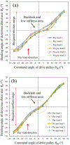
Motor drive experiment of actual-size model: command angle of drive pulley θin and bending angle of dexterous distal unit.
Fig. 20.
Fig. 20.
Command angle of drive pulley θin and bending angle of dexterous distal unit θo : (a) yaw axis, (b) pitch axis. The payload is 34 mN.

References

    1. Vander Poorten E, Riviere CN, Abbott JJ, Bergeles C, Nasseri MA, Kang JU, Sznitman R, Faridpooya K, and Iordachita I, 36 - Robotic Retinal Surgery, Handbook of Robotic and Image-Guided Surgery, Elsevier, 2019, pp. 627–672.
    1. Jensen PS, Grace KW, Attariwala R, Colgate JE, Glucksberg MR, “Toward robot-assisted vascular in the retina,” Graefe’s Arch Clin Exp Ophthalmol. Vol. 235, no. 11, pp.696–701. November. 1997. - PubMed
    1. Wei W, Popplewell C, Chang S, Fine HF, Simaan N, “Enabling technology for microvascular stenting in ophthalmic surgery,” Journal of Medical Devices, vol. 4, no. 1, March. 2010.
    1. Tanaka S, et al. “Quantitative assessment of manual and robotic microcannulation for eye surgery using new eye model,” hit J Med Robotics Comput Assist Surg, April.2014. - PubMed
    1. Fleming I, Balicki M, Koo J, Iordachita I, Mitchell B, Handa J, Hager G, Taylor R, “Cooperative robot assistant for retinal microsurgery,” Med. Image Comput. Comput. Assist. Interv, vol. 5242. 2008. pp.543–550. - PubMed

LinkOut - more resources