Review
. 2021 Oct 29;5(4):575-589.
doi: 10.1042/ETLS20210015.

Cell therapy for cartilage repair

Affiliations
Review

Cell therapy for cartilage repair

Charlotte H Hulme et al. Emerg Top Life Sci. .

Abstract

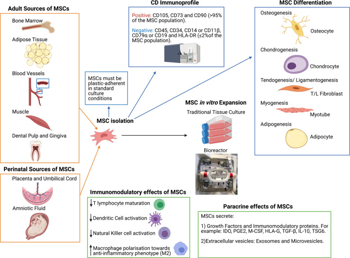
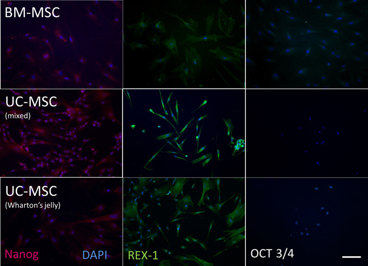
Regenerative medicine, using cells as therapeutic agents for the repair or regeneration of tissues and organs, offers great hope for the future of medicine. Cell therapy for treating defects in articular cartilage has been an exemplar of translating this technology to the clinic, but it is not without its challenges. These include applying regulations, which were designed for pharmaceutical agents, to living cells. In addition, using autologous cells as the therapeutic agent brings additional costs and logistical challenges compared with using allogeneic cells. The main cell types used in treating chondral or osteochondral defects in joints to date are chondrocytes and mesenchymal stromal cells derived from various sources such as bone marrow, adipose tissue or umbilical cord. This review discusses some of their biology and pre-clinical studies before describing the most pertinent clinical trials in this area.

Keywords: cartilage; cell therapy; chondrocytes; mesenchymal stem cell; osteoarthritis; regenerative medicine.


Conflict of interest statement

The authors declare that there are no competing interests associated with the manuscript.

Figures

Figure 1.
Figure 1.. Cell therapy repair of cartilage defects.
(AC) Representative 3-T magnetic resonance imaging (MRI) scans from a 35-year-old patient who underwent treatment with ACI. Proton density-weighted turbo spin-echo fat-suppressed (PD-TSE-FS) sequence MRI, showing (A) coronal and (B) sagittal view of the knee joint with an osteochondral defect (red arrow) prior to ACI treatment and (C) the treated defect 13 months post-ACI. Yellow arrow indicates normal healthy cartilage. D-F) Representative histological images of haematoxylin and eosin-stained sections of (D) normal, healthy articular cartilage, (E) fibrillated, degenerative cartilage and (F) repair tissue formed 12 months post-ACI. c, cartilage; b, bone. Scale bars = 500 µm.
Figure 2.
Figure 2.. Sources and characteristics of multipotent mesenchymal stromal cells (MSCs).
MSCs can be readily isolated from numerous adult and perinatal sources. The minimal criteria for MSC characterisation, published by the ISCT [35], states that MSCs must be plastic-adherent in the standard tissue culture conditions, demonstrate a specific CD immunoprofile as measure by flow cytometry (subsequently amended for adipose-derived MSCs [45]) and demonstrate a specific in vitro differentiation potential by differentiating down, osteogenic, adipogenic and chondrogenic lineages in vitro [35]. Specific stimuli can also promote MSCs to differentiate down myogenic and tenogenic lineages. Alongside traditional tissue culture for cell expansion, MSCs have been effectively up-scaled using bioreactors, thus enabling a switch from autologous to allogeneic multi-dose cell banking for therapeutic uses [59]. Evidence suggests that MSCs secrete large numbers of soluble and vesicle-bound growth factors and immunomodulatory proteins, which may not only have trophic effects on endogenous cells but also modulate the environment for repair [37]. (Created using Biorender.com.).
Figure 3.
Figure 3.. MSC expression of pluripotency markers.
The expression of pluripotency markers, Nanog, REX-1 and OCT 3/4, is more common on MSCs isolated from umbilical cords (either as a mixed population from all the whole cord (mixed) or from the Wharton's jelly) than those isolated from bone marrow (BM-MSCs). Scale bar represents 100 μm. (Reproduced from [43]).
Figure 4.
Figure 4.. Selected characteristics of animals used for cartilage repair models.
The most commonly used small animal models include the mouse, rat, guinea pig and rabbit, whilst typically the dog, sheep, goat, pig or horse are considered ‘large animal’ models. With numerous models currently available, choosing the most appropriate remains a challenge, although it is vital to note that a single model cannot encompass all of the extensive aspects involved in human cartilage repair [87]. Both small and large animals have their advantages and disadvantages for example; small animal models reach skeletal maturity faster, thereby reducing husbandry costs, experimental durations, drug and housing requirements. However, larger animals present with greater anatomical similarity in regards to the thickness of articular cartilage, joint size and biomechanics to humans. For example, in mice, the average articular cartilage thickness (mm) in the knee joint is ∼0.03 mm, whereas in horses it is ∼1.5–2.00 mm and in humans, it is ∼2.2–2.5 mm [87–91]. Both chondral and osteochondral defects of varying sizes are used in cartilage repair models; all known critical-size defects in the knee joint from the different animal species are displayed here [87,89,92]. (Created using Biorender.com.).

References

    1. Hunter, W.V.I. (1743) Of the structure and diseases of articu­lating cartilages. Philos. Trans. R. Soc. Lond. 42, 514–521 10.1098/rstl.1742.0079 - DOI
    1. Wang, Y., Ding, C., Wluka, A.E., Davis, S., Ebeling, P.R., Jones, G.et al. (2006) Factors affecting progression of knee cartilage defects in normal subjects over 2 years. Rheumatology 45, 79–84 10.1093/rheumatology/kei108 - DOI - PubMed
    1. Registry, N.J. (2020) 17th Annual Report 2020 https://reports.njrcentre.org.uk/Portals/0/PDFdownloads/NJR%2017th%20Ann...
    1. OARSI White Paper- OA as a Serious Disease | Osteoarthritis research society international (oarsi)
    1. Kraeutler, M.J., Belk, J.W., Purcell, J.M. and McCarty, E.C. (2018) Microfracture versus autologous chondrocyte implantation for articular cartilage lesions in the knee: a systematic review of 5-year outcomes. Am. J. Sports Med. 46, 995–999 10.1177/0363546517701912 - DOI - PubMed

Publication types

MeSH terms

LinkOut - more resources