Randomized Controlled Trial
. 2021 Dec;73(12):2206-2218.
doi: 10.1002/art.41953. Epub 2021 Nov 1.

The Interleukin-1 Receptor-Associated Kinase 4 Inhibitor PF-06650833 Blocks Inflammation in Preclinical Models of Rheumatic Disease and in Humans Enrolled in a Randomized Clinical Trial

Affiliations
Randomized Controlled Trial

The Interleukin-1 Receptor-Associated Kinase 4 Inhibitor PF-06650833 Blocks Inflammation in Preclinical Models of Rheumatic Disease and in Humans Enrolled in a Randomized Clinical Trial

Aaron Winkler et al. Arthritis Rheumatol. 2021 Dec.

Abstract

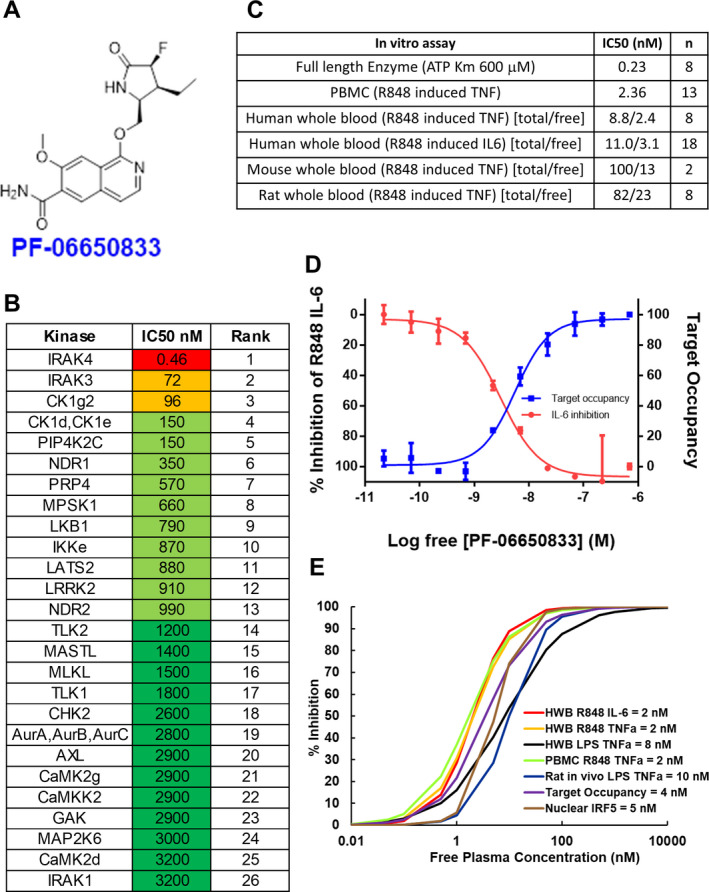
Objective: To investigate the role of PF-06650833, a highly potent and selective small-molecule inhibitor of interleukin-1-associated kinase 4 (IRAK4), in autoimmune pathophysiology in vitro, in vivo, and in the clinical setting.

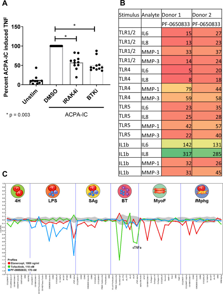
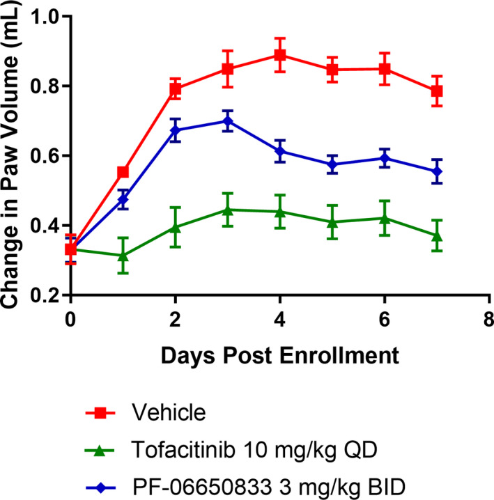
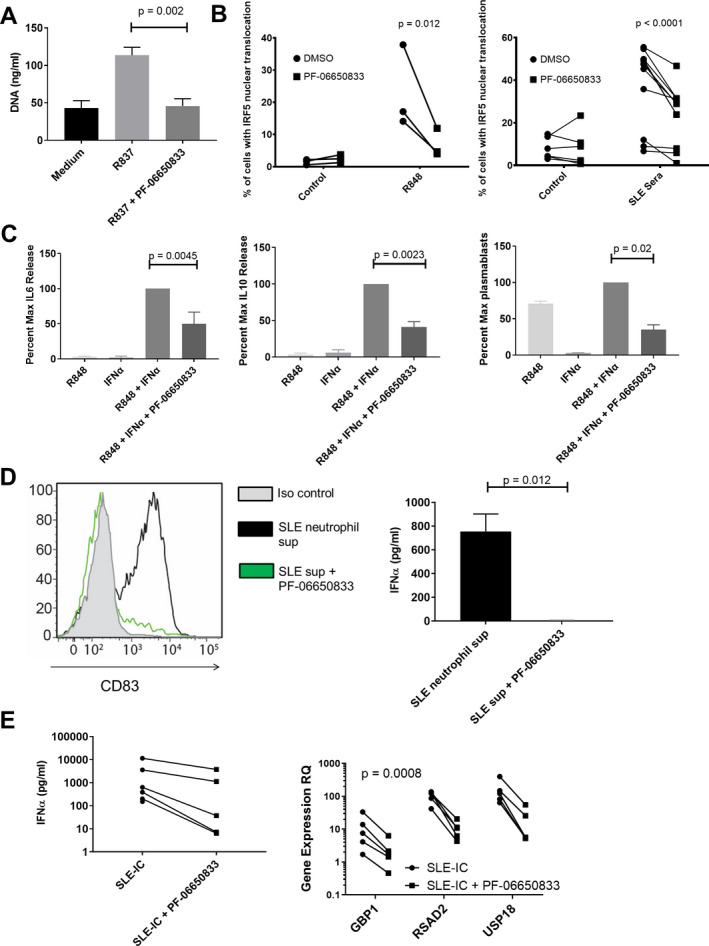
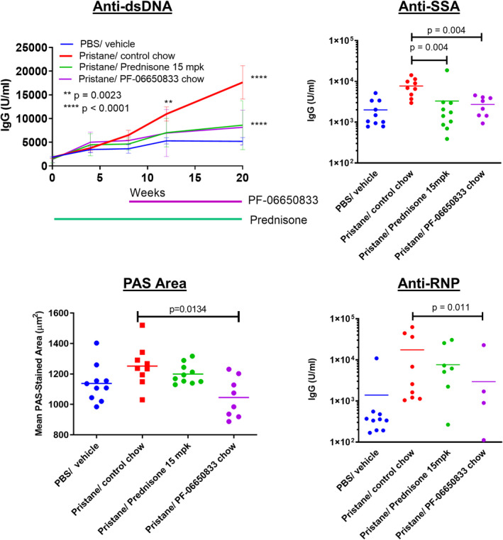
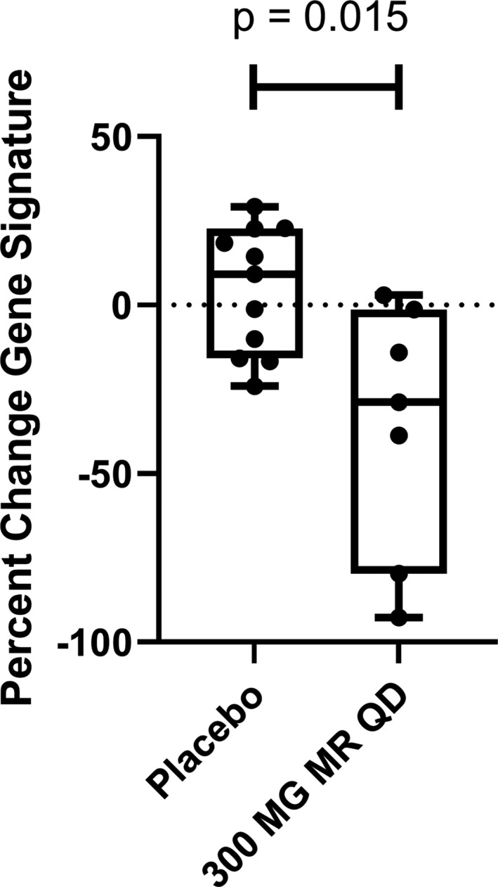
Methods: Rheumatoid arthritis (RA) inflammatory pathophysiology was modeled in vitro through 1) stimulation of primary human macrophages with anti-citrullinated protein antibody immune complexes (ICs), 2) RA fibroblast-like synoviocyte (FLS) cultures stimulated with Toll-like receptor (TLR) ligands, as well as 3) additional human primary cell cocultures exposed to inflammatory stimuli. Systemic lupus erythematosus (SLE) pathophysiology was simulated in human neutrophils, dendritic cells, B cells, and peripheral blood mononuclear cells stimulated with TLR ligands and SLE patient ICs. PF-06650833 was evaluated in vivo in the rat collagen-induced arthritis (CIA) model and the mouse pristane-induced and MRL/lpr models of lupus. Finally, RNA sequencing data generated with whole blood samples from a phase I multiple-ascending-dose clinical trial of PF-06650833 were used to test in vivo human pharmacology.

Results: In vitro, PF-06650833 inhibited human primary cell inflammatory responses to physiologically relevant stimuli generated with RA and SLE patient plasma. In vivo, PF-06650833 reduced circulating autoantibody levels in the pristane-induced and MRL/lpr murine models of lupus and protected against CIA in rats. In a phase I clinical trial (NCT02485769), PF-06650833 demonstrated in vivo pharmacologic action pertinent to SLE by reducing whole blood interferon gene signature expression in healthy volunteers.

Conclusion: These data demonstrate that inhibition of IRAK4 kinase activity can reduce levels of inflammation markers in humans and provide confidence in the rationale for clinical development of IRAK4 inhibitors for rheumatologic indications.


Figures

Figure 1
Figure 1
Pharmacologic properties of PF‐06650833, an interleukin‐1–associated kinase 4 (IRAK4) inhibitor. A, Structure of PF‐06650833. B, Selectivity of PF‐06650833 in an ActivX ATP occupancy assay using THP‐1 lysates. The top 26 of >200+ kinases are shown. Fifty percent inhibition concentrations (IC50) were determined using a 5‐point dose‐response curve (full data set shown in Supplementary Table 1, on the Arthritis & Rheumatology website at http://onlinelibrary.wiley.com/doi/10.1002/art.41953/abstract). C, Potency of PF‐06650833 in enzyme and cell‐based assays. IC50 values for enzyme and peripheral blood mononuclear cell (PBMC) assays are a single value, whereas whole blood (wb) assays for human, mouse, and rat are denoted as total concentration of compound over the non–protein‐bound (free [f]) concentration of compound, calculated as (wb IC50 × [fu/(B/P)] = IC50 free, where fraction unbound [fu] = 0.22 and blood/plasma ratio [B/P] = 0.91). D, Demonstration of the relationship between free drug concentration, IRAK4 ATP binding site occupancy, and inhibition of downstream biology by PF‐06650833 in a whole blood sample from a patient with systemic lupus erythematosus. Values are the mean ± SEM. E, Use of 50% maximum response concentration data from each preclinical assay to determine that 100 nM was the target free compound concentration required to maintain >90% IRAK4 inhibition across species and assays. CK1g2 = casein kinase 1 gamma 2; PIP4K2C = phosphatidylinositol‐5‐phosphate 4‐kinase 2C; NDR1 = nuclear Dbf2‐related kinase 1; PRP4 = pre–mRNA processing factor 4; MPSK1 = myristoylated and palmitoylated serine/threonine kinase 1; LKB1 = liver kinase B1; LATS2 = large tumor suppressor kinase 2; LRRK2 = leucine‐rich repeat kinase 2; TLK2 = tousled‐like kinase 2; MASTL = microtubule‐associated serine/threonine kinase–like; MLKL = mixed‐lineage kinase domain–like; CHK2 = checkpoint kinase 2; AurA = aurora A kinase; AXL = AXL receptor tyrosine kinase; CAMK2g = Ca2+/calmodulin‐dependent protein kinase 2g; GAK = cyclin G–associated kinase; TNF = tumor necrosis factor; IL‐6 = interleukin‐6; HWB = human whole blood; LPS = lipopolysaccharide; IRF‐5 = interferon regulatory factor 5.
Figure 2
Figure 2
PF‐06650833 inhibits rheumatoid arthritis (RA) pathophysiology. A, Human macrophages were exposed to anti–citrullinated protein antibody (ACPA) immune complexes (ICs) formed with cyclic citrullinated peptide–positive RA sera in the presence or absence of 100 nM interleukin‐1–associated kinase 4 inhibitor (IRAK4i) (PF‐06650833) or 100 nM Bruton’s tyrosine kinase inhibitor (BTKi) (PF‐303). Supernatants were analyzed by enzyme‐linked immunosorbent assay for induction of tumor necrosis factor (TNF). Values are the percent in relation to DMSO treatment (set at 100). Symbols represent individual subjects (n = 11 per group); bars show the mean. B, Human RA fibroblast‐like synoviocytes were stimulated with 10 μg/ml Pam3Cys (Toll‐like receptor 1/2 [TLR‐1/2]), 10 ng/ml lipopolysaccharide (LPS) (TLR‐4), 100 ng/ml flagellin (TLR‐5), or 0.1 ng/ml interleukin‐1β (IL‐1β) in the presence or absence of 100 nM PF‐06650833. Cytokine and matrix metalloproteinase (MMP) content in supernatants was assessed by Meso Scale Discovery assay. Values are the percent of those observed with vehicle control. C, Compounds at the noted concentrations were profiled with DiscoverRx on the BioMAP platform. Data represent the log ratio of values in compound‐treated samples to controls, with negative values indicating inhibition and points outside of the gray shading demonstrating a statistically significant difference based on assay variability. Each point on the x‐axis represents the result of a different end point in each assay system: venular endothelial cells (4H), peripheral blood mononuclear cells (PBMCs) plus venular endothelial cells stimulated with LPS (LPS), PBMCs plus venular endothelial cells stimulated with superantigen (SAg), B cells plus PBMCs (BT), lung fibroblasts (MyoF), and macrophages plus venular endothelial cells (/Mphg). Unstim = unstimulated; MCP‐1 = monocyte chemotactic factor 1; VCAM‐1 = vascular cell adhesin molecule 1; uPAR = urokinase‐type plasminogen activator receptor; SRB = sulforhodamine B staining; VEGFR‐2 = vascular endothelial growth factor receptor 2; M‐CSF = macrophage colony‐stimulating factor; sPGE2 = soluble prostaglandin E2; MIG = monokine induced by interferon‐γ; bFGF = basic fibroblast growth factor; PAI‐1 = plasminogen activator inhibitor 1; TIMP‐1 = tissue inhibitor of metalloproteinases 1.
Figure 3
Figure 3
PF‐06650833 is efficacious in rat collagen‐induced arthritis (CIA). Rats with CIA were treated for 7 days with PF‐06650833 3 mg/kg twice daily (bid), tofacitinib 10 mg/kg daily (qd), or vehicle, and paw volume was measured daily. Data are from a representative experiment (of 3 experiments performed). Values are the mean ± SEM.
Figure 4
Figure 4
PF‐06650833 inhibits pathophysiology in human systemic lupus erythematosus (SLE). A, DNA release was measured in neutrophil supernatant (sup) following exposure to R837 in the presence or absence of 100 nM PF‐06650833. Values are the mean ± SEM (n = 3 donors). B, Peripheral blood mononuclear cells (PBMCs) were exposed to either R848 or SLE sera in the presence or absence of 100 nM PF‐06650833, incubated for 2 hours at 37°C, and then interferon regulatory factor 5 (IRF‐5) nuclear translocation was analyzed by Amnis imaging cytometry. C, B cells were exposed to interferon‐α (IFNα) plus R848 in the presence or absence of 100 nM PF‐06650833. Supernatants were harvested at 24 hours to assess interleukin‐6 (IL‐6) release and at 72 hours to assess IL‐10 release. Plasmablasts were quantified by flow cytometry after 6 days. D, Neutrophils were isolated from SLE patients and NETosis induced with R837. Supernatants were used to stimulate plasmacytoid dendritic cells for 24 hours in the presence or absence of 200 nM PF‐06650833 prior to quantification of CD83 induction by flow cytometry (n = 5) or IFNα release by enzyme‐linked immunosorbent assay (ELISA) (mean ± SEM; n = 3). Iso = isotype. E, PBMCs were exposed to SLE immune complexes (ICs) in the presence or absence of 100 nM PF‐06650833. After 24 hours, supernatant was analyzed by ELISA for IFNα, and IFN‐induced gene expression in cells was measured by quantitative reverse transcription–polymerase chain reaction. RQ = relative quantification.
Figure 5
Figure 5
Effect of PF‐06650833 on pristane‐induced systemic lupus erythematosus. PF‐06650833 was administered to BALB/c mice by chow dosing during weeks 8–20 following pristane administration. Anti–double‐stranded DNA (anti‐dsDNA) was quantified by enzyme‐linked immunosorbent assay at weeks 4, 8, 12, and 20, and the mean ± SEM value at each of these time points is shown. Additional autoantibody titers (anti‐SSA, anti‐RNP) were determined, and immunohistochemical assessments (periodic acid–Schiff [PAS] staining) were performed, at week 20. Data are from a representative experiment (of 2 experiments performed). Symbols represent individual subjects; bars show the mean. PBS = phosphate buffered saline; mpk = mg/kg.
Figure 6
Figure 6
PF‐06650833 inhibits type I interferon signature in vivo in humans. Moderate‐release (MR) PF‐06650833 (300 mg/day [qd]) (n = 7) or placebo (n = 11) was administered for 14 days in a phase I multiple‐ascending‐dose trial in healthy human volunteer subjects. Whole blood was collected in a PAXgene tube on day 0 prior to administration of the first dose and on day 14 prior to administration of the last dose, RNA extracted, and a composite gene signature calculated. The percent change in the composite gene signature for each individual participant between the 2 time points is shown. Each box represents the 25th to 75th percentiles. Lines inside the boxes represent the median. Lines outside the boxes represent the full range of values. Symbols represent individual patients.

Comment in

References

    1. Malmström V, Catrina AI, Klareskog L. The immunopathogenesis of seropositive rheumatoid arthritis: from triggering to targeting [review]. Nat Rev Immunol 2016;17:60–75. - PubMed
    1. Smolen JS, Aletaha D, Barton A, Burmester GR, Emery P, Firestein GS, et al. Rheumatoid arthritis [review]. Nat Rev Dis Primers 2018;4:18001. - PubMed
    1. Aletaha D, Smolen JS. Diagnosis and management of rheumatoid arthritis: a review. JAMA 2018;320:1360–72. - PubMed
    1. Smolen JS, Aletaha D. Rheumatoid arthritis therapy reappraisal: strategies, opportunities and challenges [review]. Nat Rev Rheumatol 2015;11:276–89. - PubMed
    1. Dörner T, Furie R. Novel paradigms in systemic lupus erythematosus [review]. Lancet 2019;393:2344–58. - PubMed

Publication types

MeSH terms

Substances

Associated data