. 2021 Nov;112(11):4580-4592.
doi: 10.1111/cas.15115. Epub 2021 Sep 4.

BEX2 is required for maintaining dormant cancer stem cell in hepatocellular carcinoma

Affiliations

BEX2 is required for maintaining dormant cancer stem cell in hepatocellular carcinoma

Daisuke Fukushi et al. Cancer Sci. 2021 Nov.

Abstract

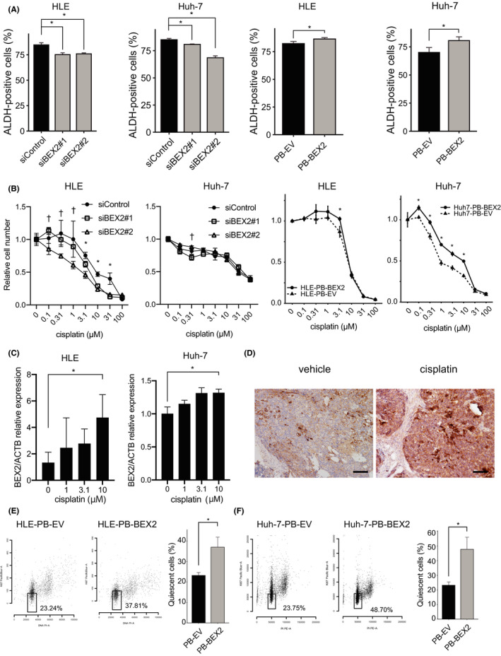
Cancer stem cells (CSCs) are responsible for therapy resistance and share several properties with normal stem cells. Here, we show that brain-expressed X-linked gene 2 (BEX2), which is essential for dormant CSCs in cholangiocarcinoma, is highly expressed in human hepatocellular carcinoma (HCC) lesions compared with the adjacent normal lesions and that in 41 HCC cases the BEX2high expression group is correlated with a poor prognosis. BEX2 localizes to Ki67-negative (nonproliferative) cancer cells in HCC tissues and is highly expressed in the dormant fraction of HCC cell lines. Knockdown of BEX2 attenuates CSC phenotypes, including sphere formation ability and aldefluor activity, and BEX2 overexpression enhances these phenotypes. Moreover, BEX2 knockdown increases cisplatin sensitivity, and BEX2 expression is induced by cisplatin treatment. Taken together, these data suggest that BEX2 induces dormant CSC properties and affects the prognosis of patients with HCC.

Keywords: BEX2; cancer stem cell; dormant; hepatocellular carcinoma; prognosis.


Conflict of interest statement

There are no financial conflicts of interest.

Figures

FIGURE 1
FIGURE 1
BEX2 is highly expressed in human hepatocellular carcinoma (HCC). A, Representative image of hematoxylin and eosin staining (left panel) and immunohistochemistry of BEX2 (right panel) in surgical resection specimens of human HCC. Brown staining indicates BEX2‐positive cells. Bar, 1 mm. B, Summary of BEX2 positivity in immunohistochemistry using the Tohoku Medical and Pharmaceutical University (TMPU) dataset. The intensity of BEX2 in BEX2high cases (17/41) was measured using ImageJ. The data are shown as the mean intensity of the tumor divided by adjacent normal area. Horizontal solid lines indicate mean ± standard deviation. C, BEX2 mRNA expression levels in The Cancer Genome Atlas Liver Hepatocellular Carcinoma (TCGA‐LIHC) dataset. A total of 50 cancer cases were collected, and the values were calculated as fragments per kilobase of transcript per million (FPKM) in tumor minus FPKM in the corresponding normal tissue. D, E, BEX2 mRNA expression levels from GSE25097 or GSE76427 from the Gene Expression Omnibus (GEO). Number of cases: GSE25097: 6 (healthy), 40 (cirrhotic), 243 (adjacent nontumor), and 267 (tumor); GSE76427: 52 (adjacent nontumor) and 115 (tumor). Red lines indicate mean value. F, Kaplan‐Meier analysis for relapse‐free survival of the BEX2high (17 cases) and BEX2low (24 cases) groups. P‐value was calculated using the log‐rank test. G, Kaplan‐Meier analysis for overall survival of the BEX2high (289 cases) and BEX2low (73 cases) groups (≥0.12 and <0.12 FPKM, respectively) in the TCGA dataset. P‐value was calculated using the log‐rank test
FIGURE 2
FIGURE 2
BEX2‐positive cells are nonproliferative. A, Surgical resection specimens in hepatocellular carcinoma (HCC) were stained with anti‐BEX2 and anti‐Ki67 antibodies, and the EGFP‐ and Ki67‐positive cells were counted. Arrowheads indicate BEX2‐ or Ki67‐positive cancer cells. Bar, 50 µm. B, HLE‐2k‐EGFP cells were stained with anti‐Ki67 antibody, and the EGFP‐ and Ki67‐positive cells were counted. A total of 11 randomly selected areas were analyzed (right panel, P < .00001, Chi‐square test). Bar, 50 µm. C, Expression patterns of BEX2 and MCM7 in HCC cells. A single‐cell RNAseq database (GSE103866, including Huh‐1, Huh‐7, and patient‐derived circulating tumor cells) was analyzed using a Seurat package
FIGURE 3
FIGURE 3
BEX2 is predominantly expressed in dormant cancer cells. A, Representative data of flow cytometry analysis of HLE cells. HLE cells were stained with pyronin Y and Hoechst33342. Sorted fractions (pyronin high and low) were reanalyzed by flow cytometry. B, Real‐time PCR of sorted HLE and Huh‐7 cells. The cells were sorted and divided into two groups, pyronin Y‐high and pyronin Y‐low, and total RNA was extracted. C, Cell cycle analysis of HLE and Huh‐7 cells grown under serum starvation conditions (24 h for HLE and 72 h for Huh‐7 cells). The open square indicates dormant cell fraction. PI, propidium iodide. D, Real‐time PCR analysis of BEX2 mRNA expression under starvation condition. The expression was normalized using ACTB (n = 3, *P < .05)
FIGURE 4
FIGURE 4
BEX2 is required for spheroid formation. A, Western blotting analysis of BEX2 expression levels in BEX2‐knockdown HLE and Huh‐7 cells and in BEX2‐overexpressing HLE and Huh‐7 cells. EV, empty vector. B, Cell proliferation capability determined by MTT assay in BEX2‐knockdown and control cells (n = 5, *P < .05 [siControl vs. siBEX2#1 and siControl vs. siBEX2#2]). C, Real‐time PCR analysis of the mRNA expression of BEX2 and SOX2. The cells were cultured in normal or sphere‐forming medium for 3 d and harvested (n = 3, *P < .05). D, Sphere formation assay after siRNA‐mediated depletion or overexpression of BEX2 in HLE and Huh‐7 cells. Total area of spheroid cells was measured using ImageJ (n = 5, *P < .05). E, Organoid formation assay in Huh‐7‐PB‐EV and Huh7‐PB‐BEX2 cells. Total area of spheroid cells was measured using ImageJ (n = 5, *P < .05)
FIGURE 5
FIGURE 5
BEX2 plays roles for aldehyde dehydrogenase (ALDH) activity, resistance to CDDP, and quiescent phase. A, ALDH activity was determined by aldefluor assay (n = 3). *P < .05. B, MTT assay analysis to measure cell viability in cells treated with cisplatin at the indicated concentration (n = 5). The duration of cisplatin treatment was 4 d (HLE, siRNA), 2 d (Huh‐7, siRNA), and 6 d (BEX2‐overexpressing cells). *P < .05 in siControl vs. siBEX2#1 and siControl vs. siBEX2#2; PB‐EV vs. PB‐BEX2. P < .05 in siControl vs. siBEX2#2. C, The BEX2 mRNA expression was also determined using real‐time PCR. The expression was normalized using ACTB. *P < .05. D, Huh‐7 cells were xenografted into NOG mice, and when the tumor was palpable, cisplatin was injected once intraperitoneally. After 7 d, mice were sacrificed and the tumors were fixed and stained using anti‐BEX2 antibody. Bar, 100 μm. E, HLE‐PB‐EV and HLE‐PB‐BEX2 cells were starved (0% FBS) for 48 h, and the quiescent population was measured using flow cytometry. F, Huh‐7‐PB‐EV and Huh‐7‐PB‐BEX2 cells were starved (0% FBS) for 48 h, and the quiescent population was measured using flow cytometry

References

    1. Forner A, Reig M, Bruix J. Hepatocellular carcinoma. Lancet. 2018;391(10127):1301‐1314. - PubMed
    1. Global Cancer Observatory [Internet]. [cited 2020 Aug 17]. Available from: https://gco.iarc.fr/
    1. Wang T, Shigdar S, Gantier MP, et al. Cancer stem cell targeted therapy: progress amid controversies. Oncotarget. 2015;6(42):44191‐44206. - PMC - PubMed
    1. Peiris‐Pagès M, Martinez‐Outschoorn UE, Pestell RG, Sotgia F, Lisanti MP. Cancer stem cell metabolism. Breast Cancer Res. 2016;18(1):55. - PMC - PubMed
    1. Yang ZF, Ho DW, Ng MN, et al. Significance of CD90+ cancer stem cells in human liver cancer. Cancer Cell. 2008;13(2):153‐166. - PubMed

MeSH terms