. 2022 Jan;22(1):28-45.
doi: 10.1111/ajt.16809. Epub 2021 Sep 20.

Pig-to-baboon lung xenotransplantation: Extended survival with targeted genetic modifications and pharmacologic treatments

Affiliations

Pig-to-baboon lung xenotransplantation: Extended survival with targeted genetic modifications and pharmacologic treatments

Lars Burdorf et al. Am J Transplant. 2022 Jan.

Abstract

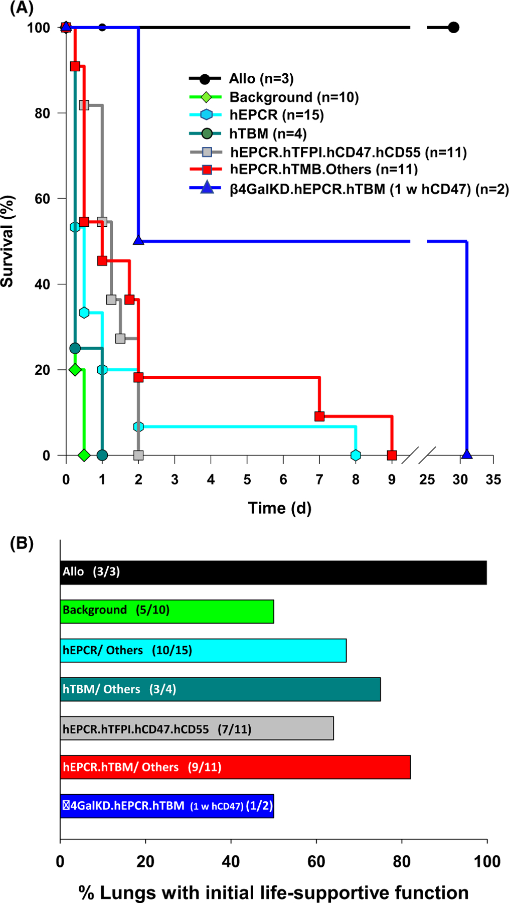
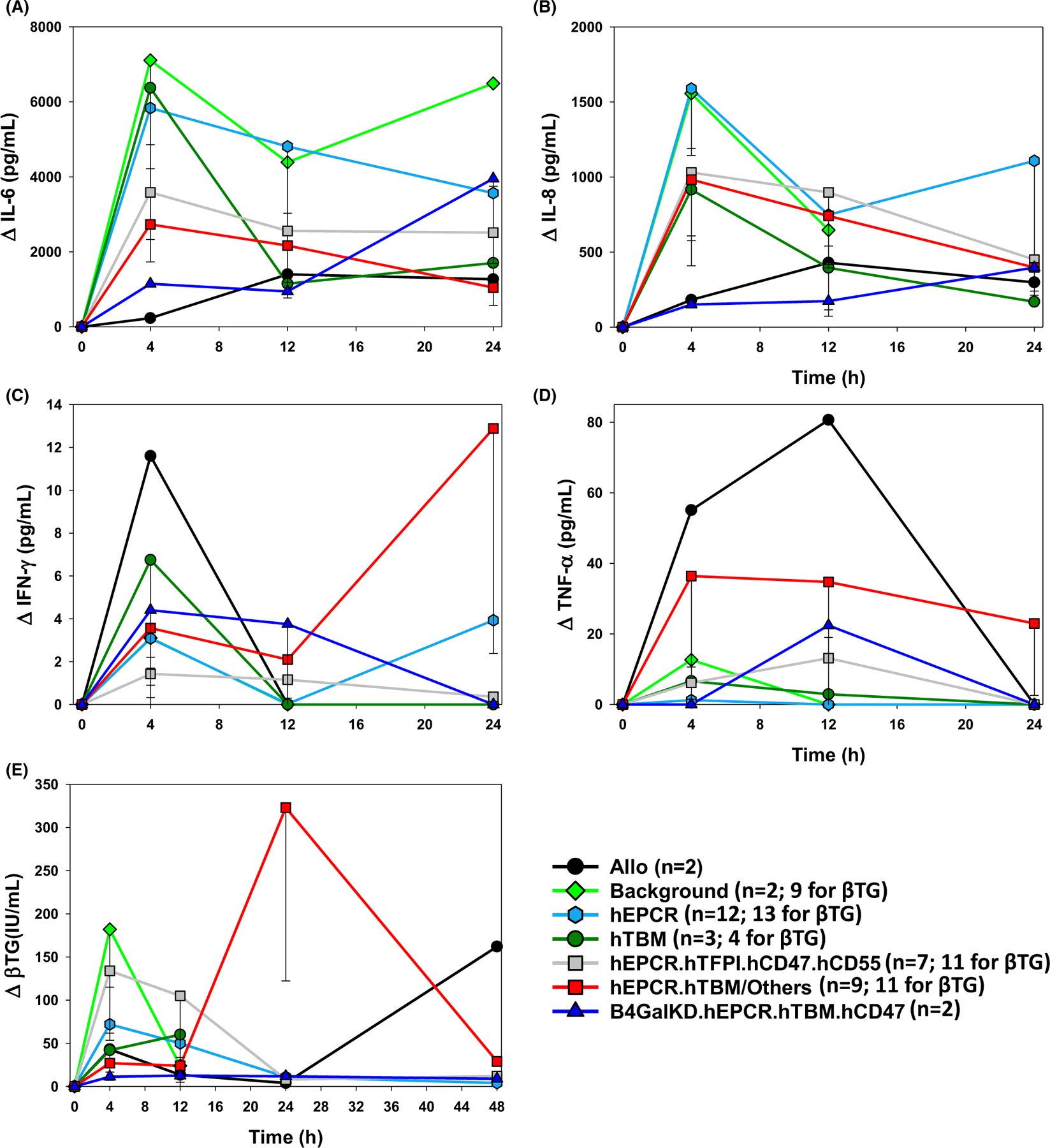
Galactosyl transferase knock-out pig lungs fail rapidly in baboons. Based on previously identified lung xenograft injury mechanisms, additional expression of human complement and coagulation pathway regulatory proteins, anti-inflammatory enzymes and self-recognition receptors, and knock-down of the β4Gal xenoantigen were tested in various combinations. Transient life-supporting GalTKO.hCD46 lung function was consistently observed in association with either hEPCR (n = 15), hTBM (n = 4), or hEPCR.hTFPI (n = 11), but the loss of vascular barrier function in the xenograft and systemic inflammation in the recipient typically occurred within 24 h. Co-expression of hEPCR and hTBM (n = 11) and additionally blocking multiple pro-inflammatory innate and adaptive immune mechanisms was more consistently associated with survival >1 day, with one recipient surviving for 31 days. Combining targeted genetic modifications to the lung xenograft with selective innate and adaptive immune suppression enables prolonged initial life-supporting lung function and extends lung xenograft recipient survival, and illustrates residual barriers and candidate treatment strategies that may enable the clinical application of other organ xenografts.

Keywords: animal models: nonhuman primate; basic (laboratory) research/science; genetics; graft survival; immunobiology; lung transplantation/pulmonology; translational research/science; xenoantibody; xenotransplantation.


Conflict of interest statement

DISCLOSURE

The authors of this manuscript have no conflicts of interest to disclose as described by the American Journal of Transplantation.

Figures

FIGURE 1
FIGURE 1
Lung xenograft recipient clinical outcomes. Survival of GalTKO.hCD46 lung xenograft recipients (A), grouped by coagulation pathway regulatory gene expression phenotype, categorized as detailed in Table 1 and Table S1. Survival to 24 h and beyond was observed more frequently with expression of hEPCR with either hTFPI or hTMB relative to hTMB or hEPCR alone. Occasional survivors beyond 2 days were associated with a variety of treatments designed to modulate inflammation as described in the text and Table S2. The incidence (B) and duration (not illustrated) of life-supporting lung function was increased with lungs expressing one or more thromboregulatory genes
FIGURE 2
FIGURE 2
Representative findings after lung xenotransplantation. Chest radiograph illustrating findings associated with interstitial lung edema accumulation (A) in a representative left lung xenograft (ILB18) failing within 12–24 h due to loss of vascular barrier function, manifesting in its late stages as tracheal flooding with edema fluid visualized through a flexible bronchoscope (B). In contrast, the lung parenchyma appears normal (C) in association with life-supporting left lung function in ILB45, which exhibited minimal postoperative interstitial changes in the mid-lung zone (D). Graft opacification after 7 days (not shown) correlated with detection of increasing titers of anti-pig antibody prior to euthanasia for deteriorating clinical condition on day 9
FIGURE 3
FIGURE 3
ILB64 chest x-rays. In the recipient with 31-day survival, partial graft consolidation progressed between day 1 (Figure 3A) and day 3 (Figure 3B) without TE on bronchoscopy or recipient respiratory distress. Left upper lung zone aeration improved by day 14 (Figure 3C), with additional partial re-expansion of the left lower lung field by day 28 (Figure 3D)
FIGURE 4
FIGURE 4
Cytokine and βTG elaboration by lung xenograft phenotype. Expressions of IL-6, IL-8, TNFα, IFNγ, and βTG were measured by Luminex at intervals over the first 12–48 h in an arbitrarily selected subset of experiments and expressed as mean with standard error of the mean. Prolific elaboration of IL-6 (A) during the first 12 hours after GalTKO.hCD46 lung xenotransplantion was attenuated in association with the expression of hEPCR with hTFPI or hTBM and with β4GalKD.hEPCR.hTBM, but not with hTBM or hEPCR alone, whereas IL-8 was lower only with allografts and with β4GalKD.hEPCR.hTBM xenografts (B). TNFα and IFNγ were highest in 2 lung allografts and did not vary statistically significantly in association with the combinations of thromboregulatory genes and drug treatments tested here (B,C). βTG elaboration 4 h after transplant was lower, and similar to lung allografts, in association with hTBM expression alone (42 ± 17 IU/ml; n too small for p-value calculation) or with hEPCR (27 ± 10 IU/ml; p = .035) relative to high levels with GalTKO.hCD46 background genetics (182 ± 67 IU/ml), while βTG elevations were intermediate with hEPCR (72 ± 19 IU/ml; p = .08). Of note, except for TNFα, pro-inflammatory cytokine and βTG elaboration were generally lowest in association with β4GalKD.hEPCR.hTBM lung xenografts, including ILB64 (31 d)
FIGURE 5
FIGURE 5
Anti-non-Gal antibody titers after lung xenotransplantation. Levels of baboon-anti-pig IgM (A) and IgG (B) antibody were measured by flow cytometry in the serum or plasma of four baboons that exhibited graft survival ≥7 days using a GalTKO endothelial cell line, expressed as a fold increase in MFI relative to pretransplant serum. Initial decline in IgM and IgG titers presumably reflected adsorption of preformed non-Gal anti-pig antibody to the lung xenograft. In association with calcineurin-based IS (ILB28), increased IgM and IgG were detected on POD 7 around the time of euthanasia for graft failure on POD 8. In three animals treated with αCD40, IgM rise was appreciated by day 7–8, but IgG elaboration appeared to be attenuated, consistent with prevention of class-switching that would be expected in association with effective blockade of the CD40/CD154 costimulatory pathway
FIGURE 6
FIGURE 6
CBC data by lung phenotype after lung xenotransplantation. Platelet (A), neutrophil (B), and monocyte (C) counts as well as hemoglobin levels (D) during intervals after lung transplantation (nadir within the first 4 h and between 4 and 8 h; and peak at 12, 24, and 48 h) are expressed as a percent of pre-transplant (t = 0). Absolute counts and calculations are shown in Table S2
FIGURE 7
FIGURE 7
Representative lung histology. Lung samples obtained within hours after implantation (A, B) or at xenograft explant and recipient euthanasia (C–F) were fixed, processed, and stained as described in in vitro methods. Alveolar septal cellularity and capillaries engorged by red blood cells were consistent findings in failing lung xenografts (Figure C–E) and to a lesser degree in the native right lung at experimental termination, but absent in normal baboon (Figure A) or pig lung (Figure B). When experiments were terminated due to high pulmonary vascular resistance in the transplant, lung xenograft architecture was otherwise minimally perturbed (Figure C), whereas proteinaceous pulmonary edema and alveolar hemorrhage (Figure D) became prominent when tracheal flooding due to loss of barrier function was observed. Lung endothelium activation with prominent endothelial cell nuclei and adherent leukocytes (Figure E) was prominent in pig pulmonary arterial and arteriolar vessels; both white (presumed platelet-rich fibrin aggregates) and red thrombi (clots with entrapped erythrocytes) were identified more commonly in lungs lacking thromboregulatory gene expression (Figure F)

Similar articles

Cited by

References

    1. Kim SC, Mathews DV, Breeden CP, et al. Long-term survival of pig-to-rhesus macaque renal xenografts is dependent on CD4 T cell depletion. Am J Transplant 2019;19(8):2174–2185. - PMC - PubMed
    1. Längin M, Mayr T, Reichart B, et al. Consistent success in life-supporting porcine cardiac xenotransplantation. Nature 2018;564(7736):430–433. - PubMed
    1. Nguyen BN, Azimzadeh AM, Zhang T, et al. Life-supporting function of genetically modified swine lungs in baboons. J Thorac Cardiovasc Surg 2007;133(5):1354–1363. - PubMed
    1. Burdorf L, Azimzadeh AM, Pierson RN 3rd. Progress and challenges in lung xenotransplantation: an update. Curr Opin Organ Transplant 2018;23(6):621–627. - PubMed
    1. Nguyen BN, Azimzadeh AM, Schroeder C, et al. Absence of Gal epitope prolongs survival of swine lungs in an ex vivo model of hyperacute rejection. Xenotransplant 2011;18(2):94–107. - PMC - PubMed

Publication types