. 2021 Oct 15;89(11):e0025621.
doi: 10.1128/IAI.00256-21. Epub 2021 Aug 16.

NanI Sialidase Contributes to the Growth and Adherence of Clostridium perfringens Type F Strain F4969 in the Presence of Adherent Mucus

Affiliations

NanI Sialidase Contributes to the Growth and Adherence of Clostridium perfringens Type F Strain F4969 in the Presence of Adherent Mucus

Jihong Li et al. Infect Immun. .

Abstract

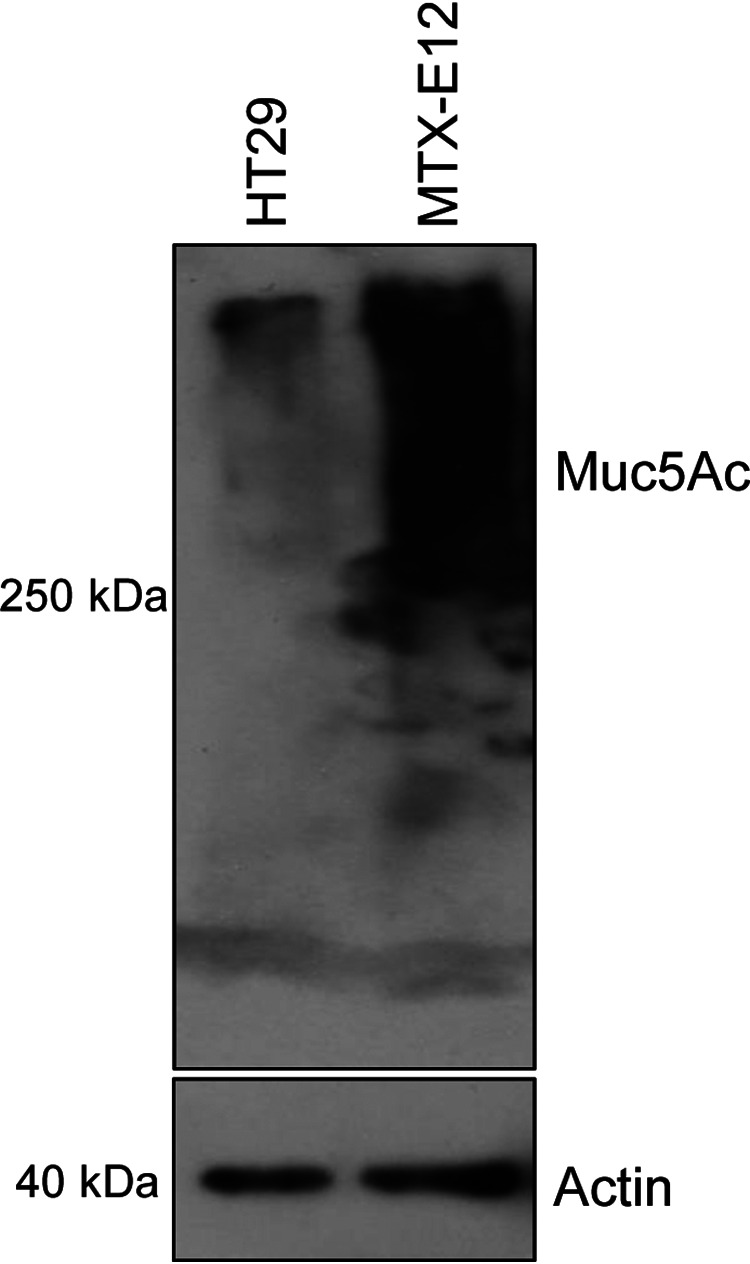
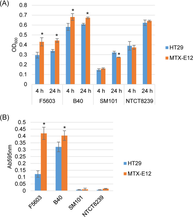
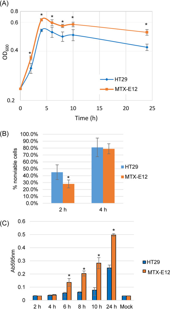
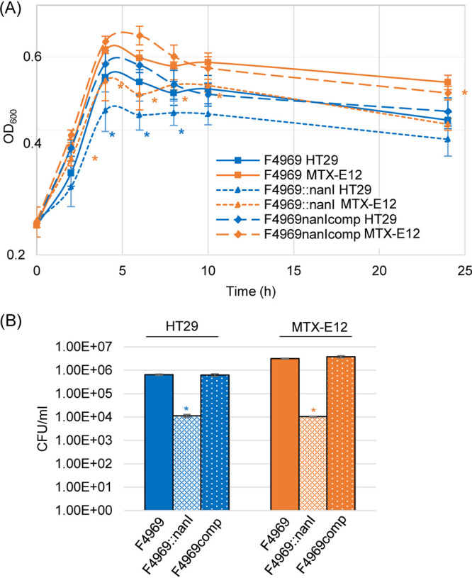
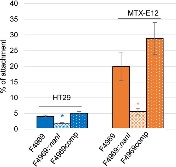
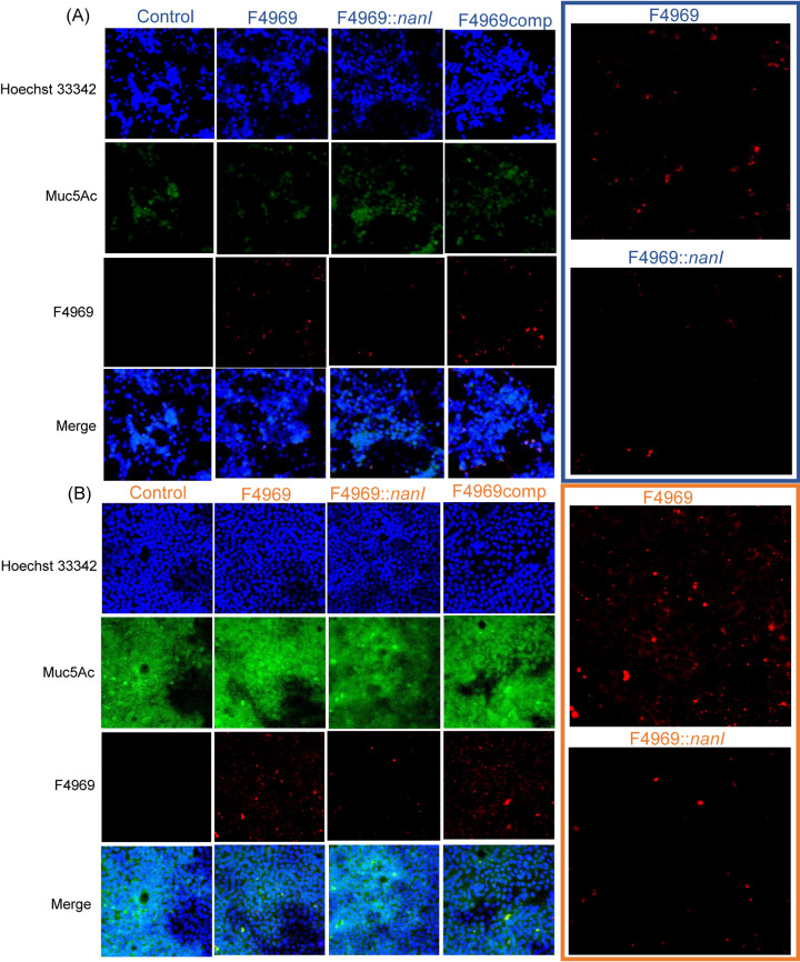
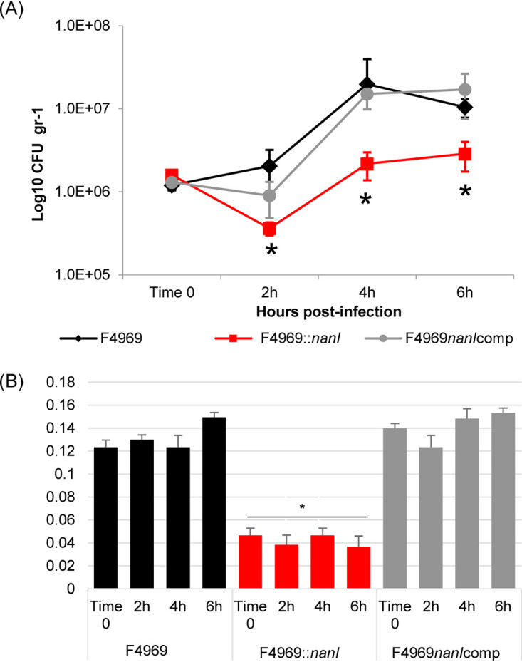
Clostridium perfringens type F strains causing nonfoodborne human gastrointestinal diseases (NFD) typically produce NanI sialidase as their major secreted sialidase. Type F NFDs can persist for several weeks, indicating their pathogenesis involves intestinal colonization, including vegetative cell growth and adherence, with subsequent sporulation that fosters enterotoxin production and release. We previously reported that NanI contributes to type F NFD strain adherence and growth using Caco-2 cells. However, Caco-2 cells make minimal amounts of mucus, which is significant because the intestines are coated with adherent mucus. Therefore, it was important to assess if NanI contributes to the growth and adherence of type F NFD strains in the presence of adherent mucus. Consequently, the current study first demonstrated greater growth of nanI-carrying versus non-nanI-carrying type F strains in the presence of HT29-MTX-E12 cells, which produce an adherent mucus layer, versus their parental HT29 cells, which make minimal mucus. Demonstrating the specific importance of NanI for this effect, type F NFD strain F4969 or a complementing strain grew and adhered better than an isogenic nanI null mutant in the presence of HT29-MTX-E12 cells versus HT29 cells. Those effects involved mucus production by HT29-MTX-E12 cells since mucus reduction using N-acetyl cysteine reduced F4969 growth and adherence. Consistent with those in vitro results, NanI contributed to growth of F4969 in the mouse small intestine. By demonstrating a growth and adherence role for NanI in the presence of adherent mucus, these results further support NanI as a potential virulence factor during type F NFDs.

Keywords: Clostridium perfringens; NanI sialidase; bacterial attachment; bacterial growth; intestinal disease; mucus.


Figures

FIG 1
FIG 1
Muc5Ac Western blot analysis for HT29 and MTX-E12 cell lines. (Top) A 50-µg protein aliquot of each cell lysate supernatant (see Materials and Methods) was separated on an SDS-containing 8% polyacrylamide gel. After transfer to a PVDF membrane, a Muc5Ac Western blot analysis was performed. (Bottom) The same amount of each protein sample was loaded onto an SDS-containing 12% polyacrylamide gel and probed with anti-beta actin antibody for use as a loading control. Size of proteins in kilodaltons is shown at left. The results shown are representative of three repetitions.
FIG 2
FIG 2
Comparison of C. perfringens type F strain growth in the presence of HT29 or MTX-E12 cells. (A) Postinoculation changes in the OD600 for type F strains B40, F5603, SM101, and NCTC8239 grown in PBS buffer and the presence of HT29 or MTX-E12 cells at 37°C for 4 h or 24 h. (B) Sialidase activity measured for 24-h cocultures using supernatants of samples from panel A. All panels show the mean values from three independent experiments. Error bars indicate the SD. *, P < 0.05 relative to culture with the HT29 cell line.
FIG 3
FIG 3
Comparison of growth, sialidase activity, and NanI activity levels when F4969 is cocultured in PBS buffer with the HT29 or MTX-E12 cell lines. (A) Postinoculation changes in the OD600 for cultures of wild-type F4969 growing in PBS buffer with HT29 or MTX-E12 cells at 37°C. Every 2 h up to 10 h of coculture, and then again at 24 h, a 0.5-ml aliquot of the coculture was removed, and the OD600 was determined. (B) HT29 and MTX-E12 cell cytotoxicity caused by coincubation with F4969 for 2-h and 4-h culture at 37°C. (C) F4969 sialidase activity measured every 2 h of coculture up to 10 h, and then at 24 h, using the supernatants of samples from panel A. All panels show the mean values from three independent experiments. Error bars indicate the SD. *, P < 0.05 relative to culture with HT29 cell line.
FIG 4
FIG 4
Sialidase activity, sialic acid release, and nanI gene expression of F4969, the F4969 nanI null mutant, and complementing strain when cocultured in PBS buffer with the HT29 or MTX-E12 cell lines. (A) Culture supernatant sialidase activity measured for 4, 8, or 24 h coculture of F4969 or its derivatives in PBS buffer and HT29 or MTX-E12 cells. Results shown are the average of three repetitions; error bars indicate the SD. Blue asterisks indicate a P value of <0.05 relative to nanI null mutant cultured with HT29 cell line. Orange asterisks indicate a P value of <0.05 relative to nanI null mutant cultured with MTX-E12 cell line. #, P < 0.05 relative to culture with HT29 cell line. (B) Sialic acid release by NanI when F4969 cocultures in PBS buffer with the HT29 or MTX-E12 cell lines at 6 h and 24 h. Sialic acid release due to NanI was calculated as the difference between the concentration of sialic acid released by coculture with F4969 versus the F4969 nanI null mutant strain. Shown is the mean of three repetitions. Error bars indicate SD. *, P < 0.05 relative to culture with HT29 cells. (C) Quantitative qRT-PCR analyses of nanI transcription was performed using 20 ng of F4969 RNA isolated from culture with HT29 or MTX-E12 cells for 4 h at 37°C. Average threshold cycle (CT) values were normalized to the value for the housekeeping 16S RNA gene, and the fold differences were calculated using the comparative CT (2−ΔΔCT) method. Shown is the mean of three repetitions. Error bars indicate SD. *, P < 0.05 relative to culture with HT29 cells.
FIG 5
FIG 5
Comparison of growth and levels of vegetative cell survival by F4969, its isogenic nanI null mutant, and a complemented strain cocultured in PBS with HT29 or MTX-E12 cell lines. (A) F4969, the nanI null mutant, and a complemented strain were grown to 24 h at 37°C in PBS buffer with the presence of HT29 or MTX-E12 cells. At designated time points, the OD600 of each culture was determined. (B) Quantification of viable vegetative cells of F4969, its nanI null mutant, and a complemented strain when grown in PBS with HT29 or MTX-E12 cells. After the bacteria were cocultured with the host cells for 24 h at 37°C, those cultures were then plated onto BHI agar plates with 10-fold serial dilutions using PBS buffer. These BHI agar cultures were grown anaerobically overnight at 37°C for colony counting (log10 scale). Shown are the mean values from three independent experiments. Error bars indicate the SD. Blue asterisks indicate a P value of <0.05 relative to wild type in PBS cultured with the HT29 cell line. Orange asterisks represent a P value of <0.05 relative to wild type in PBS cultured with MTX-E12 cell line.
FIG 6
FIG 6
C. perfringens vegetative cell adherence to HT29 and MTX-E12 cell lines. HT29 or MTX-E12 cell monolayers were incubated for 2 h with washed vegetative cells of F4969, its nanI null mutant, or the complementing strain at 37°C under anaerobic conditions. Monolayers were then washed three times with PBS. After collection, the adherent bacteria were plated onto BHI agar plates for counting. Adherence was expressed as the percentage of attached bacteria relative to the total number of input bacteria. All data showed the mean values from three independent experiments. Error bars indicate the SD. Blue asterisks indicate a P value of <0.05 relative to wild-type adherence to the HT29 cell line. Orange asterisks represent a P value of <0.05 relative to wild-type adherence to the MTX-E12 cell line.
FIG 7
FIG 7
Microscopic comparison of mucin production by the HT29 and MTX-E12 cell lines, as well as detection of C. perfringens attachment to HT29 (A) and MTX-E12 (B) cells. Attachment of C. perfringens F4969, its nanI null mutant, and complementing strain to HT29 or MTX-E12 cell lines as detected by Olympus confocal laser scanning biological microscope (FluoView FV1000) with FV10-ASW (version 1.4) software. All pictures were taken at a magnification of ×400. Red, C. perfringens; green, Muc5Ac; blue, mammalian cell nuclei. Panels at right show enlarged view of attached wild-type F4969 and nanI null mutant in panels at left.
FIG 8
FIG 8
Reduction of mucus in MTX-E12 cultures using a NAC pretreatment to reduce adherent mucus affects F4969 growth, sialidase production, and adherence. (A) Muc5Ac Western blot analysis performed on an SDS-containing 8% polyacrylamide gel for MTX-E12 cells and MTX-E12 cells pretreated with NAC (see Materials and Methods). Size of proteins in kilodaltons is shown on the left. The same amount of each protein sample was loaded onto a SDS-containing 12% polyacrylamide gel or Western blotted with an anti-beta actin antibody for a loading control. Size of proteins in kilodaltons is shown at right. The gel results shown are representative of three repetitions. (B) Postinoculation changes in the OD600 for cultures of wild-type F4969 growing in PBS buffer with MTX-E12 or NAC-pretreated MTX-E12 cells at 37°C for 24 h. A 0.5-ml aliquot of each culture was removed, and the OD600 was determined. (C) Sialidase activity measured for F4969 growing in PBS buffer with MTX-E12 or NAC-pretreated MTX-E12 cells at 37°C for 24 h. Sialidase activity was measured in culture supernatants of samples from panel B. (D) Attachment of MTX-E12 or NAC-pretreated MTX-E12 cell monolayers incubated for 2 h with washed vegetative cells of F4969 at 37°C under anaerobic conditions. Monolayers were then washed three times with PBS. After collection, the adherent bacteria were plated onto BHI agar plates for counting. All panels show the mean values from three independent experiments. The error bars indicate the SD. *, P < 0.05 relative to coculture with MTX-E12 cell line.
FIG 9
FIG 9
C. perfringens grows and produces sialidase in the intestines. (A) Shown are CFU per gram of intestinal tissue for F4969, its nanI null mutant, and the complementing strain as measured by plate counting at 2, 4, and 6 h postinfection in mouse small intestinal loops. (B) Sialidase activity present in supernatants of the intestinal contents of panel A loops at 2, 4, and 6 h postinfection. All data showed the mean values from 6 mice. Error bars indicate the SD. *, P < 0.05 relative to F4969 wild-type strain.

References

    1. Navarro MA, McClane BA, Uzal FA. 2018. Mechanisms of action and cell death associated with Clostridium perfringens toxins. Toxins (Basel) 10:212. 10.3390/toxins10050212. - DOI - PMC - PubMed
    1. McDonel JL. 1986. Toxins of Clostridium perfringens types A, B, C, D, and E, p 477–517. In Dorner F, Drews H (ed), Pharmacology of bacterial toxins, Pergamon Press, Oxford, UK.
    1. Uzal FA, Freedman JC, Shrestha A, Theoret JR, Garcia J, Awad MM, Adams V, Moore RJ, Rood JI, McClane BA. 2014. Towards an understanding of the role of Clostridium perfringens toxins in human and animal disease. Future Microbiol 9:361–377. 10.2217/fmb.13.168. - DOI - PMC - PubMed
    1. McClane BA, Uzal FA, Miyakawa MF, Lyerly D, Wilkins TD. 2006. The enterotoxic Clostridia, p 688–752. In Dworkin M, Falkow S, Rosenburg E, Schleifer H, Stackebrandt E (ed), The prokaryotes, 3rd ed, Springer Press, New York, NY.
    1. Rood JI, Adams V, Lacey J, Lyras D, McClane BA, Melville SB, Moore RJ, Popoff MR, Sarker MR, Songer JG, Uzal FA, Van Immerseel F. 2018. Expansion of the Clostridium perfringens toxin-based typing scheme. Anaerobe 53:5–10. 10.1016/j.anaerobe.2018.04.011. - DOI - PMC - PubMed

Publication types