Review
. 2021 Aug 31;12(4):e0140921.
doi: 10.1128/mBio.01409-21. Epub 2021 Aug 24.

Organelle Dynamics in Apicomplexan Parasites

Affiliations
Review

Organelle Dynamics in Apicomplexan Parasites

Julie M J Verhoef et al. mBio. .

Abstract

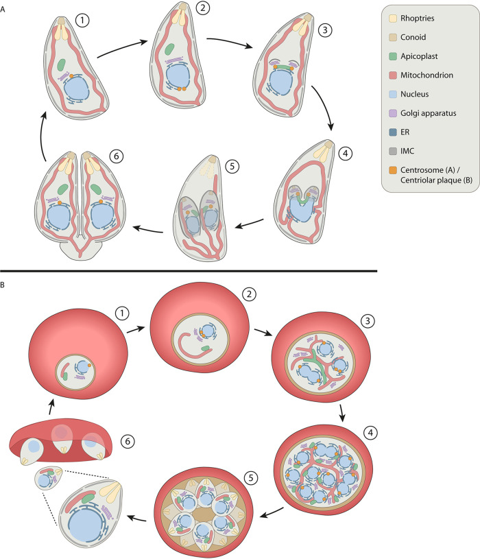
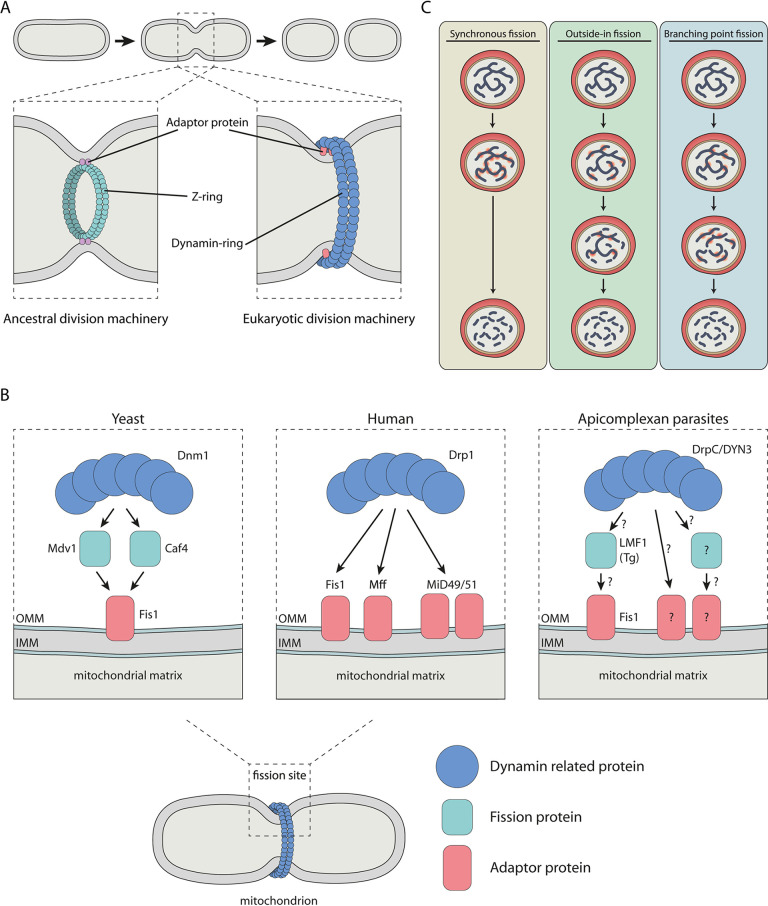
Apicomplexan parasites, such as Toxoplasma gondii and Plasmodium falciparum, are the cause of many important human and animal diseases. While T. gondii tachyzoites replicate through endodyogeny, during which two daughter cells are formed within the parental cell, P. falciparum replicates through schizogony, where up to 32 parasites are formed in a single infected red blood cell and even thousands of daughter cells during mosquito- or liver-stage development. These processes require a tightly orchestrated division and distribution over the daughter parasites of one-per-cell organelles such as the mitochondrion and apicoplast. Although proper organelle segregation is highly essential, the molecular mechanism and the key proteins involved remain largely unknown. In this review, we describe organelle dynamics during cell division in T. gondii and P. falciparum, summarize the current understanding of the molecular mechanisms underlying organelle fission in these parasites, and introduce candidate fission proteins.

Keywords: Plasmodium; Toxoplasma; dynamins; mitochondrion; organelle segregation.


Figures

FIG 1
FIG 1
Schematic overview of organelle morphology during endodyogeny in T. gondii and schizogony in P. falciparum. (A) Replication cycle of T. gondii tachyzoites. (1) Mature parasite. (2) Lateral elongation of the Golgi and migration and duplication of the centrosome at the basal site of the nucleus. (3) Centrosomes migrate back to the apical side of the nucleus and associate with the Golgi, which undergoes medial fission. The apicoplast also associates with the centrosomes and undergoes lateral extension. Budding is initiated with the formation of the IMC of daughter parasites. (4) Further formation of the IMC scaffold. Apicoplast remains associated with the centrosomes resulting in a U-shape. Nucleus and surrounding ER start to divide and enter the daughter parasites. (5) Fission of the apicoplast and nucleus with the ER. IMC scaffold encapsulates divided organelles. Extensions of the mitochondrion enter the daughter parasites. Degradation of parental secretory organelles and IMC. (6) Daughter parasites emerge, formation of the secretory organelles, establishment of the mitochondrial lasso, formation of the basal body. Only at the very last moment of division, mitochondria are separated at the basal end. (B) Asexual replication of P. falciparum in red blood cells. (1) Ring-stage parasite. (2) Elongation of the mitochondrion and division of the centriolar plaque (CP) and Golgi. ER forms extensions into the cytosol. (3) Further elongation and branching of the mitochondrion and apicoplast. Further replication of the Golgi and CP. Replication and expansion of the ER surrounding the dividing nuclei. (4) Apicoplast divides and associates with mitochondrial branches. Last round of nuclear division. (5) Mitochondrial division and formation of the daughter parasites. (6) Egress of merozoites from the red blood cell.
FIG 2
FIG 2
Schematic representations of organelle division mechanisms. (A) Endosymbiotic organelle division machineries. Endosymbiotic organelles are divided by the ancestral FtsZ-based division machinery where the Z-ring forms beneath the inner organelle membrane and/or the eukaryotic dynamin-based division machinery in which the dynamin ring forms at the cytosolic side of the outer organelle membrane. (B) Adaptor proteins recruit the mitochondrial division machinery in yeast, human, and apicomplexan parasites. In yeast, the membrane-anchored Fis1 recruits adaptor proteins Mdv1 and Caf4, which in turn recruit Dnm1 to form the constrictive ring. In human cells, multiple membrane-anchored adaptor proteins, including Fis1, Mff, and MiD49/51, are able to recruit Drp1 and form the division machinery. In apicomplexan parasites, the function of Fis1 in the recruitment of the division machinery is dispensable, indicating the existence of other essential adaptor proteins. Additionally, in T. gondii, LMF1 seems to bind to Fis1 and might be directly or indirectly involved in the recruitment of the division machinery. (C) Three possible scenarios for mitochondrial and apicoplast fission during schizogony. In the synchronous fission scenario, many fission points will occur simultaneously, resulting in an instant division of the organelle in daughter organelles. In the outside-in fission scenario, the fission points will be formed at the endings of the network-like organelle and daughter organelles will be formed by fission from the endings to the center. In the branching point fission scenario, fission points occur at the branching points of the organelle network, generating smaller fragments.

References

    1. Nilsson SK, Childs LM, Buckee C, Marti M. 2015. Targeting human transmission biology for malaria elimination. PLoS Pathog 11:e1004871. doi:10.1371/journal.ppat.1004871. - DOI - PMC - PubMed
    1. Francia ME, Striepen B. 2014. Cell division in apicomplexan parasites. Nat Rev Microbiol 12:125–136. doi:10.1038/nrmicro3184. - DOI - PubMed
    1. Rudlaff RM, Kraemer S, Marshman J, Dvorin JD. 2020. Three-dimensional ultrastructure of Plasmodium falciparum throughout cytokinesis. PLoS Pathog 16:e1008587. doi:10.1371/journal.ppat.1008587. - DOI - PMC - PubMed
    1. Vaishnava S, Morrison DP, Gaji RY, Murray JM, Entzeroth R, Howe DK, Striepen B. 2005. Plastid segregation and cell division in the apicomplexan parasite Sarcocystis neurona. J Cell Sci 118:3397–3407. doi:10.1242/jcs.02458. - DOI - PubMed
    1. Nishi M, Hu K, Murray JM, Roos DS. 2008. Organellar dynamics during the cell cycle of Toxoplasma gondii. J Cell Sci 121:1559–1568. doi:10.1242/jcs.021089. - DOI - PMC - PubMed

Publication types

Substances

LinkOut - more resources