. 2021 Oct;96(10):2561-2575.
doi: 10.1016/j.mayocp.2021.07.001. Epub 2021 Jul 15.

Acute Kidney Injury in Severe COVID-19 Has Similarities to Sepsis-Associated Kidney Injury: A Multi-Omics Study

Affiliations

Acute Kidney Injury in Severe COVID-19 Has Similarities to Sepsis-Associated Kidney Injury: A Multi-Omics Study

Mariam P Alexander et al. Mayo Clin Proc. 2021 Oct.

Abstract

Objective: To compare coronavirus disease 2019 (COVID-19) acute kidney injury (AKI) to sepsis-AKI (S-AKI). The morphology and transcriptomic and proteomic characteristics of autopsy kidneys were analyzed.

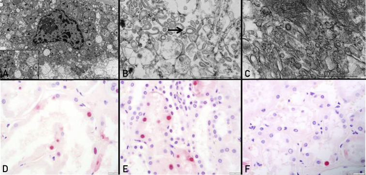
Patients and methods: Individuals 18 years of age and older who died from COVID-19 and had an autopsy performed at Mayo Clinic between April 2020 to October 2020 were included. Morphological evaluation of the kidneys of 17 individuals with COVID-19 was performed. In a subset of seven COVID-19 cases with postmortem interval of less than or equal to 20 hours, ultrastructural and molecular characteristics (targeted transcriptome and proteomics analyses of tubulointerstitium) were evaluated. Molecular characteristics were compared with archived cases of S-AKI and nonsepsis causes of AKI.

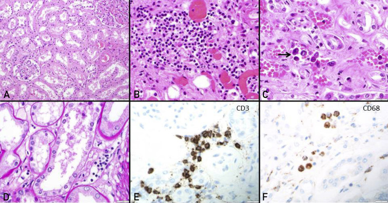
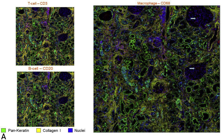
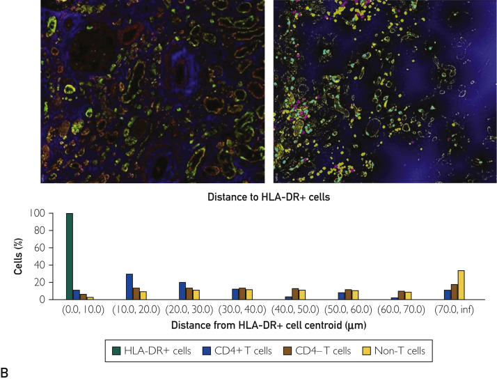
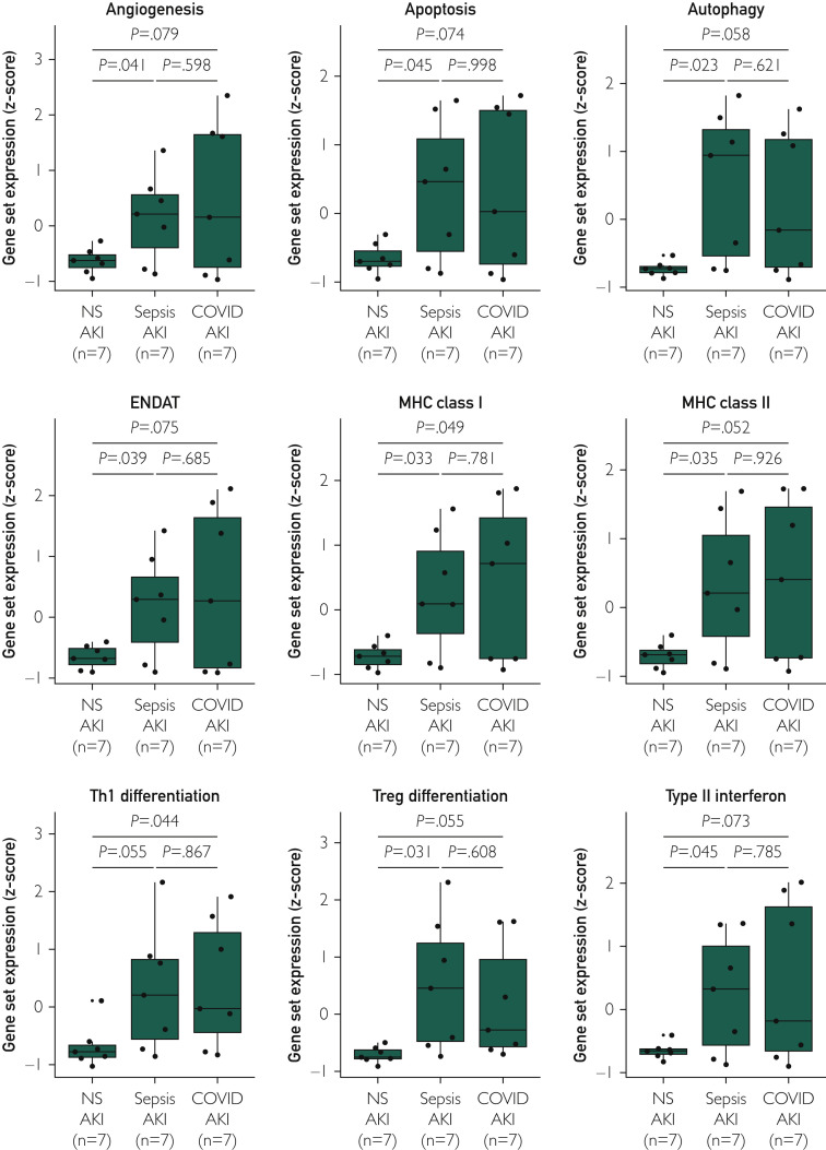
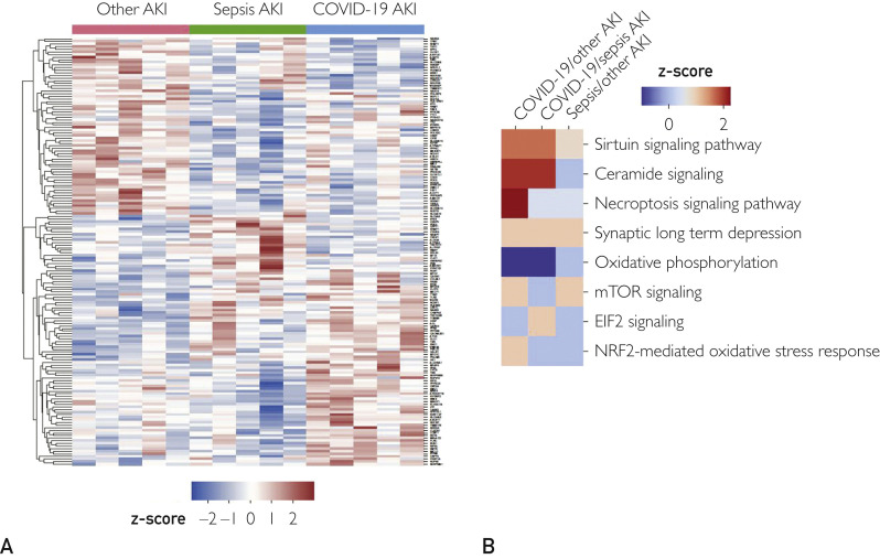
Results: The spectrum of COVID-19 renal pathology included macrophage-dominant microvascular inflammation (glomerulitis and peritubular capillaritis), vascular dysfunction (peritubular capillary congestion and endothelial injury), and tubular injury with ultrastructural evidence of mitochondrial damage. Investigation of the spatial architecture using a novel imaging mass cytometry revealed enrichment of CD3+CD4+ T cells in close proximity to antigen-presenting cells, and macrophage-enriched glomerular and interstitial infiltrates, suggesting an innate and adaptive immune tissue response. Coronavirus disease 2019 AKI and S-AKI, as compared to nonseptic AKI, had an enrichment of transcriptional pathways involved in inflammation (apoptosis, autophagy, major histocompatibility complex class I and II, and type 1 T helper cell differentiation). Proteomic pathway analysis showed that COVID-19 AKI and to a lesser extent S-AKI were enriched in necroptosis and sirtuin-signaling pathways, both involved in regulatory response to inflammation. Upregulation of the ceramide-signaling pathway and downregulation of oxidative phosphorylation in COVID-19 AKI were noted.

Conclusion: This data highlights the similarities between S-AKI and COVID-19 AKI and suggests that mitochondrial dysfunction may play a pivotal role in COVID-19 AKI. This data may allow the development of novel diagnostic and therapeutic targets.


Figures

Figure 1
Figure 1
Acute tubular injury and immune cell infiltration in coronavirus disease 2019 kidneys. (A) The renal cortex on light microscopy shows acute tubular injury with lumina ectasia (hematoxylin and eosin [HE] stain, original magnification ×20). (B) Mild mononuclear interstitial infiltration and peritubular capillary congestion is noted (HE stain, original magnification ×40). (C and D) Peritubular capillaritis (arrow) is a notable feature within congested capillaries (C). The microvascular inflammation is composed of CD3-positive T cells (E) and CD68-positive macrophages (F) (original magnification ×40 for E and F).
Figure 2
Figure 2
Evidence of tubular injury with mitochondrial insult and apoptosis in coronavirus disease 2019 kidneys. (A) Ultrastructural evaluation of proximal tubular epithelial cells show cytoplasmic vacuolization and swollen mitochondria with distorted cristae (see inset). (B) The mitochondrial injury includes ring- or donut-shaped mitochondria (arrow). (C) Clathrin-coated vesicles, easily mistaken for the virus, are also noted (arrow). Terminal deoxynucleotidyl transferase of dUTP (TUNEL) assay allows detection of TUNEL-positive cells (black arrow) in tubular epithelial cells of coronavirus disease 2019 kidneys (D), sepsis-associated acute kidney injury (E), and non-sepsis–related acute kidney injury (F).
Figure 3
Figure 3
Hyperion imaging mass cytometry showing simultaneous immunophenotyping of inflammatory cells and renal structural components with exploration of their spatial relation. (A) Presence of T cells (left, top) and macrophages (right) in the tubulointerstitial compartment with a conspicuous absence of B cells (left, bottom). Glomerulitis is appreciated in the right panel with margination of macrophages (arrows). All cells of interest have been tagged red. (B) Computational image neighborhood analysis was performed after labeling tubules (PanK and E-cadherin), intertubular stroma (collagen and alpha smooth muscle actin) and glomeruli (vimentin) (left). After cells were classified using a supervised approach (CD4+ T cells, CD4- T cells, and non-T cells), a distance map measuring the closest distance from the HLA-DR+ cells to each of the other cells was generated (middle, HLA-DR in yellow, distance from HLA-DR shown in increasing intensity of blue color). The percent of cells within each defined distance bin is displayed (right).
Figure 3
Figure 3
Hyperion imaging mass cytometry showing simultaneous immunophenotyping of inflammatory cells and renal structural components with exploration of their spatial relation. (A) Presence of T cells (left, top) and macrophages (right) in the tubulointerstitial compartment with a conspicuous absence of B cells (left, bottom). Glomerulitis is appreciated in the right panel with margination of macrophages (arrows). All cells of interest have been tagged red. (B) Computational image neighborhood analysis was performed after labeling tubules (PanK and E-cadherin), intertubular stroma (collagen and alpha smooth muscle actin) and glomeruli (vimentin) (left). After cells were classified using a supervised approach (CD4+ T cells, CD4- T cells, and non-T cells), a distance map measuring the closest distance from the HLA-DR+ cells to each of the other cells was generated (middle, HLA-DR in yellow, distance from HLA-DR shown in increasing intensity of blue color). The percent of cells within each defined distance bin is displayed (right).
Figure 4
Figure 4
Transcriptomic analysis. Box plots show the relative expression of select functional annotation gene sets between coronavirus disease 2019 (COVID-19) acute kidney injury (AKI), sepsis-related AKI (S-AKI) and nonsepsis-related AKI (NS-AKI). ENDAT, endothelial cell-associated transcripts; MHC, major histocompatibility complex; Th1, type 1 T helper cell; Treg, regulatory T cell.
Figure 5
Figure 5
Proteomic analysis. (A) The heat map gives a global overview of the differential expression of protein levels for each individual gene in the three acute kidney injury (AKI) cohorts. Proteins that were significantly over- (red) or under-expressed (blue) at P<.05 are shown. (B) The Ingenuity Pathway Analysis of differential protein expression in coronavirus disease 2019 (COVID-19) AKI, sepsis-associated AKI, and other AKI controls is shown. EIF2, Eukaryotic initiation factor 2; mTOR, mammalian target of rapamycin; NRF2, nuclear factor erythroid 2–related factor 2.

Similar articles

Cited by

References

    1. Hirsch J.S., Ng J.H., Ross D.W. Acute kidney injury in patients hospitalized with COVID-19. Kidney Int. 2020;98(1):209–218. - PMC - PubMed
    1. Pei G., Zhang Z., Peng J. Renal involvement and early prognosis in patients with COVID-19 pneumonia. J Am Soc Nephrol. 2020;31(6):1157–1165. - PMC - PubMed
    1. Cheng Y., Luo R., Wang K. Kidney disease is associated with in-hospital death of patients with COVID-19. Kidney Int. 2020;97(5):829–838. - PMC - PubMed
    1. Kudose S., Batal I., Santoriello D. Kidney biopsy findings in patients with COVID-19. J Am Soc Nephrol. 2020;31(9):1959–1968. - PMC - PubMed
    1. Su H., Yang M., Wan C. Renal histopathological analysis of 26 postmortem findings of patients with COVID-19 in China. Kidney Int. 2020;98(1):219–227. - PMC - PubMed

Publication types