. 2021 Oct 15:287:117283.
doi: 10.1016/j.envpol.2021.117283. Epub 2021 May 7.

Pharmacological inhibition of PAI-1 alleviates cardiopulmonary pathologies induced by exposure to air pollutants PM2.5

Affiliations

Pharmacological inhibition of PAI-1 alleviates cardiopulmonary pathologies induced by exposure to air pollutants PM2.5

Asish K Ghosh et al. Environ Pollut. .

Abstract

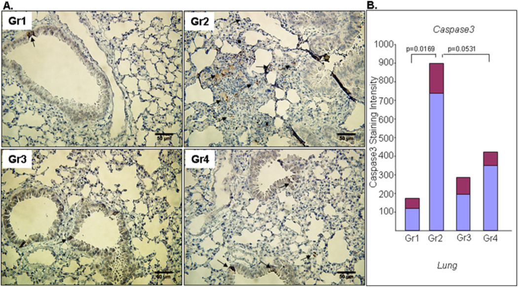
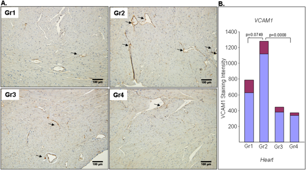
Numerous studies have established that acute or chronic exposure to environmental pollutants like particulate matter (PM) leads to the development of accelerated aging related pathologies including pulmonary and cardiovascular diseases, and thus air pollution is one of the major global threats to human health. Air pollutant particulate matter 2.5 (PM2.5)-induced cellular dysfunction impairs tissue homeostasis and causes vascular and cardiopulmonary damage. To test a hypothesis that elevated plasminogen activator inhibitor-1 (PAI-1) levels play a pivotal role in air pollutant-induced cardiopulmonary pathologies, we examined the efficacy of a drug-like novel inhibitor of PAI-1, TM5614, in treating PM2.5-induced vascular and cardiopulmonary pathologies. Results from biochemical, histological, and immunohistochemical studies revealed that PM2.5 increases the circulating levels of PAI-1 and thrombin and that TM5614 treatment completely abrogates these effects in plasma. PM2.5 significantly augments the levels of pro-inflammatory cytokine interleukin-6 (IL-6) in bronchoalveolar lavage fluid (BALF), and this also can be reversed by TM5614, indicating its efficacy in amelioration of PM2.5-induced increases in inflammatory and pro-thrombotic factors. TM5614 reduces PM2.5-induced increased levels of inflammatory markers cluster of differentiation 107 b (Mac3) and phospho-signal transducer and activator of transcription-3 (pSTAT3), adhesion molecule vascular cell adhesion molecule 1 (VCAM1), and apoptotic marker cleaved caspase 3. Longer exposure to PM2.5 induces pulmonary and cardiac thrombosis, but TM5614 significantly ameliorates PM2.5-induced vascular thrombosis. TM5614 also reduces PM2.5-induced increased blood pressure and heart weight. In vitro cell culture studies revealed that PM2.5 induces the levels of PAI-1, type I collagen, fibronectin (Millipore), and sterol regulatory element binding protein-1 and 2 (SREBP-1 and SREBP-2), transcription factors that mediate profibrogenic signaling, in cardiac fibroblasts. TM5614 abrogated that stimulation, indicating that it may block PM2.5-induced PAI-1 and profibrogenic signaling through suppression of SREBP-1 and 2. Furthermore, TM5614 blocked PM2.5-mediated suppression of nuclear factor erythroid related factor 2 (Nrf2), a major antioxidant regulator, in cardiac fibroblasts. Pharmacological inhibition of PAI-1 with TM5614 is a promising therapeutic approach to control air pollutant PM2.5-induced cardiopulmonary and vascular pathologies.

Keywords: Air pollutants; PAI-1; Particulate matter PM(2.5); TM5614; Vascular thrombosis.


Conflict of interest statement

Declaration of competing interest

The authors declare that they have no known competing financial interests or personal relationships that could have appeared to influence the work reported in this paper.

Figures

Fig. 1.
Fig. 1.. PAI-1 inhibitor TM5614 inhibits the levels of PM2.5-induced PAI-1 and TAT in plasma and IL-6 in BALF.
Plasma collected from 4 groups of mice (n = 4–8) was used in duplicate for PAI-1 and TAT assays. Day 1–6: TM5614 (10 mg/kg/day); Day 7: PM2.5 (50 μg/mouse) or PBS intratracheal instillation; Day 8: Plasma collected and processed for PAI-1 (A) or TAT (B) assay using an ELISA kit. (C) TAT levels in plasma from mice after 72 h treatment with PM2.5 (200 μg/mouse) and TM5614 (10 mg/kg/day) (n = 4–7). (D) IL-6 levels in BALF from mice after 24 h treatment with PM2.5 (50 μg/mouse) and TM5614 (10 mg/kg/day) were determined in duplicate using an ELISA kit (n = 7–8). Data presented as Mean (blue bar) ± SEM (red bar). Gr1: PBS; Gr2: PM2.5; Gr3: TM5614; Gr4: TM5614 + PM2.5.
Fig. 2.
Fig. 2.. Effect of PAI-1 inhibitor TM5614 on PM2.5-induced inflammation in lungs.
Lungs collected from 4 groups of mice (n = 4–6) were processed for immunohistochemistry using anti-pSTAT3 antibody (A,B) and antiMac3 antibody (C,D). Day 1–6: TM5614 (10 mg/kg/day); Day 7: PM2.5 (200 μg/mouse) or PBS instillation; Day 10: Lungs were collected and processed for immunohistochemistry. Representative images of pSTAT3 stained lung sections (A) and Mac3 stained lung sections (C) are shown. Images are reduced form of original 20X images. The levels of nuclear pSTAT3 in several fields of each lung section were determined by ImageJ software followed by statistical analysis. Quantitative data are shown in (B) for pSTAT3 and (D) for Mac3. Data presented as Mean (blue bar) ± SEM (red bar). Gr1: PBS; Gr2: PM2.5; Gr3: TM5614; Gr4: TM5614 + PM2.5. Arrows indicate the presence of pSTAT3 (A) and Mac3 (B) positive cells.
Fig. 3.
Fig. 3.. Effect of TM5614 on PM2.5-induced cellular apoptosis in lungs.
Lungs collected from 4 groups of mice (n = 4–6) were processed for immunohistochemistry using an anti-cleaved caspase 3 antibody. Mice were fed with TM5614 (10 mg/kg/day) for 6 days and on day 7, PM2.5 (200 μg/mouse) instillation or PBS was used as control. After 72 h, lungs were processed for immunohistochemistry. Representative images are reduced form of original 40X images (A). The levels of cleaved caspase 3 in several fields of each lung were determined by ImageJ software. Quantitative data are shown in (B). Data are presented as Mean (blue bar) ± SEM (red bar). Gr1: PBS; Gr2: PM2.5; Gr3: TM5614; Gr4: TM5614 + PM2.5. Arrows indicate the presence of cleaved caspase 3 positive cells.
Fig. 4.
Fig. 4.. TM5614 reduces PM2.5-induced adhesion molecule VCAM1 in lungs.
Lungs collected from 4 groups of mice (n = 4–6) were processed for immunohistochemistry using an anti-VCAM1 antibody. Day 1–6: TM5614 (10 mg/kg/day); Day 7: PM2.5 (200 μg/mouse) or PBS instillation; Day 10: Lungs were processed for immunohistochemistry. Representative images are reduced form of original 20X images (A). The levels of VCAM1 in several fields of each lung were determined by ImageJ. Quantitative data are shown (B). Data are presented as Mean (blue bar) ± SEM (red bar). Gr1: PBS; Gr2: PM2.5; Gr3: TM5614; Gr4: TM5614 + PM2.5. Arrows indicate the presence of VCAM1 positive sites.
Fig. 5.
Fig. 5.. Effect of PAI-1 inhibitor TM5614 on PM2.5-induced cellular apoptosis and inflammation in hearts.
Hearts collected from 4 groups of mice (n = 4–6) were processed for immunohistochemistry using an anti-cleaved caspase 3 antibody (A) and anti-Mac3 antibody (C). Day 1–6: TM5614 (10 mg/kg/day); Day 7: PM2.5 (200 μg/mouse) or PBS instillation. After 72 h, hearts were collected and processed for immunohistochemistry. Representative images are reduced form of original 40X images. The levels of cleaved caspase 3 and Mac3 in several fields of each heart section were determined by ImageJ. Quantitative data are shown in (B) for cleaved caspase 3 and (D) for Mac3. Data are presented as Mean (blue bar) ± SEM (red bar). Gr1: PBS; Gr2: PM2.5; Gr3: TM5614; Gr4: TM5614 + PM2.5. Arrows indicate the presence of cleaved caspase 3 (A) and Mac3 (C) positive cells.
Fig. 6.
Fig. 6.. TM5614 reduces PM2.5-induced adhesion molecule VCAM1 in hearts.
Hearts collected from 4 groups of mice (n = 4–6) as indicated were processed for immunohistochemistry using an anti-VCAM1 antibody. Day 1–6: TM5614 (10 mg/kg/day); Day 7: PM2.5 (200 μg/mouse) or PBS instillation; Day 10: Heart sections were processed for immunohistochemistry. Representative images are shown in (A). Images are reduced form of original 20X images. The levels of VCAM1 in several fields of each heart were determined by ImageJ software. Quantitative data are shown in (B). Data are presented as Mean (blue bar) ± SEM (red bar). Gr1: PBS; Gr2: PM2.5; Gr3: TM5614; Gr4: TM5614 + PM2.5. Arrows indicate the presence of VCAM1 positivity.
Fig. 7.
Fig. 7.. PM2.5 induces pulmonary thrombosis and PAI-1 inhibitor TM5614 prevents lung thrombogenesis.
Mice were divided into 4 groups (n = 4–7). In Group1, mice were fed with regular chow and received PBS. In Group 2, mice were fed with regular chow and received PM2.5 (100 μg/mouse/once per week for 4 weeks). In Group 3, mice were fed with chow containing TM5614 (10 mg/kg/day) for 6 days and then received PBS. In Group 4, mice were fed with chow containing TM5614 (10 mg/kg/day) for 6 days and then received PM2.5 (100 μg/mouse/once per week for 4 weeks). Representative image from each group are shown. Images are reduced form of original 20X images. An arrow indicates the presence of a thrombus.
Fig. 8.
Fig. 8.. TM5614 blocks PM2.5-induced cellular stress and profibrogenic responses.
Human cardiac fibroblasts (HCF) were cultured in 12-well clusters. After 24 h, media were replaced with 0.1% FBS containing DMEM for 3 h. Cells were pretreated with TM5614 (10 μM) or DMSO (vehicle control) for 2 h followed by treatment with PM2.5 (50 μg/ml) in triplicate for another 2 h. Supernatants and cell lysates were used for western blotting using antibodies as indicated (A). In (B) and (C), cells were pretreated with TM5614 (10 μM) or DMSO in triplicate. After 24 h, media were replaced with 0.1% FBS containing DMEM and treated with TM5614 (10 μM) or DMSO and PM2.5 (50 μg/ml) in triplicate for another 24 h. Supernatants and cell lysates were processed for western blotting using antibodies as indicated (B, C). Fbn: fibronectin; COL1: Type 1 collagen; α-Tub: α-Tubulin.
Fig. 9.
Fig. 9.. Possible pathological pathways driven by PM2.5-induced PAI-1 and the beneficial effect of PAI-1 inhibitor TM5614.
Air pollution stressors increase the levels of proinflammatory and prothrombotic mediators/regulators that cause cellular and vascular dysfunction and contribute to cardiopulmonary vascular pathologies. Neutralization of PM2.5-induced PAI-1 with TM5614 reduces PM2.5-induced cardiopulmonary vascular pathologies. In the depicted model, the individual step and feed-back loop are supported by the present study and or previous published works by other investigators. 1. Air-pollutant: PM2.5 increases the level of PAI-1 (present study and Upadhyay et al., 2010; Budinger et al., 2011); 2. PAI-1 increases inflammation and apoptosis (This study and Kubala et al., 2018); 3. Inflammation increases ROS (Mittal et al., 2014); 4. Inflammation induces thrombin (Margetic S. 2012, Foley and Conway, 2016); 5. Thombin induces the levels of ROS (Carrim et al., 2015); 6. Thrombin increases inflammation/apoptosis (Lopez et al., 2007; Chen and Dorling, 2009; Danckwardt et al., 2013; Foley and Conway, 2016); 7. Thrombin increases PAI-1 level (Hsieh et al., 2019); 8. Thrombin induces cellular and vascular abnormalities (present study and Rabiet et al., 1994); 9. Increased cellular dysfunction, elevation of thrombin-induced fibrinogen to fibrin deposition leads to cardiopulmonary pathologies, hypertension, thrombosis (present study and Savoia et al., 2011), 10. The results of the present biochemical, histological, immunohistological and cellular studies provide evidence on the pivotal role of air-pollutant PM2.5-induced PAI-1 in cardiopulmonary vascular pathologies, and the efficacy of a novel PAI-1 inhibitor TM5614 in improving air pollutant-induced cardiopulmonary vascular pathologies. Red upward arrow indicates induction of pathological factors by air-pollutants. Red downward arrow indicate downregulation of anti-pathological factor. Blue downward arrow indicates amelioration of pathological events by PAI-1 inhibitor TM5614. Blue upward arrow indicates upregulation of anti-pathological factor.

References

    1. Bourdrel T, Bind MA, Béjot Y, Morel O, Argacha JF, 2017. Cardiovascular effects of air pollution. Arch. Cardiovasc. Dis. 110, 634–642. - PMC - PubMed
    1. Brook RD, Franklin B, Cascio W, Hong Y, Howard G, Lipsett M, Luepker R, Mittleman M, Samet J, Smith SC Jr., Tager I, 2004. Expert panel on population and prevention science of the American heart association. Air pollution and cardiovascular disease: a statement for healthcare professionals from the expert panel on population and prevention science of the American heart association. Circulation 109, 2655–2671. - PubMed
    1. Brook RD, Rajagopalan S, 2009. Particulate matter, air pollution, and blood pressure. J Am Soc Hypertens 3, 332–350. - PubMed
    1. Brook RD, Rajagopalan S, Pope CA 3rd, Brook JR, Bhatnagar A, Diez-Roux AV, Holguin F, Hong Y, Luepker RV, Mittleman MA, Peters A, Siscovick D, Smith SC Jr., Whitsel L, Kaufman JD, 2010. American heart association council on epidemiology and prevention, council on the kidney in cardiovascular disease, and council on nutrition, physical activity and metabolism. Particulate matter air pollution and cardio-vascular disease: an update to the scientific statement from the American heart association. Circulation 121, 2331–2378. - PubMed
    1. Budinger GR, McKell JL, Urich D, Foiles N, Weiss I, Chiarella SE, Gonzalez A, Soberanes S, Ghio AJ, Nigdelioglu R, Mutlu EA, Radigan KA, Green D, Kwaan HC, Mutlu GM, 2011. Particulate matter-induced lung inflammation increases systemic levels of PAI-1 and activates coagulation through distinct mechanisms. PloS One 6, e18525. - PMC - PubMed