Review
. 2021 Oct 22;118(42):713-719.
doi: 10.3238/arztebl.m2021.0309.

Imaging of Prostate Cancer

Affiliations
Review

Imaging of Prostate Cancer

Heinz-Peter Schlemmer et al. Dtsch Arztebl Int. .

Abstract

Background: Prostate cancer is the most common type of solid tumor in men and the second most common cause of cancer-related death in males in Germany. The conventional strategy for its primary detection, i.e., systematic ultrasound-guided prostate biopsy in men who have elevated PSA levels and/or positive findings on digital rectal examination, fails to reveal all cases. The same is true of the use of conventional computed tomography (CT), magnetic resonance imaging (MRI), and skeletal scintigraphy for the early detection of recurrences and distant metastases.

Methods: This review is based on pertinent publications retrieved by a selective search, including the German clinical practice guideline on prostate cancer and systematic review articles.

Results: Prospective multicenter trials have shown that the detection of clinically significant prostate cancer is markedly improved with multiparametric MRI (mpMRI) and MR/TRUS fusion biopsy (TRUS = transrectal ultrasonography), compared to conventional systematic biopsy. A recent Cochrane review showed that the rate of overdiagnosis of low-risk prostate cancer was reduced with mpMRI and MR/TRUS fusion biopsy compared with conventional systematic biopsy (95/1000 vs. 139/1000), and that clinically significant prostate cancer was more reliably detected (sensitivity 72% vs. 63%), albeit with slightly lower specificity (96% vs. 100%). Prostate- specific membrane antigen (PSMA) hybrid imaging improves the detection of lymphogenic and bony metastases in patients with high-risk prostate cancer. PSMA hybrid imaging is most commonly used to detect biochemical recurrences. A metaanalysis showed that the detection rate depends on the PSA concentration: 74.1% overall, 33.7% with PSA <0.2 ng/mL, and 91.7% with PSA ≥= 2.0 ng/mL.

Conclusion: The appropriate use of mpMRI and MR/TRUS fusion biopsy improves the initial detection of prostate cancer as well as the assessment of the prognosis. PSMA hybrid imaging is useful for the staging of high-risk patients and for the detection of recurrences. These methods are now recommended in the German clinical practice guideline on prostate cancer as well as in guidelines from other countries.


Figures
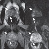
Figure 1
Figure 1
Multiparametric magnetic resonance tomography (mpMRI) of the prostate of a 77 year old patient with PSA 4.4 ng/mL, 43 ml prostate volume, PSA density 0.1 ng/mL2. No prior biopsy. Top row from left to right: axial T2 weighted sequence and diffusion weighted imaging DWI (b value 1500 s/mm2). Bottom row: dynamic contrast medium enhanced MRI (DCE) in the early arterial phase and apparent diffusion coefficient card. A 9 mm focus configured in an oval shape in the peripheral zone (PZ) posteromedially on the right in the center with notable diffusion impairment and early contrast medium accumulation (long arrow). Corresponds to a suspect PI-RADS 4 finding. Furthermore visible in the PZ are bilateral flat signal attentuations with only slight diffusion impairment and weak contrast medium accumulation, consistent with unspecific (post)inflammatory/regressive changes. A partially diffusion impaired nodule of benign prostatic hyperplasia (BHP) in the transitional zone (TZ) in the middle on the right indicates stromal components. Targeted MRI/TRUS fusion biopsy of the PI-RADS 4 lesion found in up to 60% of punch cylinders a clinically significant carcinoma of the prostate (Gleason 7a, 80 % Gleason pattern 3, 20 % Gleason pattern 4). The systematic biopsies also found a Gleason 6 prostate cancer in up to 15% of a punch cylinder specimen in the sextant located posteriorly to the right.
Figure 2
Figure 2
Real-time fusion of the transrectal ultrasounds of the prostate with multiparametric magnetic resonance tomography (mpMRI) imaging data in the context of MR-TRUS fusion biopsy, in a 51-year-old patient with a suspect lesion on mpMRI. On the morphological, T2 weighted MRI sequences, the contours of the prostate (purple) and the lesion detected on mpMRI (here in the peripheral zone, posterolateral, center left, green) are outlined and then transferred to the images from real-time sonography, or fused with these (left-sagittal aspect of the ultrasound). The fused images enable a precise biopsy of the lesions previously seen on mpMRI (marked in green) and the real-time documentation of the successful positioning of the biopsy needle in the target trajectory (left-yellow).
Figure 3
Figure 3
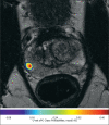
Illustration of the deep learning process: radiologically (not shown), a small 5 mm subcapsular focus was found in the peripheral zone (PZ), located posterolaterally to the right in the center with focal obvious diffusion impairment and small focal early contrast medium accumulation, consistent with a PI-RADS 4 finding. Furthermore, unspecific (post-)inflammatory/regressive changes were seen in the PZ. In the transitional zone left, center, an encapsulated BPH nodule appears (benign prostatic hyperplasia) with diffusion impairment. The image includes a T2-weighted axial MR image with an overlay of deep learning based tumor probability according to the color scale at the bottom. The automatic deep learning process detects the suspect PI-RADS 4 finding and quantifies a predicted tumor probability, in this case of up to a maximum of 43%. The targeted biopsy found a Gleason Grade Group 3 significant prostate carcinoma in 55% of the punch biopsies.
Figure 4
Figure 4
Ga-68-PSMA-PET/CT of a patient with prostate cancer (pT2c, pN1 L1 V0 R0, Gleason score 3+4=7). The primary treatment consisted of radical prostatectomy without any further therapies. A year after the surgery the patient’s PSA value started to rise slightly, from 0.08 ng/mL to 0.69 ng/mL. The latter value was followed by Ga-68-PSMA-PET/CT and pathological PSMA concentrations with a high contrast were seen in several para iliac lymph nodes bilaterally, in the sense of lymph node metastases of the prostate cancer. This was decisive for planning further treatment, as only the prostatic fossa is targeted by standard radiotherapy, whereas if lymph nodes are affected the lymph drainage channels in the little pelvis are included in the radiation field.

Comment in

  • Qualitative Data Are Lacking.
    Bruder W. Bruder W. Dtsch Arztebl Int. 2022 Apr 15;119(15):277. doi: 10.3238/arztebl.m2022.0114. Dtsch Arztebl Int. 2022. PMID: 35811351 Free PMC article. No abstract available.
  • In Reply.
    Krause BJ. Krause BJ. Dtsch Arztebl Int. 2022 Apr 15;119(15):277. doi: 10.3238/arztebl.m2022.0115. Dtsch Arztebl Int. 2022. PMID: 35811352 Free PMC article. No abstract available.

References

    1. Robert Koch-Institut e.V. GdeKiD. Krebs in Deutschland 2015/2016. www.krebsdaten.de/Krebs/DE/Content/Publikationen/Krebs_in_Deutschland/ki... (last accessed on 3 September 2021)
    1. Rider JR, Sandin F, Andren O, Wiklund P, Hugosson J, Stattin P. Long-term outcomes among noncuratively treated men according to prostate cancer risk category in a nationwide, population-based study. Eur Urol. 2013;63:88–96. - PubMed
    1. D‘Amico AV, Whittington R, Malkowicz SB, et al. Biochemical outcome after radical prostatectomy, external beam radiation therapy, or interstitial radiation therapy for clinically localized prostate cancer. JAMA. 1998;280:969–974. - PubMed
    1. Catalona WJ, Smith DS, Ratliff TL, et al. Measurement of prostate-specific antigen in serum as a screening test for prostate cancer. N Engl J Med. 1991;324:1156–1161. - PubMed
    1. Corcoran NM, Hong MK, Casey RG, et al. Upgrade in gleason score between prostate biopsies and pathology following radical prostatectomy significantly impacts upon the risk of biochemical recurrence. BJU Int. 2011;108(8) Pt 2 E202-10. - PubMed