. 2022 Mar 2;28(3):434-446.
doi: 10.1093/ibd/izab193.

Antitumor Necrosis Factor-like Ligand 1A Therapy Targets Tissue Inflammation and Fibrosis Pathways and Reduces Gut Pathobionts in Ulcerative Colitis

Affiliations

Antitumor Necrosis Factor-like Ligand 1A Therapy Targets Tissue Inflammation and Fibrosis Pathways and Reduces Gut Pathobionts in Ulcerative Colitis

Mina Hassan-Zahraee et al. Inflamm Bowel Dis. .

Abstract

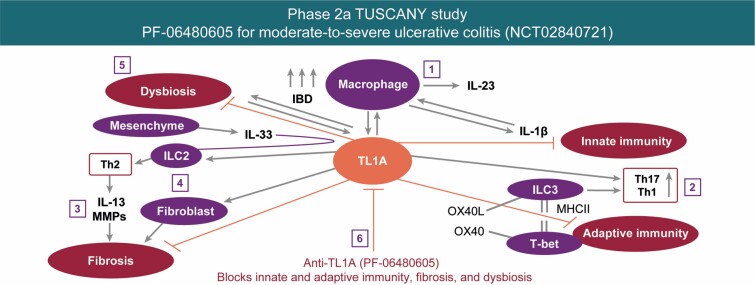
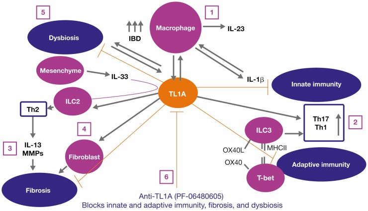
Background: The first-in-class treatment PF-06480605 targets the tumor necrosis factor-like ligand 1A (TL1A) molecule in humans. Results from the phase 2a TUSCANY trial highlighted the safety and efficacy of PF-06480605 in ulcerative colitis. Preclinical and in vitro models have identified a role for TL1A in both innate and adaptive immune responses, but the mechanisms underlying the efficacy of anti-TL1A treatment in inflammatory bowel disease (IBD) are not known.

Methods: Here, we provide analysis of tissue transcriptomic, peripheral blood proteomic, and fecal metagenomic data from the recently completed phase 2a TUSCANY trial and demonstrate endoscopic improvement post-treatment with PF-06480605 in participants with ulcerative colitis.

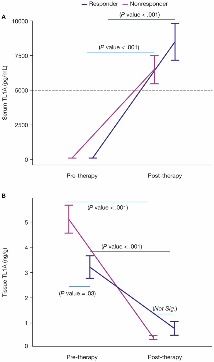
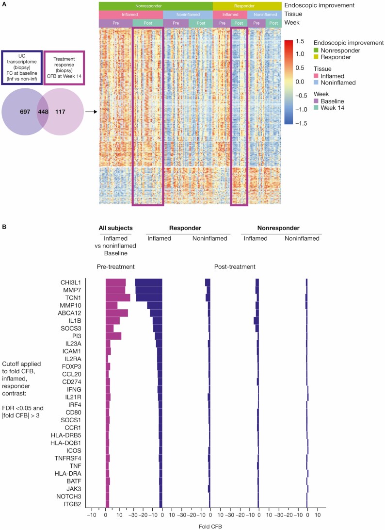
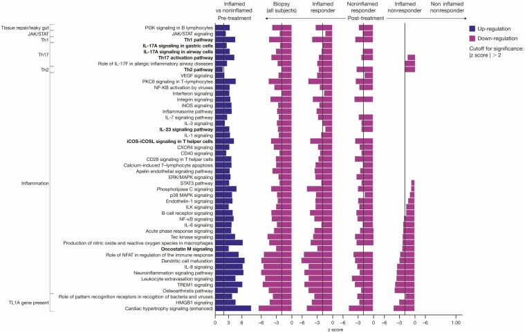
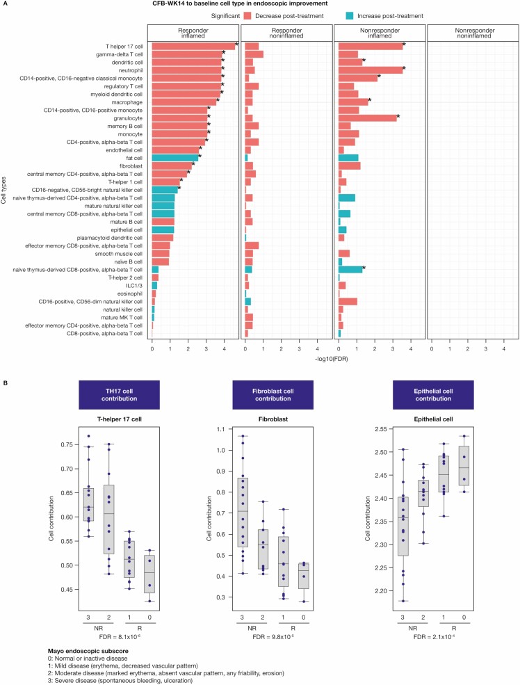
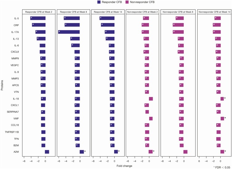
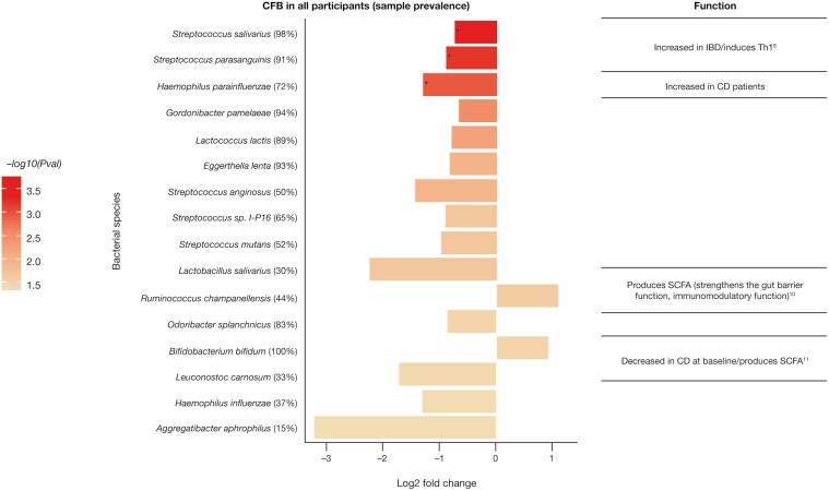
Results: Our results revealed robust TL1A target engagement in colonic tissue and a distinct colonic transcriptional response reflecting a reduction in inflammatory T helper 17 cell, macrophage, and fibrosis pathways in patients with endoscopic improvement. Proteomic analysis of peripheral blood revealed a corresponding decrease in inflammatory T-cell cytokines. Finally, microbiome analysis showed significant changes in IBD-associated pathobionts, Streptococcus salivarius, S. parasanguinis, and Haemophilus parainfluenzae post-therapy.

Conclusions: The ability of PF-06480605 to engage and inhibit colonic TL1A, targeting inflammatory T cell and fibrosis pathways, provides the first-in-human mechanistic data to guide anti-TL1A therapy for the treatment of IBD.

Keywords: TL1A inhibition; inflammatory bowel disease; transcriptional response; ulcerative colitis.


Conflict of interest statement

M.H.Z., Z.Y., L.X., M.L.B., X.L., C.L.H., J.Z., N.R., F.K., J.Q., D.Z., S.N., C.L., D.E.C., U.S., M.S.V., and K.E.H. are employees and stockholders of Pfizer Inc. J.R.A has received consultancy fees from Finch Therapeutics, Janssen, Pfizer Inc, Merck, and Takeda; has received grants from Merck; has served as a site Principal Investigator for Pfizer Inc for this trial. J.R. has no conflicts of interest to declare. E.J.S. has received grants from Abbott (AbbVie), AstraZeneca, Crohn’s and Colitis Foundation of America (CCFA), Celgene, Genentech, Janssen Research & Development, Johns Hopkins University, National Institute of Diabetes and Digestive and Kidney, National Institute of Health, New York Crohn’s Foundation, Pfizer Inc, Seres Therapeutics, UCB, and UCSF-CCFA Clinical Research Alliance; has received corporation consultant/advisory board fees from AbbVie, CCFA, Entera Health, Evidera, GI Health Foundation, Janssen, Protagonist Therapeutics, Seres Health, and Takeda Pharmaceuticals; holds stock in Gilead; has received honoraria from GI Health Foundation and Janssen for nonbranded speaker’s bureau. M.K. has received lecture fees from Alfa-Sigma, Ferring Pharmaceuticals, Janssen, Pharmabest, and Takeda; has received travel/accommodation/meeting expenses from Alfa-Sigma, Ferring Pharmaceuticals, Janssen, Pharmabest, Polish Foundation for Gastroenterology, and Takeda. S.D. has received consultancy fees from AbbVie, Allergan, Amgen, AstraZeneca, Biogen, Boehringer Ingelheim, Celgene, Celltrion, Ely Lilly, Enthera, Ferring Pharmaceuticals, Gilead, Hospira, Janssen, Johnson & Johnson, MSD, Mundipharma, Mylan, Pfizer Inc, Roche, Sandoz, Sublimity Therapeutics, Takeda, TiGenix, UCB, and Vifor. R.L. has received consultancy fees from Janssen, Pfizer Inc, and Takeda.

Figures

Graphical Abstract
Graphical Abstract
Figure 1.
Figure 1.
Anti-TL1A demonstrates target engagement in serum and tissue. A, Serum total TL1A levels were measured at baseline in endoscopic improvement responders (n = 18) and nonresponders (n = 32) and at week 14 in endoscopic improvement responders (n = 18) and nonresponders (n = 29). Serum total TL1A increased post-treatment in both endoscopic improvement responders (P < .001) and nonresponders (P < .001). Serum total TL1A was not significantly different between responders and nonresponders at both pre-treatment (P = .14) and post-treatment (P = .23). B, Tissue TL1A levels were measured at baseline in endoscopic improvement responders (n = 18) and nonresponders (n = 32) and at week 14 in endoscopic improvement responders (n = 16) and nonresponders (n = 30). Tissue TL1A decreased post-treatment in both endoscopic improvement responders (P < .001) and nonresponders (P < .001). Tissue TL1A was significantly different between responders and nonresponders pretreatment (P = .03) and was not significantly different post-treatment (P = 1). The responders and nonresponders with averages and standard errors were intentionally displayed side by side for both serum and tissue TL1A measured pre- and post-treatment to provide easy and clear comparison. Abbreviation: TL1A, tumor necrosis factor-like ligand 1A.
Figure 2.
Figure 2.
Anti-TL1A transcriptomics demonstrates colonic molecular disease resolution. A, Left: the UC transcriptome shown in purple represents the FC of inflamed vs noninflamed biopsies at baseline; a total of 1115 genes were identified. The anti-TL1A transcriptome shows the genes that are modulated post-treatment (light purple) and represents the CFB at week 14 in the biopsy; a total of 565 genes were identified. In total, 448 disease-specific anti-TL1A treatment-modulated genes were identified. The genes represented in the Venn diagram met a significant cutoff FDR < 0.05 and |FC| >2. Right: a heatmap of differential genes identified in the Venn diagram is presented in clusters by endoscopic improvement responders and nonresponders in inflamed and noninflamed biopsies pre- and post-treatment. B, Shows that mechanistic and cellular pathways impacting inflammation and fibrosis genes were associated with anti-TL1A treatment in tissue. Significant genes in mechanistic and cellular pathways that were modulated in endoscopic improvement inflamed biopsies post-treatment with anti-TL1A were identified by CFB with FDR <0.05 and |FC| >3. Abbreviations: CFB, change from baseline; FC, fold change; FDR, false discovery rate; IL, interleukin; TL1A, tumor necrosis factor-like ligand 1A; UC, ulcerative colitis.
Figure 3.
Figure 3.
The Th1, Th2, and IL-23 pathway downregulation was enriched in responders post-treatment with TL1A. Pathway analysis was performed using IPA with a significance cutoff |z score| >2. Abbreviations: IL, interleukin; IPA, Ingenuity Pathway Analysis; TL1A, tumor necrosis factor-like ligand 1A.
Figure 4.
Figure 4.
Deconvolution of biopsy transcriptomics identifies adaptive and innate cellular components after anti-TL1A treatment. A, Cell-type estimation of the CFB at week 14 in inflamed and noninflamed biopsies using bulk RNA sequencing in endoscopic improvement responders and nonresponders using the CytoReason deconvolution method. Cell types shown with (*) have an FDR <0.05. B, Strong correlation of lower Th17 and fibroblast cell-type estimates with better clinical outcomes measured by Mayo endoscopic subscore at week 14. Higher epithelial cell estimates also show a corresponding correlation. The FDR was based on a linear model of cell-type estimates at week 14 with endoscopic subscores and was adjusted for all 37 estimated cell types. All cell types deconvolved and analyzed in Figure 4 passed the cell signature quality assessment (Supplementary Table 1). Abbreviations: CFB, change from baseline; FDR, false discovery rate; ILC, innate lymphoid cell; TL1A, tumor necrosis factor-like ligand 1A.
Figure 5.
Figure 5.
Proteomics identified inflammatory blood biomarkers of endoscopic improvement. A total of 52 out of 63 proteins were evaluated and significant proteins were selected based on the CFB at week 14 in endoscopic improvement responders with FDR <0.05. All proteins with (*) have an FDR <0.05. Abbreviations: CFB, change from baseline; FDR, false discovery rate; IL, interleukin.
Figure 6.
Figure 6.
Reduction in the intestinal pathobiont post-treatment with anti-TL1A. Bacterial species identified that the CFB (P value ≤ .05), –log10(P value) = 1.3 in fecal samples in all participants. The top 3 bacterial species (*) are based on the threshold using FDR ≤0.05. Abbreviations: CFB, change from baseline; FDR, false discovery rate; TL1A, tumor necrosis factor-like ligand 1A.
Figure 7.
Figure 7.
Precision medicine proposed mechanism of action for PF-06480605. We hypothesize the following mechanisms of action for PF-06480605: (1) inflammatory MΦ are increased in IBD and produce IL-23, IL-1B, and TL1A. Interleukin-1B can feed back in an autocrine fashion to promote cytokine production. (2) TL1A stimulates pathogenic Th17 and mediates ILC3 and regulates Th1 through either OX40/OX40L or via the transitioning of ILC3 to ILC1 and the production of interferon gamma. (3) Additionally, TL1A and IL-33 regulate ILC2, which contributes to the Th2-driven IL-13 response, matrix metalloproteinase activation, tissue remodeling, and fibrosis. (4) TL1A stimulates fibroblast proliferation and contributes to fibrosis. (5) By blocking TL1A, we inhibit inflammation, inflammatory macrophage requirement, and fibrosis. Abbreviations: IBD, inflammatory bowel disease; IL, interleukin; ILC, innate lymphoid cell; Inf, inflamed; MΦ, macrophages; TL1A, tumor necrosis factor-like ligand 1A.

References

    1. Plichta DR, Graham DB, Subramanian S, Xavier RJ. Therapeutic opportunities in inflammatory bowel disease: mechanistic dissection of host-microbiome relationships. Cell. 2019;178:1041–1056. - PMC - PubMed
    1. Kashani A, Schwartz DA. The expanding role of anti-IL-12 and/or anti-IL-23 antibodies in the treatment of inflammatory bowel disease. Gastroenterol Hepatol (N Y). 2019;15:255–265. - PMC - PubMed
    1. Rubin DT, Ananthakrishnan AN, Siegel CA, et al. ACG clinical guideline: ulcerative colitis in adults. Am J Gastroenterol. 2019;114:384–413. - PubMed
    1. de Lange KM, Moutsianas L, Lee JC, et al. Genome-wide association study implicates immune activation of multiple integrin genes in inflammatory bowel disease. Nat Genet. 2017;49:256–261. - PMC - PubMed
    1. Siakavellas SI, Bamias G. Tumor necrosis factor-like cytokine TL1A and Its receptors DR3 and DcR3: important new factors in mucosal homeostasis and inflammation. Inflamm Bowel Dis. 2015;21:2441–2452. - PubMed

Publication types

MeSH terms

Substances